Chapter 11 Meiosis review

0.0(0)
Studied by 0 people
call kaiCall Kai
learnLearn
examPractice Test
spaced repetitionSpaced Repetition
heart puzzleMatch
flashcardsFlashcards
GameKnowt Play
Card Sorting

1/83

encourage image

There's no tags or description

Looks like no tags are added yet.

84 Terms

1
New cards

Meiosis overview

Meiosis is a specialized form of cell division that reduces a diploid cell (2n) into four genetically unique haploid gametes (n). It involves one round of DNA replication followed by two consecutive divisions—Meiosis I and Meiosis II—generating genetic diversity through crossing over and independent assortment

  • Sexual reproduction combines gametes from 2 parents through fertilization

  • Mixing chromosomes from two parents increases genetic diversity


<p><span>Meiosis is a specialized form of cell division that reduces a diploid cell </span>(2n) into four genetically unique haploid gametes (n). It involves one round of DNA replication followed by two consecutive divisions—Meiosis I and Meiosis II—generating genetic diversity through crossing over and independent assortment</p><ul><li><p>Sexual reproduction combines gametes from 2 parents through fertilization</p></li><li><p>Mixing chromosomes from two parents increases genetic diversity</p></li></ul><p></p>
2
New cards

The Process of Meiosis

  • Animals have two sets of chromosomes: one set from each parent: they are called diploid (2n)

  • Reducing chromosomes by half is essential to avoid doubling in every generation

  • Gametes (egg and sperm) have one set of chromosomes. They are called haploid cells (1n)


3
New cards

Gametes

A haploid (1n) reproductive or sex cell that contains only one set of chromosomes (half the number of a normal somatic/body cell) and fuses with another gamete during sexual reproduction to form a diploid zygote

  • Haploid (1n) In humans, gametes contain 23 chromosomes (compared to 46 in somatic cells)

  • Egg and sperm

  • Gametes (egg and sperm) have one set of chromosomes. They are called haploid cells (1n)


<p>A <strong>haploid (1n) reproductive or sex cell</strong> that contains only one set of chromosomes (half the number of a normal somatic/body cell) and fuses with another gamete during sexual reproduction to form a diploid zygote</p><ul><li><p>Haploid (1n) In humans, gametes contain <strong>23 chromosomes</strong> (compared to 46 in somatic cells)</p></li><li><p>Egg and sperm</p></li><li><p>Gametes (egg and sperm) have one set of chromosomes. They are called haploid cells (1n)</p></li></ul><p></p>
4
New cards

Meiosis reduces the number of chromosomes by one half

  • Reductional Division (Meiosis I): Homologous chromosome pairs separate from each other, cutting the number of unique chromosome sets in half

  • Equational Division (Meiosis II): Sister chromatids separate (similar to mitosis), producing a total of four haploid (n) daughter cells from one diploid (2n) parent cell

  • The process through two rounds of division: Meiosis I
    and Meiosis II

<ul><li><p><span><strong>Reductional Division (Meiosis I):</strong> Homologous chromosome pairs separate from each other, cutting the number of unique chromosome sets in half</span></p></li><li><p><span><strong>Equational Division (Meiosis II):</strong> Sister chromatids separate (similar to mitosis), producing a total of <strong>four haploid (n) daughter cells</strong> from one diploid (2n) parent cell</span></p></li><li><p><span>The process through two rounds of division: Meiosis I</span><br><span>and Meiosis II</span></p></li></ul><p></p>
5
New cards

Egg (ovum)

An egg (ovum) is a haploid female gamete produced through the process of oogenesis via meiosis

  • Haploid (n): Contains a single set of chromosomes (23) chromosomes in humans), half the number of a somatic diploid (2n) cell

  • Unlike spermatogenesis (which yields four functional sperm), meiosis in females is asymmetric. It produces one large, functional egg cell and non-functional polar bodies that eventually degrade, conserving cytoplasm and nutrients for the potential zygote


<p><span>An </span>egg (ovum) is a haploid female gamete produced through the process of oogenesis via meiosis</p><ul><li><p>Haploid (n): Contains a single set of chromosomes (23) chromosomes in humans), half the number of a somatic diploid (2n) cell</p></li><li><p>Unlike spermatogenesis (which yields four functional sperm), meiosis in females is asymmetric. It produces one large, functional egg cell and non-functional polar bodies that eventually degrade, conserving cytoplasm and nutrients for the potential zygote</p></li></ul><p></p>
6
New cards

Sperm

A haploid male gamete produced via meiosis that fuses with a female egg (ovum) during sexual reproduction to restore a diploid zygote

  • Haploid (n = 23 chromosomes in humans), carrying half the somatic cell genome.

  • Determines genetic sex in mammals: carries either an X chromosome (resulting in an XX female zygote) or a Y chromosome (resulting in an XY male zygote)

  • Contributes nuclear DNA to the zygote, while the egg contributes nuclear DNA, mitochondria, and most cytoplasmic organelle


7
New cards

Meiosis I

The first of two consecutive nuclear divisions in eukaryotic cell division. It is a reductional division that takes a diploid cell (with two sets of chromosomes) and separates homologous chromosome pairs to form two haploid daughter cells, each with half the original chromosome count

  • Chromosomes are replicated to make identical sister chromatids: DNA Replication (Interphase): Chromosomes copy their DNA to form two identical sister chromatids

  • Sister chromatids are attached at the centromere


<p>The first of two consecutive nuclear divisions in eukaryotic cell division. It is a reductional division that takes a diploid cell (with two sets of chromosomes) and separates homologous chromosome pairs to form two haploid daughter cells, each with half the original chromosome count</p><ul><li><p>Chromosomes are replicated to make identical sister chromatids: DNA Replication (Interphase): Chromosomes copy their DNA to form two identical sister chromatids</p></li><li><p>Sister chromatids are attached at the centromere</p></li></ul><p></p>
8
New cards

S phase

The second stage of interphase, occurring before meiosis I, during which the cell replicates its DNA

  • DNA Duplication: each single chromosome is copied to form two identical sister chromatids joined together at the centromere by cohesin proteins

  • Genetic Content Change: the DNA amount doubles (from (2C) to (4C), while the actual number of chromosomes remains the same (2n)

  • Centrosome Replication: the centrosomes (which organize the meiotic spindle) also duplicate during this time

  • Chromosomes are duplicated during interphase (S phase)

  • The resulting sister chromatids are held together at the centromere

  • The centrosomes are also duplicated


<p>The second stage of interphase, occurring before meiosis I, during which the cell replicates its DNA</p><ul><li><p><span><strong>DNA Duplication:</strong> each single chromosome is copied to form <strong>two identical sister chromatids</strong> joined together at the <strong>centromere</strong> by cohesin proteins</span></p></li><li><p><span><strong>Genetic Content Change:</strong> the DNA amount doubles (from (2C) to (4C), while the actual number of chromosomes remains the same (2n)</span></p></li><li><p><span><strong>Centrosome Replication:</strong> the centrosomes (which organize the meiotic spindle) also duplicate during this time</span></p></li><li><p>Chromosomes are duplicated during interphase (S phase)</p></li><li><p>The resulting sister chromatids are held together at the centromere </p></li><li><p>The centrosomes are also duplicated </p></li></ul><p></p>
9
New cards

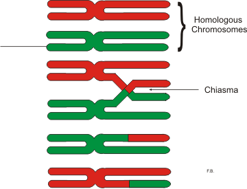
Chiasmata

The X-shaped regions where crossing over has occurred between non-sister chromatids of homologous chromosomes

  • They form specifically between non-sister chromatids belonging to a tetrad (a pair of homologous chromosomes)

  • They represent the exact sites where genetic recombination (crossing over) occurred, allowing for the exchange of genetic material between maternal and paternal chromosomes

  • Crossing over occurs at the chiasmata


<p>The X-shaped regions where crossing over has occurred between non-sister chromatids of homologous chromosomes</p><ul><li><p>They form specifically between non-sister chromatids belonging to a tetrad (a pair of homologous chromosomes)</p></li><li><p>They represent the exact sites where genetic recombination (crossing over) occurred, allowing for the exchange of genetic material between maternal and paternal chromosomes</p></li><li><p>Crossing over occurs at the chiasmata</p></li></ul><p></p>
10
New cards

Tetrad

A group of four sister chromatids formed when two homologous chromosomes pair up side-by-side during prophase I of meiosis

  • a group of four closely associated chromatids formed when two homologous chromosomes pair up during prophase I of meiosis

  • Each of the two homologous chromosomes has already replicated into two sister chromatids, creating a total of four chromatids

  • They form via a process called synapsis, held together by a protein structure known as the synaptonemal complex

  • Tetrads are essential for crossing over (recombination), where non-sister chromatids exchange segments of maternal and paternal DNA


<p>A group of four sister chromatids formed when two homologous chromosomes pair up side-by-side during prophase I of meiosis</p><ul><li><p>a group of four closely associated chromatids formed when two <span>homologous chromosomes</span> pair up during prophase I of meiosis</p></li><li><p>Each of the two homologous chromosomes has already replicated into two sister chromatids, creating a total of four chromatids</p></li><li><p>They form via a process called <strong>synapsis</strong>, held together by a protein structure known as the synaptonemal complex</p></li><li><p>Tetrads are essential for <strong>crossing over</strong> (recombination), where non-sister chromatids exchange segments of maternal and paternal DNA</p></li></ul><p></p>
11
New cards

Crossing Over

The exchange of genetic material between non-sister chromatids of homologous chromosomes during prophase I of meiosis

  • occurs during Meiosis I (specifically in Prophase I)

  • At points called chiasmata, where homologous chromosomes physically connect (synapsis)

  • Non-sister chromatids (one from the maternal homologue and one from the paternal homologue), not sister chromatids (which are identical copies attached at the centromere)

  • It creates recombinant chromosomes with new allele combinations, significantly increasing genetic diversity in gametes


<p>The exchange of genetic material between non-sister chromatids of homologous chromosomes during prophase I of meiosis</p><ul><li><p><span>occurs during </span>Meiosis I (specifically in Prophase I)</p></li></ul><ul><li><p><span>At points called chiasmata, where homologous chromosomes physically connect (synapsis) </span></p></li><li><p><span>Non-sister chromatids (one from the maternal homologue and one from the paternal homologue), <em>not</em> sister chromatids (which are identical copies attached at the centromere)</span></p></li><li><p>It creates recombinant chromosomes with new allele combinations, significantly increasing genetic diversity in gametes</p></li></ul><p></p>
12
New cards

Prophase I

The first and longest stage of meiosis I, where homologous chromosomes pair up and exchange genetic material to create genetic diversity

  • Chromosomes condense and the nuclear envelope fragments

  • Homologous chromosomes bind firmly together along their length, forming a tetrad

  • Chiasmata form between non-sister chromatids

  • Crossing over occurs at the chiasmata

  • Spindle fibers emerge from the centrosomes


<p>The first and longest stage of meiosis I, where homologous chromosomes pair up and exchange genetic material to create genetic diversity</p><ul><li><p>Chromosomes condense and the nuclear envelope fragments </p></li><li><p>Homologous chromosomes bind firmly together along their length, forming a tetrad</p></li><li><p>Chiasmata form between non-sister chromatids</p></li><li><p>Crossing over occurs at the chiasmata</p></li><li><p>Spindle fibers emerge from the centrosomes</p></li></ul><p></p>
13
New cards

Synapsis

The physical pairing and alignment of homologous chromosomes that occurs exclusively during prophase I of meiosis I

  • During this process, maternal and paternal versions of the same chromosome find each other and tightly zip together lengthwise. This precise alignment is physically held together by a protein lattice called the synaptonemal complex

  • When homologous chromosomes undergo synapsis, they form a group of four closely associated sister chromatids known as a tetrad (or bivalent)

  • Synapsis aligns matching genes directly opposite one another. This alignment is required for crossing over (genetic recombination) to take place


<p>The physical pairing and alignment of homologous chromosomes that occurs exclusively during prophase I of meiosis I</p><ul><li><p>During this process, maternal and paternal versions of the same chromosome find each other and tightly zip together lengthwise. This precise alignment is physically held together by a protein lattice called the synaptonemal complex</p></li><li><p>When homologous chromosomes undergo synapsis, they form a group of four closely associated sister chromatids known as a <strong>tetrad</strong> (or bivalent)</p></li><li><p>Synapsis aligns matching genes directly opposite one another. This alignment is required for <strong>crossing over</strong> (genetic recombination) to take place</p></li></ul><p></p>
14
New cards

Synapsis

The physical pairing and alignment of homologous chromosomes that occurs exclusively during prophase I of meiosis I

  • During this process, maternal and paternal versions of the same chromosome align precisely gene-for-gene. They are held tightly together by a zipper-like protein structure called the synaptonemal complex, forming a structure known as a tetrad (or bivalent)


<p>The physical pairing and alignment of homologous chromosomes that occurs exclusively during prophase I of meiosis I</p><ul><li><p>During this process, maternal and paternal versions of the same chromosome align precisely gene-for-gene. They are held tightly together by a zipper-like protein structure called the <strong>synaptonemal complex</strong>, forming a structure known as a <strong>tetrad</strong> (or bivalent)</p></li></ul><p></p>
15
New cards

Synaptonemal complex

A zipper-like protein structure that physically connects homologous chromosomes gene-by-gene during prophase I of meiosis I

  • Its primary function is to facilitate synapsis (the tight pairing of homologous chromosomes) and crossing over (homologous recombination), which allows non-sister chromatids to trade genetic material and create genetic diversity


<p>A zipper-like protein structure that physically connects homologous chromosomes gene-by-gene during prophase I of meiosis I</p><ul><li><p>Its primary function is to facilitate <strong>synapsis</strong> (the tight pairing of homologous chromosomes) and <strong>crossing over</strong> (homologous recombination), which allows non-sister chromatids to trade genetic material and create genetic diversity</p></li></ul><p></p>
16
New cards

Prometaphase I

The transitional phase of meiosis I that occurs between prophase I and metaphase

  • Homologous chromosomes are attached to spindle microtubules at the fused kinetochore shared by the sister chromatids

  • Chromosomes continue to condense

  • The nuclear envelope completely disappears


<p>The transitional phase of <strong>meiosis I</strong> that occurs between <strong>prophase I</strong> and <strong>metaphase</strong></p><ul><li><p>Homologous chromosomes are attached to spindle microtubules at the fused kinetochore shared by the sister chromatids</p></li><li><p>Chromosomes continue to condense</p></li><li><p>The nuclear envelope completely disappears</p></li></ul><p></p>
17
New cards

Metaphase I

The stage in meiosis I where homologous pairs of chromosomes (tetrads) align randomly along the equator (metaphase plate) of the cell, with spindle fibers attached to the kinetochores of each homolog

  • Homologous chromosomes randomly assemble at the metaphase plate, where they have been manuevered into place by the microtubules

  • The random orientation of each maternal and paternal pair along the equator is the physical basis of independent assortment, which drives genetic diversity in the resulting gametes

  • Homologous chromosome pairs (tetrads) line up randomly (indepdendant assortment) at the cell's equator (the metaphase plate)


<p>The stage in meiosis I where homologous pairs of chromosomes (tetrads) align randomly along the equator (metaphase plate) of the cell, with spindle fibers attached to the kinetochores of each homolog</p><ul><li><p>Homologous chromosomes randomly assemble at the metaphase plate, where they have been manuevered into place by the microtubules</p></li><li><p>The random orientation of each maternal and paternal pair along the equator is the physical basis of <strong>independent assortment</strong>, which drives genetic diversity in the resulting gametes</p></li><li><p>Homologous chromosome pairs (tetrads) line up randomly (indepdendant assortment) at the cell's equator (the metaphase plate) </p></li></ul><p></p>
18
New cards

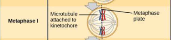
What lines up at the metaphase plate during metaphase I?

During metaphase I of meiosis, homologous pairs of chromosomes (also called tetrads or bivalents) line up along the metaphase plate (the equator of the cell)

  • HOMOLOGOUS CHROMOSOME PAIRS LINE UP AT THE METAPHASE PLATE SIDE-BY-SIDE

  • What lines up: paired homologous chromosomes (each made up of two sister chromatids), rather than individual chromosomes

  • How they align: They line up side-by-side or stacked as pairs along the equator

<p><span>During </span>metaphase I of meiosis, homologous pairs of chromosomes (also called tetrads or bivalents) line up along the metaphase plate (the equator of the cell)</p><ul><li><p>HOMOLOGOUS CHROMOSOME PAIRS LINE UP AT THE METAPHASE PLATE SIDE-BY-SIDE</p></li><li><p><span>What lines up: paired homologous chromosomes (each made up of two sister chromatids), rather than individual chromosomes</span></p></li><li><p><span>How they align: They line up side-by-side or stacked as pairs along the equator</span></p></li></ul><p></p>
19
New cards

What lines up at the metaphase plate during metaphase I?

During metaphase I of meiosis, homologous pairs of chromosomes (also known as tetrads or bivalents) line up along the metaphase plate (the equator of the cell)

  • What aligns: pairs of homologous chromosomes (each made up of two sister chromatids, for a total of four chromatids per tetrad) rather than individual chromosomes

  • In mitosis and meiosis II, individual replicated chromosomes line up in a single file line—not in homologous pairs


<p><span>During metaphase I of meiosis, </span>homologous pairs of chromosomes (also known as tetrads or bivalents) line up along the metaphase plate (the equator of the cell)</p><ul><li><p><strong>What aligns:</strong> pairs of homologous chromosomes (each made up of two sister chromatids, for a total of four chromatids per tetrad) rather than individual chromosomes</p></li><li><p>In mitosis and meiosis II, <em>individual</em> replicated chromosomes line up in a single file line—not in homologous pairs</p></li></ul><p></p>
20
New cards

Difference between metaphase I and II

The main difference is how chromosomes align along the metaphase plate and the ploidy (chromosome number) of the cell

Metaphase I

  • Homologous pairs (tetrads) line up side-by-side along the metaphase plate (equator of the cell

  • Spindle fibers attach to the kinetochores of each homologous chromosome (one from each parent)

  • Next Step (Anaphase I): Homologous pairs separate, while sister chromatids stay attached

Metaphase II

  • Individual chromosomes (single file) line up along the metaphase plate, which looks identical to metaphase in mitosis

  • Spindle fibers attach to individual kinetochores on opposite sides of each chromosome's centromere

  • Next Step (Anaphase II): Sister chromatids separate and move to opposite poles, just like in mitosis


<p>The main difference is how chromosomes align along the metaphase plate and the ploidy (chromosome number) of the cell</p><p>Metaphase I</p><ul><li><p><strong>Homologous pairs</strong> (tetrads) line up side-by-side along the metaphase plate (equator of the cell</p></li><li><p>Spindle fibers attach to the kinetochores of each homologous chromosome (one from each parent)</p></li><li><p><strong>Next Step (Anaphase I):</strong> Homologous pairs separate, while <strong>sister chromatids stay attached</strong></p></li></ul><p>Metaphase II</p><ul><li><p><strong>Individual chromosomes</strong> (single file) line up along the metaphase plate, which looks identical to metaphase in mitosis</p></li><li><p>Spindle fibers attach to individual kinetochores on opposite sides of each chromosome's centromere</p></li><li><p><strong>Next Step (Anaphase II):</strong> <strong>Sister chromatids separate</strong> and move to opposite poles, just like in mitosis</p></li></ul><p></p>
21
New cards

When does indepdendent assortment occur?

Independent assortment occurs during metaphase I of meiosis I

  • Random Orientation: during Metaphase I, homologous pairs of chromosomes line up in random, independent orientations along the metaphase plate (the middle of the cell)

  • This random alignment is a major contributor to genetic variation in sexual reproduction, along with crossing over (in Prophase I) and random fertilization

  • The 2^n Rule: The total number of possible chromosome combinations in the resulting gametes is calculated as 2^n, where n is the haploid number of chromosomes

  • 23 chromosomes = 2²³


22
New cards

Indepdendent assortment

The principle that the alleles of two or more different genes get sorted into gametes independently of one another. In other words, the allele a gamete receives for one gene does not influence the allele it receives for another gene

  • It applies to genes located on different chromosomes (or far apart on the same chromosome). Genes that are very close together are "linked" and do not assort independently

  • It is a major source of genetic variation in sexually reproducing organisms. For example, humans have 23 pairs of chromosomes, leading to over 8 million (2²³) possible chromosomal combinations just from independent assortment alone


<p>The principle that the alleles of two or more different genes get sorted into gametes independently of one another. In other words, the allele a gamete receives for one gene does not influence the allele it receives for another gene</p><ul><li><p><span>It applies to genes located on <em>different</em> chromosomes (or far apart on the same chromosome). Genes that are very close together are "linked" and do not assort independently</span></p></li><li><p>It is a major source of genetic variation in sexually reproducing organisms. For example, humans have 23 pairs of chromosomes, leading to over 8 million (2²³) possible chromosomal combinations just from independent assortment alone</p></li></ul><p></p>
23
New cards

Independent assortment

The principle that the alleles of two (or more) different genes get sorted into gametes independently of one another. In other words, the allele a gamete receives for one gene does not influence the allele it receives for another gene

  • It occurs during Metaphase I of meiosis, when homologous chromosome pairs line up randomly at the cell's equator (metaphase plate)

  • It is one of the primary drivers of genetic variation in sexually reproducing organisms (along with crossing over in Prophase I and random fertilization)


<p>The principle that the alleles of two (or more) different genes get sorted into gametes independently of one another. In other words, the allele a gamete receives for one gene does not influence the allele it receives for another gene</p><ul><li><p>It occurs during <strong>Metaphase I of meiosis</strong>, when homologous chromosome pairs line up randomly at the cell's equator (metaphase plate)</p></li><li><p>It is one of the primary drivers of genetic variation in sexually reproducing organisms (along with crossing over in Prophase I and random fertilization)</p></li></ul><p></p>
24
New cards

Anaphase I

The stage of meiosis I in which homologous chromosome pairs separate and move toward opposite poles of the cell, while sister chromatids remain attached to each other at their centromeres

  • Spindle microtubules pull the homologous chromosomes apart

  • The sister chromatids are still attached at the centromere

  • Sister chromatids remain joined by cohesin proteins at the centromere

  • Reduction division: this is the step that reduces the cell from diploid (2n) to haploid (1n) because each pole receives only one chromosome from each homologous pair


<p>The stage of meiosis I in which homologous chromosome pairs separate and move toward opposite poles of the cell, while sister chromatids remain attached to each other at their centromeres</p><ul><li><p>Spindle microtubules pull the homologous chromosomes apart</p></li><li><p>The sister chromatids are still attached at the centromere</p></li><li><p><span>Sister chromatids remain joined by cohesin proteins at the centromere</span></p></li><li><p><span><strong>Reduction division:</strong> this is the step that reduces the cell from diploid (2n) to haploid (1n) because each pole receives only one chromosome from each homologous pair</span></p></li></ul><p></p>
25
New cards

Telophase I and Cytokinesis

Mark the end of the first meiotic division, converting a single diploid cell into two haploid cells

  • Sister chromatids arrive at the poles of the cells and begin to decondense

  • A nuclear envelope forms around each nucleus, and the cytoplasm is divided by a cleavage furrow

  • This results in two haploid cells

  • Each cell contains one duplicated copy of each homologous chromosome pair


<p>Mark the end of the first meiotic division, converting a single diploid cell into two haploid cells</p><ul><li><p>Sister chromatids arrive at the poles of the cells and begin to decondense</p></li><li><p>A nuclear envelope forms around each nucleus, and the cytoplasm is divided by a cleavage furrow </p></li><li><p>This results in two haploid cells</p></li><li><p>Each cell contains one duplicated copy of each homologous chromosome pair </p></li></ul><p></p>
26
New cards

End result of Meiosis I

Two haploid (n) daughter cells, each with duplicated chromosomes (consisting of two sister chromatids held together at the centromere)

  • The chromosome number is cut in half. Starting from one diploid (2n) parent cell, meiosis I produces cells that are now haploid (n) because homologous chromosomes separated, not sister chromatids

  • Even though the cells are haploid regarding the number of unique chromosome types, each individual chromosome is still composed of two sister chromatids (meaning DNA has not yet split into single-chromatid strands

  • Because of crossing over (synapsis and exchange of genetic material in Prophase I) and independent assortment (random orientation and separation of maternal and paternal homologs at the metaphase plate), these two daughter cells are genetically unique from each other and from the original parent cell


<p><span>T</span>wo haploid (n) daughter cells, each with duplicated chromosomes (consisting of two sister chromatids held together at the centromere)</p><ul><li><p>The chromosome number is cut in half. Starting from one diploid (2n) parent cell, meiosis I produces cells that are now haploid (n) because <strong>homologous chromosomes</strong> separated, not sister chromatids</p></li><li><p>Even though the cells are haploid regarding the number of unique chromosome types, each individual chromosome is still composed of two sister chromatids (meaning DNA has not yet split into single-chromatid strands</p></li><li><p>Because of <strong>crossing over</strong> (synapsis and exchange of genetic material in Prophase I) and <strong>independent assortment</strong> (random orientation and separation of maternal and paternal homologs at the metaphase plate), these two daughter cells are genetically unique from each other and from the original parent cell</p></li></ul><p></p>
27
New cards

Prophase I

Homologous chromosomes pair. The synaptonemal complex holds them close together in synapsis

  • A pair of homologous chromosomes lined up next to each other during Prophase I of meiosis is called a tetrad or a bivalent

  • Non-sister chromatids of homologous chromosomes exchanged through crossing over (at the chiasmata, causing genetic diversity)

  • Crossing over occurs here


<p><span>Homologous chromosomes pair. The synaptonemal complex holds them close together in synapsis</span></p><ul><li><p><span>A pair of homologous chromosomes lined up next to each other during Prophase I of meiosis is called </span>a tetrad or a bivalent</p></li></ul><ul><li><p><strong>Non-sister chromatids of homologous chromosomes </strong><span>exchanged through crossing over (at the chiasmata, causing genetic diversity)</span></p></li><li><p>Crossing over occurs here</p></li></ul><p></p>
28
New cards

Prophase I structures

  • Structures at cross over points are called chiasmata

  • The four chromatids held together by chiasmata are called a tetrad

  • Chiasmata: the X-shaped regions where crossing over occurs between non-sister chromatids of homologous chromosomes during prophase I of meiosis

  • Tetrad: a group of four sister chromatids formed when two homologous chromosomes pair up side-by-side during prophase I of meiosis


<ul><li><p><span>Structures at cross over points are called chiasmata</span></p></li><li><p><span>The four chromatids held together by chiasmata are called a tetrad</span></p></li><li><p>Chiasmata: the X-shaped regions where <strong>crossing over</strong> occurs between non-sister chromatids of homologous chromosomes during prophase I of meiosis</p></li><li><p>Tetrad: a group of <strong>four sister chromatids</strong> formed when two homologous chromosomes pair up side-by-side during <strong>prophase I of meiosis</strong></p></li></ul><p></p>
29
New cards

Synapsis

The pairing of homologous chromosomes during prophase I of meiosis. This precise alignment allows for crossing over, where homologous chromosomes exchange genetic material to create genetic diversity

  • the lengthwise pairing of homologous chromosomes that occurs exclusively during prophase I of meiosis

  • Homologous chromosomes (one maternal and one paternal) physically align gene-for-gene. They are held tightly together by a protein structure called the synaptonemal complex


<p>The pairing of homologous chromosomes during prophase I of meiosis. This precise alignment allows for crossing over, where homologous chromosomes exchange genetic material to create genetic diversity</p><ul><li><p>the <strong>lengthwise pairing of homologous chromosomes</strong> that occurs exclusively during <strong>prophase I of meiosis</strong></p></li><li><p>Homologous chromosomes (one maternal and one paternal) physically align gene-for-gene. They are held tightly together by a protein structure called the <strong>synaptonemal complex</strong></p></li></ul><p></p>
30
New cards

Synaptonemal complex

A zipper-like lattice of proteins that physically connects homologous chromosomes gene-for-gene during prophase I of meiosis

  • Facilitates Synapsis: It acts like a molecular zipper to bind maternal and paternal homologous chromosomes tightly together into a structure called a tetrad (or bivalent)


<p>A zipper-like lattice of proteins that physically connects homologous chromosomes gene-for-gene during prophase I of meiosis</p><ul><li><p><strong>Facilitates Synapsis:</strong> It acts like a molecular zipper to bind maternal and paternal homologous chromosomes tightly together into a structure called a <strong>tetrad</strong> (or bivalent)</p></li></ul><p></p>
31
New cards

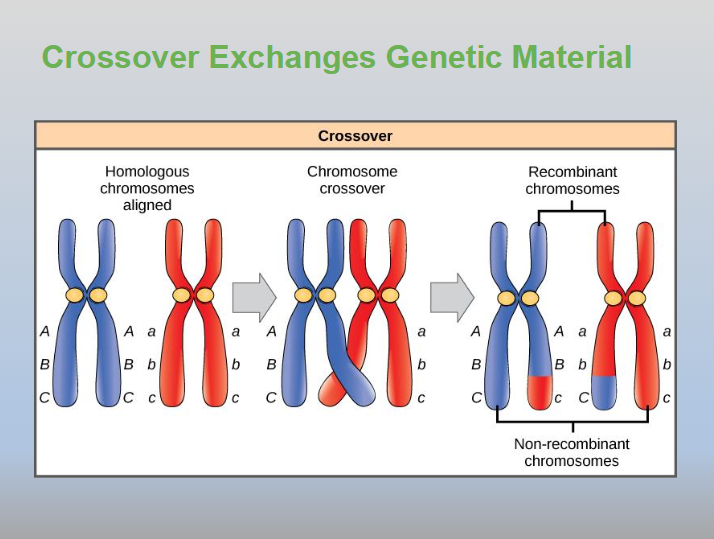
Crossover

Or chromosomal crossover, is defined as the exchange of genetic material between non-sister chromatids of homologous chromosomes

  • It takes place exclusively during prophase I of meiosis I.

  • The physical points where the chromatids overlap and intertwine are called chiasmata

  • Homologous chromosomes pair up gene-by-gene in a process called synapsis, forming a four-chromatid structure known as a tetrad

  • Crossover exchanges genetic material, increasing genetic diversity


<p>Or chromosomal crossover, is defined as the exchange of genetic material between non-sister chromatids of homologous chromosomes</p><ul><li><p>It takes place exclusively during <strong>prophase I of meiosis I</strong>.</p></li><li><p>The physical points where the chromatids overlap and intertwine are called <strong>chiasmata</strong></p></li><li><p>Homologous chromosomes pair up gene-by-gene in a process called <strong>synapsis</strong>, forming a four-chromatid structure known as a <strong>tetrad</strong></p></li><li><p>Crossover exchanges genetic material, increasing genetic diversity</p></li></ul><p></p>
32
New cards

Recombinant chromosomes

A chromosome that carries a new combination of alleles created when genetic material is exchanged between non-sister chromatids of homologous chromosomes

  • The mechanism: Crossing over (homologous chromosomes pair up as a tetrad and exchange matching segments of DNA at points called chiasmata)

  • Parental chromosomes: Keep the original combination of alleles inherited from the organism's mother or father

  • Recombinant chromosomes: Contain a mix of maternal and paternal alleles on a single chromatid


<p>A chromosome that carries a new combination of alleles created when genetic material is exchanged between non-sister chromatids of homologous chromosomes</p><ul><li><p><strong>The mechanism:</strong> <strong>Crossing over</strong> (homologous chromosomes pair up as a tetrad and exchange matching segments of DNA at points called chiasmata)</p></li></ul><ul><li><p><span><strong>Parental chromosomes:</strong> Keep the original combination of alleles inherited from the organism's mother or father</span></p></li><li><p><span><strong>Recombinant chromosomes:</strong> Contain a mix of maternal and paternal alleles on a single chromatid</span></p></li></ul><p></p>
33
New cards

Non-recombinant chromosome

Also called a parental-type chromosome - is a chromosome that does not undergo any exchange of genetic material via crossing over during meiosis

  • Do not participate in crossing over (unaffected by a chiasma)

  • retains the exact combination of alleles (genes) that was originally inherited from one of the individual's parents

<p>Also called a <strong>parental-type chromosome</strong> - is a chromosome that does not undergo any exchange of genetic material via crossing over during meiosis</p><ul><li><p>Do not participate in crossing over (unaffected by a chiasma)</p></li><li><p>retains the exact combination of alleles (genes) that was originally inherited from one of the individual's parents</p></li></ul><p></p>
34
New cards

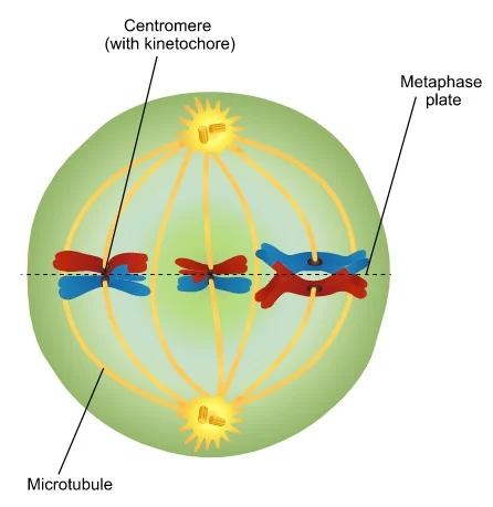
Metaphase I

  • Homologous chromosomes are arranged at the cell equator with kinetochores facing opposite poles

  • Maternal and paternal chromatids orient randomly and are mixed when they migrate to the poles

  • This creates additional genetic variation among daughter cells

  • Indepdendent assortment occurs here

  • The homologous pairs (one chromosome from the mother and one from the father) align side-by-side or in pairs along the cell's equator (the metaphase plate), rather than single file

<ul><li><p>Homologous chromosomes are arranged at the cell equator with kinetochores facing opposite poles</p></li><li><p>Maternal and paternal chromatids orient randomly and are mixed when they migrate to the poles</p></li><li><p>This creates additional genetic variation among daughter cells</p></li><li><p>Indepdendent assortment occurs here</p></li><li><p>The homologous pairs (one chromosome from the mother and one from the father) align side-by-side or in pairs along the cell's equator (the metaphase plate), rather than single file</p></li></ul><p></p>
35
New cards

Anaphase I

The stage of meiosis I in which paired homologous chromosomes separate and move toward opposite poles of the cell, while sister chromatids remain attached to each other at their centromeres

  • The microtubules pull tetrads apart

  • Chiasmata are broken but sister chromatids remain attached at centromere

  • This phase reduces the chromosome number from diploid (2n) to haploid (n)

  • Unlike mitosis or anaphase II where sister chromatids split, anaphase I separates whole homologous pairs

  • Nondisjunction can occur here


<p>The stage of meiosis I in which paired <strong>homologous chromosomes</strong> separate and move toward opposite poles of the cell, while <strong>sister chromatids</strong> remain attached to each other at their centromeres</p><ul><li><p><span>The microtubules pull tetrads apart</span></p></li><li><p><span>Chiasmata are broken but sister chromatids remain attached at centromere</span></p></li><li><p><span>This phase reduces the chromosome number from <strong>diploid (2n)</strong> to <strong>haploid (n)</strong></span></p></li><li><p><span>Unlike mitosis or anaphase II where sister chromatids split, anaphase I separates whole homologous pairs</span></p></li><li><p>Nondisjunction can occur here</p></li></ul><p></p>
36
New cards

Nondisjunction

The failure of homologous chromosomes (during Meiosis I) or sister chromatids (during Meiosis II or mitosis) to separate properly during anaphase

  • Improper separation leads to daughter cells with abnormal chromosome numbers

  • Meiosis I Error: Homologous chromosomes fail to separate, resulting in 100% abnormal gametes

  • Meiosis II Error: Sister chromatids fail to separate, resulting in 50% abnormal gametes and 50% normal gametes


<p>The failure of <strong>homologous chromosomes</strong> (during Meiosis I) or <strong>sister chromatids</strong> (during Meiosis II or mitosis) to separate properly during anaphase</p><ul><li><p><span>Improper separation leads to daughter cells with abnormal chromosome numbers</span></p></li><li><p>Meiosis I Error: Homologous chromosomes fail to separate, resulting in 100% abnormal gametes</p></li><li><p>Meiosis II Error: Sister chromatids fail to separate, resulting in 50% abnormal gametes and 50% normal gametes</p></li></ul><p></p>
37
New cards

When can nondisjunction occur?

Nondisjunction can occur during anaphase in meiosis I, meiosis II, or mitosis when chromosomes or sister chromatids fail to separate properly

  • the failure of homologous chromosomes (during meiosis I) or sister chromatids (during meiosis II or mitosis) to separate properly during cell division

  • Trisomy: three copies of a chromosome (down syndrome/trisomy 21)

  • Monosomy: only one copy of a chromosome (Turner syndrome)


38
New cards

Telophase I and Cytokinesis

In meiosis I mark the final stage of the first reductional division, where chromosomes arrive at opposite poles of the cell, nuclear envelopes may reform, and the cytoplasm splits to produce two haploid daughter cells

  • Separated chromosomes arrive at opposite poles

  • Cytokinesis is separation of cytoplasmic contents into daughter cells and the nuclei reform

  • The cell's cytoplasm divides (via a cleavage furrow in animal cells or a cell plate in plant cells), physically separating the single cell into two distinct entities

  • Two haploid (n) daughter cells are formed. Crucially, each chromosome still consists of two sister chromatids, meaning the genetic material is not yet reduced to single-chromatid state (that happens later in Meiosis II)


<p>In meiosis I mark the final stage of the first reductional division, where chromosomes arrive at opposite poles of the cell, nuclear envelopes may reform, and the cytoplasm splits to produce <strong>two haploid daughter cells</strong></p><ul><li><p>Separated chromosomes arrive at opposite poles</p></li><li><p>Cytokinesis is separation of cytoplasmic contents into daughter cells and the nuclei reform</p></li><li><p>The cell's cytoplasm divides (via a cleavage furrow in animal cells or a cell plate in plant cells), physically separating the single cell into two distinct entities</p></li><li><p>Two haploid (n) daughter cells are formed. Crucially, each chromosome still consists of two sister chromatids, meaning the genetic material is <strong>not</strong> yet reduced to single-chromatid state (that happens later in Meiosis II)</p></li></ul><p></p>
39
New cards

End result of Meiosis I

At the end of Meiosis I, the single diploid (2n) parent cell has divided into two haploid (n) daughter cells

<p><span>At the end of </span><strong>Meiosis I</strong>, the single diploid (2n) parent cell has divided into <strong>two haploid (n) daughter cells</strong></p>
40
New cards

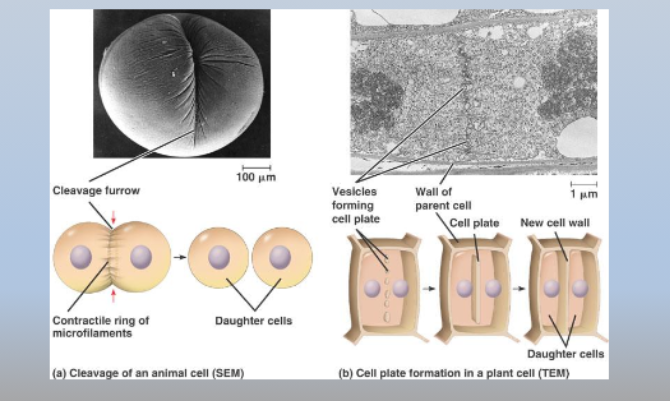
Cytokinesis in plants vs. animals

  • Cytokinesis in animal occurs via a cleavage furrow that
    constricts

  • Plant cells form a cell plate that ultimately produces cell
    walls that separate the daughter cells

<ul><li><p><span>Cytokinesis in animal occurs via a cleavage furrow that<br>constricts</span></p></li><li><p><span>Plant cells form a cell plate that ultimately produces cell<br>walls that separate the daughter cells</span></p></li></ul><p></p>
41
New cards

Cytokinesis in plant cells

The physical division of the cytoplasm that follows nuclear division (telophase I and telophase II), using a cell plate mechanism due to the plant cell's rigid cell wall

  • Plant cells form a cell plate that ultimately produces cell
    walls that separate the daughter cells


<p>The physical division of the cytoplasm that follows nuclear division (telophase I and telophase II), using a <strong>cell plate</strong> mechanism due to the plant cell's rigid cell wall</p><ul><li><p>Plant cells form a cell plate that ultimately produces cell<br>walls that separate the daughter cells</p></li></ul><p></p>
42
New cards

Cytokinesis in animal cells

The physical splitting of the cytoplasm that divides one cell into two distinct daughter cells. It occurs twice during meiosis (after Telophase I and after Telophase II)

  • Contractile Ring: a ring of microfilaments made of actin and myosin proteins forms just beneath the plasma membrane around the cell's equator

  • Cleavage Furrow: as the actin-myosin ring contracts (pulling like a drawstring), it pinches the plasma membrane inward from the outside-in

  • The indentation deepens into a cleavage furrow until the parent cell is completely pinched into two separate daughter cells


<p>The physical splitting of the cytoplasm that divides one cell into two distinct daughter cells. It occurs twice during meiosis (after Telophase I and after Telophase II)</p><ul><li><p><span><strong>Contractile Ring:</strong> a ring of microfilaments made of <strong>actin</strong> and <strong>myosin</strong> proteins forms just beneath the plasma membrane around the cell's equator</span></p></li><li><p><span><strong>Cleavage Furrow:</strong> as the actin-myosin ring contracts (pulling like a drawstring), it pinches the plasma membrane inward from the outside-in</span></p></li><li><p><span><strong>T</strong>he indentation deepens into a <strong>cleavage furrow</strong> until the parent cell is completely pinched into two separate daughter cells</span></p></li></ul><p></p>
43
New cards

Meiosis II

The second round of cell division in meiosis, where sister chromatids separate to produce four genetically distinct haploid (n) gametes from the two haploid cells created in Meiosis I

  • Cells start haploid and end haploid.

  • No DNA Replication: there is no intervening S phase or DNA replication between Meiosis I and Meiosis II (only a brief interkinesis)

  • Sister Chromatids Split: unlike Meiosis I (which separates homologous chromosomes), Meiosis II splits the centromeres and pulls sister chromatids apart during anaphase II


<p>The second round of cell division in meiosis, where sister chromatids separate to produce four genetically distinct haploid (n) gametes from the two haploid cells created in Meiosis I</p><ul><li><p><span>Cells start haploid and end haploid.</span></p></li><li><p><span><strong>No DNA Replication:</strong> there is no intervening S phase or DNA replication between Meiosis I and Meiosis II (only a brief interkinesis)</span></p></li><li><p><span><strong>Sister Chromatids Split:</strong> unlike Meiosis I (which separates homologous chromosomes), Meiosis II splits the centromeres and pulls sister chromatids apart during anaphase II</span></p></li></ul><p></p>
44
New cards

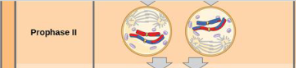
Prophase II

The first stage of meiosis II, where the nuclear envelope breaks down, the nucleolus disappears, chromosomes condense again, and a new spindle apparatus begins to form in each of the two haploid daughter cells produced by meiosis I

  • Sister chromatids condense

  • A new spindle begins to form

  • The nuclear envelope starts to fragment

  • Functionally, Prophase II behaves very much like mitotic prophase, setting up the separation of sister chromatids in Anaphase II

  • Crossing over does not occur here (only prophase I)

  • Each cell entering prophase II is already haploid (n)


<p>The first stage of meiosis II, where the nuclear envelope breaks down, the nucleolus disappears, chromosomes condense again, and a new spindle apparatus begins to form in each of the two haploid daughter cells produced by meiosis I</p><ul><li><p>Sister chromatids condense</p></li><li><p>A new spindle begins to form</p></li><li><p>The nuclear envelope starts to fragment</p></li><li><p>Functionally, Prophase II behaves very much like mitotic prophase, setting up the separation of sister chromatids in Anaphase II</p></li><li><p>Crossing over does not occur here (only prophase I)</p></li><li><p>Each cell entering prophase II is already haploid (n)</p></li></ul><p></p>
45
New cards

Prometaphase II

The transitional phase of cell division (occurring right after prophase and before metaphase) where the nuclear envelope breaks down and spindle fibers attach to the chromosomes

  • The nuclear envelope disappears

  • The spindle fibers engage the individual kinetochores on the sister chromatids


<p>The transitional phase of cell division (occurring right after prophase and before metaphase) where the nuclear envelope breaks down and spindle fibers attach to the chromosomes</p><ul><li><p>The nuclear envelope disappears</p></li><li><p>The spindle fibers engage the individual kinetochores on the sister chromatids </p></li></ul><p></p>
46
New cards

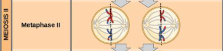
Metaphase II

The stage of meiosis II where individual chromosomes align single-file along the middle of the cell (at the metaphase plate), with spindle fibers attached to the kinetochores of each sister chromatid from opposite poles

  • Sister chromatids line up at the metaphase plate

  • Individual chromosomes (consisting of two sister chromatids) line up single-file, unlike Metaphase I where homologous pairs line up together

  • Haploid State: The cells entering metaphase II are haploid (n), meaning they have only one set of chromosomes rather than the diploid (2n) set found in mitosis or meiosis I

  • Microtubules attach to the opposite kinetochores of each centromere, setting up the separation of sister chromatids in the next phase

<p>The stage of meiosis II where individual chromosomes align single-file along the middle of the cell (at the <strong>metaphase plate</strong>), with spindle fibers attached to the kinetochores of each sister chromatid from opposite poles</p><ul><li><p>Sister chromatids line up at the metaphase plate</p></li><li><p><span>Individual chromosomes (consisting of two sister chromatids) line up single-file, unlike Metaphase I where homologous pairs line up together</span></p></li><li><p><span><strong>Haploid State:</strong> The cells entering metaphase II are haploid (n), meaning they have only one set of chromosomes rather than the diploid (2n) set found in mitosis or meiosis I</span></p></li><li><p>Microtubules attach to the opposite kinetochores of each centromere, setting up the separation of sister chromatids in the next phase</p></li></ul><p></p>
47
New cards

Anaphase II

The stage in meiosis II where the sister chromatids of each chromosome separate and move toward opposite poles of the cell

  • The proteins (cohesins) holding sister chromatids together break down, allowing them to pull apart

  • New status: once separated, each individual chromatid is officially classified as a distinct, single-stranded chromosome

  • Sister chromatids are pulled apart by the shortening of the kinetochore microtubules


<p>The stage in <strong>meiosis II</strong> where the <strong>sister chromatids</strong> of each chromosome separate and move toward opposite poles of the cell</p><ul><li><p>The proteins (cohesins) holding sister chromatids together break down, allowing them to pull apart</p></li><li><p><strong>New status:</strong> once separated, each individual chromatid is officially classified as a distinct, single-stranded chromosome</p></li><li><p>Sister chromatids are pulled apart by the shortening of the kinetochore microtubules</p></li></ul><p></p>
48
New cards

Telophase II and Cytokinesis

The final stages of meiosis II. They complete the reduction and separation of genetic material, turning two haploid cells (with duplicated sister chromatids) into four genetically unique haploid gametes

  • Complete the second round of meiotic division, resulting in four genetically unique haploid (1n) cells (gametes)

  • Chromosomes arrive at the poles of the cell and decondense

  • Nuclear envelopes surround the four nuclei

  • Cleavage furrows divide the two cells into four gentically different haploid cells (1n)


<p>The final stages of meiosis II. They complete the reduction and separation of genetic material, turning two haploid cells (with duplicated sister chromatids) into four genetically unique haploid gametes</p><ul><li><p>Complete the second round of meiotic division, resulting in <strong>four genetically unique haploid (1n) cells</strong> (gametes)</p></li><li><p>Chromosomes arrive at the poles of the cell and decondense </p></li><li><p>Nuclear envelopes surround the four nuclei</p></li><li><p>Cleavage furrows divide the two cells into four gentically different haploid cells (1n)</p></li></ul><p></p>
49
New cards

End product of meiosis II

The end product of meiosis II is four genetically distinct haploid (n) daughter cells (gametes like sperm or egg cells)

  • The cells remain haploid throughout meiosis II. They start and end with half the chromosome number (\(n\)) of the original parent cell

  • Variations by Sex (Gametogenesis):

    • In spermatogenesis, it yields four functional haploid sperm cells.

    • In oogenesis, uneven cytokinesis typically yields one functional haploid egg (ovum) and polar bodies


<p><span>The end product of </span>meiosis II is four genetically distinct haploid (n) daughter cells (gametes like sperm or egg cells)</p><ul><li><p>The cells remain haploid throughout meiosis II. They start and end with half the chromosome number (\(n\)) of the original parent cell</p></li><li><p><span>Variations by Sex (Gametogenesis):</span></p><ul><li><p><span>In spermatogenesis, it yields four functional haploid sperm cells.</span></p></li><li><p><span>In oogenesis, uneven cytokinesis typically yields one functional haploid egg (ovum) and polar bodies</span></p></li></ul></li></ul><p></p>
50
New cards

End product of meiosis II

The end product of meiosis II is four genetically distinct haploid (n) daughter cells (gametes like sperm or egg cells)

  • The end product of meiosis II is four genetically distinct haploid (n) daughter cells (gametes like sperm or egg cells, or spores depending on the organism)


<p><span>The end product of </span>meiosis II is four genetically distinct haploid (n) daughter cells (gametes like sperm or egg cells)</p><ul><li><p><span>The end product of meiosis II is </span>four genetically distinct haploid (n) daughter cells (gametes like sperm or egg cells, or spores depending on the organism)</p></li></ul><p></p>
51
New cards

Prophase II

The first stage of Meiosis II, occurring in two haploid daughter cells left over from Meiosis I

  • Centrosomes migrate to opposite poles and new spindles form

  • Chromosome Condensation: chromatin re-condenses into visible, distinct chromosomes (each still composed of two sister chromatids).

  • Nuclear Envelope Breakdown: the nuclear envelope and nucleolus dissolve in each daughter cell.

  • Spindle Apparatus Formation: centrosomes (or MTOCs) duplicate and move toward opposite poles, starting to grow new spindle fibers.

  • No Crossing Over


<p>The first stage of <strong>Meiosis II</strong>, occurring in two haploid daughter cells left over from Meiosis I</p><ul><li><p>Centrosomes migrate to opposite poles and new spindles form</p></li><li><p><strong>Chromosome Condensation:</strong> chromatin re-condenses into visible, distinct chromosomes (each still composed of two sister chromatids).</p></li><li><p><strong>Nuclear Envelope Breakdown:</strong> the nuclear envelope and nucleolus dissolve in each daughter cell.</p></li><li><p><strong>Spindle Apparatus Formation:</strong> centrosomes (or MTOCs) duplicate and move toward opposite poles, starting to grow new spindle fibers.</p></li><li><p><strong>No Crossing Over</strong></p></li></ul><p></p>
52
New cards

Prometaphase II

A transitional sub-phase of Meiosis II that occurs after prophase II and right before metaphase II

  • Nuclear envelopes completely disappear

  • Spindle is fully formed

  • Each sister chromatid forms a kinetochore and attaches
    to microtubules from opposite poles

  • The meiotic spindle apparatus finishes forming, and spindle microtubules capture the chromosomes by attaching to the kinetochores (protein structures located at the centromere of each sister chromatid)


<p>A transitional sub-phase of <strong>Meiosis II</strong> that occurs after prophase II and right before metaphase II</p><ul><li><p><span>Nuclear envelopes completely disappear</span></p></li><li><p><span>Spindle is fully formed</span></p></li><li><p><span>Each sister chromatid forms a kinetochore and attaches<br>to microtubules from opposite poles</span></p></li><li><p>The meiotic spindle apparatus finishes forming, and spindle microtubules capture the chromosomes by attaching to the <strong>kinetochores</strong> (protein structures located at the centromere of each sister chromatid)</p></li></ul><p></p>
53
New cards

Chromosome alignment differs between meiosis I and meiosis II

Chromosome alignment differs fundamentally between Meiosis I and Meiosis II in what is lining up at the metaphase plate and how the spindle fibers attach

Meiosis I

  • Homologous chromosome pairs (tetrads or bivalents) line up side-by-side along the metaphase plate during Metaphase I; maternal and paternal homologues face opposite poles randomly (independent assortment), creating genetic diversity

  • What separates: homologous chromosomes separate during Anaphase I, reducing the cell from diploid (2n) to haploid (n)

Meiosis II

  • Individual, replicated chromosomes (single-file) line up along the metaphase plate during Metaphase II—much like in mitosis

  • Attachment: sister chromatids attach to spindle fibers from opposite poles (bipolar attachment), just as they do during standard mitosis

  • Sister chromatids separate during Anaphase II, moving to opposite poles

  • The number of chromosomes per cell stays haploid (n→n), resulting in four unique haploid daughter cells total


<p><span>Chromosome alignment differs fundamentally between </span>Meiosis I and Meiosis II in what is lining up at the metaphase plate and how the spindle fibers attach</p><p><strong>Meiosis I</strong></p><ul><li><p>Homologous chromosome pairs (tetrads or bivalents) line up side-by-side along the metaphase plate during Metaphase I; maternal and paternal homologues face opposite poles randomly (independent assortment), creating genetic diversity</p></li><li><p><strong>What separates:</strong> <strong>homologous chromosomes separate</strong> during <strong>Anaphase I</strong>, reducing the cell from diploid (2n) to haploid (n)</p></li></ul><p><strong>Meiosis II</strong></p><ul><li><p><span><strong>Individual, replicated chromosomes</strong> (single-file) line up along the metaphase plate during <strong>Metaphase II</strong>—much like in mitosis</span></p></li><li><p><span><strong>Attachment:</strong> sister chromatids attach to spindle fibers from <em>opposite</em> poles (bipolar attachment), just as they do during standard mitosis</span></p></li><li><p><span><strong>Sister chromatids separate</strong> during <strong>Anaphase II</strong>, moving to opposite poles</span></p></li><li><p><span>The number of chromosomes per cell stays haploid (n→n), resulting in four unique haploid daughter cells total</span></p></li></ul><p></p>
54
New cards

Metaphase II and Anaphase II

Sister chromatids separate and move toward opposite
poles (similar to Mitosis)

Metaphase II

  • Alignment: The chromosomes (each consisting of two sister chromatids) line up individually in a single file along the cell's equator, known as the metaphase plate

  • Spindle Attachment: Spindle fibers (microtubules) from opposite poles attach to the kinetochores located at the centromeres of each chromosome

Anaphase II

  • Separation: the protein complexes holding sister chromatids together (cohesins) break down, causing the centromeres to split

  • Movement: the individual sister chromatids are pulled apart by shortening spindle fibers toward opposite poles of the cell


<p><span>Sister chromatids separate and move toward opposite</span><br><span>poles (similar to Mitosis)</span></p><p><span><strong>Metaphase II</strong></span></p><ul><li><p><span><strong>Alignment:</strong> The chromosomes (each consisting of two sister chromatids) line up individually in a single file along the cell's equator, known as the <strong>metaphase plate</strong></span></p></li><li><p><span><strong>Spindle Attachment:</strong> Spindle fibers (microtubules) from opposite poles attach to the <strong>kinetochores</strong> located at the centromeres of each chromosome</span></p></li></ul><p><strong>Anaphase II</strong></p><ul><li><p><span><strong>Separation:</strong> the protein complexes holding sister chromatids together (cohesins) break down, causing the centromeres to split</span></p></li><li><p><span><strong>Movement:</strong> the individual sister chromatids are pulled apart by shortening spindle fibers toward opposite poles of the cell</span></p></li></ul><p></p>
55
New cards

Telophase II and Cytokinesis

  • The chromosomes at opposite poles

  • Nuclear envelopes form around chromosomes

  • Cytokinesis separates the two cells into four unique haploid cells

Telophase II

  • Nuclear envelope reformation

  • Chromosomes decondense

  • Spindle breakdown

Cytokinesis II

  • Cytoplasm division: the physical division of the cytoplasm occurs simultaneously with or immediately following telophase II


<ul><li><p><span>The chromosomes at opposite poles</span></p></li><li><p><span>Nuclear envelopes form around chromosomes</span></p></li><li><p><span>Cytokinesis separates the two cells into four unique haploid cells</span></p></li></ul><p><strong>Telophase II</strong></p><ul><li><p>Nuclear envelope reformation</p></li><li><p>Chromosomes decondense</p></li><li><p>Spindle breakdown</p></li></ul><p><strong>Cytokinesis II</strong></p><ul><li><p><strong>Cytoplasm division:</strong> the physical division of the cytoplasm occurs simultaneously with or immediately following telophase II</p></li></ul><p></p>
56
New cards

Cytokinesis in plants vs. animals

The physical division of the cytoplasm occurs simultaneously with or immediately following telophase II

  • Cellular mechanism:

    • In animal cells, a contractile ring of microfilaments pinches the plasma membrane inward, forming a cleavage furrow

    • In plant cells, vesicles derived from the Golgi apparatus fuse along the metaphase plate to form a cell plate, which eventually develops into a new cell wall


<p><span>The physical division of the cytoplasm occurs simultaneously with or immediately following telophase II</span></p><ul><li><p><span><strong>Cellular mechanism:</strong></span></p><ul><li><p><span>In <strong>animal cells</strong>, a contractile ring of microfilaments pinches the plasma membrane inward, forming a <strong>cleavage furrow</strong></span></p></li><li><p><span>In <strong>plant cells</strong>, vesicles derived from the Golgi apparatus fuse along the metaphase plate to form a <strong>cell plate</strong>, which eventually develops into a new cell wall</span></p></li></ul></li></ul><p></p>
57
New cards

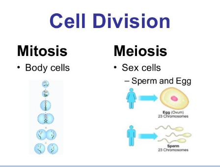
Comparing meiosis and mitosis

  • Both processes divide the nucleus in eukaryotic cells

  • Mitosis is a single division; meiosis has two

  • Homologous chromosomes pair to form tetrads in meiosis
    but not in mitosis

  • In meiosis, crossing over between sister chromatids and
    random assortment of chromosomes in anaphase I create
    genetic variation – mitosis produces genetically identical
    clones

  • Meiosis reduces chromosomes (2n → 1n), mitosis does not


<ul><li><p><span>Both processes divide the nucleus in eukaryotic cells</span></p></li><li><p><span>Mitosis is a single division; meiosis has two</span></p></li><li><p><span>Homologous chromosomes pair to form tetrads in meiosis<br>but not in mitosis</span></p></li><li><p><span>In meiosis, crossing over between sister chromatids and<br>random assortment of chromosomes in anaphase I create<br>genetic variation – mitosis produces genetically identical<br>clones</span></p></li><li><p><span>Meiosis reduces chromosomes (2n → 1n), mitosis does not</span></p></li></ul><p></p>
58
New cards

Comparing meiosis and mitosis

  • Both processes divide the nucleus in eukaryotic cells

  • Mitosis is a single division; meiosis has two

  • Homologous chromosomes pair to form tetrads in meiosis
    but not in mitosis

  • In meiosis, crossing over between sister chromatids and
    random assortment of chromosomes in anaphase I create
    genetic variation – mitosis produces genetically identical
    clones

  • Meiosis reduces chromosomes (2n → 1n), mitosis does not


<ul><li><p>Both processes divide the nucleus in eukaryotic cells</p></li><li><p>Mitosis is a single division; meiosis has two</p></li><li><p>Homologous chromosomes pair to form tetrads in meiosis<br>but not in mitosis</p></li><li><p>In meiosis, crossing over between sister chromatids and<br>random assortment of chromosomes in anaphase I create<br>genetic variation – mitosis produces genetically identical<br>clones</p></li><li><p>Meiosis reduces chromosomes (2n → 1n), mitosis does not</p></li></ul><p></p>
59
New cards

Comparing meiosis and mitosis

  • Both processes divide the nucleus in eukaryotic cells

  • Mitosis is a single division; meiosis has two

  • Homologous chromosomes pair to form tetrads in meiosis
    but not in mitosis

  • In meiosis, crossing over between sister chromatids and
    random assortment of chromosomes in anaphase I create
    genetic variation – mitosis produces genetically identical
    clones

  • Meiosis reduces chromosomes (2n → 1n), mitosis does not


<ul><li><p>Both processes divide the nucleus in eukaryotic cells</p></li><li><p>Mitosis is a single division; meiosis has two</p></li><li><p>Homologous chromosomes pair to form tetrads in meiosis<br>but not in mitosis</p></li><li><p>In meiosis, crossing over between sister chromatids and<br>random assortment of chromosomes in anaphase I create<br>genetic variation – mitosis produces genetically identical<br>clones</p></li><li><p>Meiosis reduces chromosomes (2n → 1n), mitosis does not</p></li></ul><p></p>
60
New cards

End product of mitosis vs. meiosis

Mitosis End Products

  • Number of cells: two daughter cells.

  • Chromosome number: diploid (2n) — identical to the parent cell (e.g., in humans, each cell has 46 chromosomes or 23 pairs)

  • Genetic identity: genetically identical to each other and to the original parent cell (barring rare mutations)

  • Purpose: somatic cell growth, tissue repair, asexual reproduction, and cell replacement

Meiosis End Products

  • Number of cells: four daughter cells

  • (Chromosome number): haploid (n) — half the number of chromosomes of the parent cell (e.g., in humans, each gamete has 23 single chromosomes)

  • Genetic identity: genetically unique/distinct from one another and from the parent cell


<p><strong>Mitosis End Products</strong></p><ul><li><p><span><strong>Number of cells:</strong> two daughter cells.</span></p></li><li><p><span><strong>Chromosome number:</strong> <strong>diploid</strong> (2n) — identical to the parent cell (e.g., in humans, each cell has 46 chromosomes or 23 pairs)</span></p></li><li><p><span><strong>Genetic identity:</strong> genetically <strong>identical</strong> to each other and to the original parent cell (barring rare mutations)</span></p></li><li><p><span><strong>Purpose:</strong> somatic cell growth, tissue repair, asexual reproduction, and cell replacement</span></p></li></ul><p><strong>Meiosis End Products</strong></p><ul><li><p><span><strong>Number of cells:</strong> four daughter cells</span></p></li><li><p><span><strong>(Chromosome number):</strong> <strong>haploid</strong> (n) — half the number of chromosomes of the parent cell (e.g., in humans, each gamete has 23 single chromosomes)</span></p></li><li><p><span><strong>Genetic identity:</strong> genetically <strong>unique/distinct</strong> from one another and from the parent cell</span></p></li></ul><p></p>
61
New cards

Meiosis Vs. Mitosis end products

  • Meiosis: four genetically unique haploid (n) daughter cells (gametes such as sperm or eggs) derived from a single diploid (2n) parent cell

  • Mitosis: two genetically identical diploid (2n) daughter cells


<ul><li><p><span>Meiosis: </span><strong>four genetically unique haploid (n) daughter cells</strong> (gametes such as sperm or eggs) derived from a single diploid (2n) parent cell</p></li><li><p>Mitosis: <strong>two genetically identical diploid (2n) daughter cells</strong></p></li></ul><p></p>
62
New cards

What causes genetic diversity during meiosis?

  1. Crossing over (recombination) during prophase I of meiosis

  2. Independent assortment during metaphase I

  3. Random fertilization (occurs after meiosis, during sexual reproduction)


63
New cards

Nondisjunction

The failure of homologous chromosomes to separate properly during meiosis I or sister chromatids to separate properly during meiosis II (or mitosis)

  • Resulting Condition: leads to aneuploidy, a condition where cells have an abnormal number of chromosomes (such as 2n + 1 trisomy or 2n - 1 monosomy)

  • Nondisjunction occurs during meiosis I (anaphase I) or meiosis II (anaphase II), and can also occasionally happen during mitosis


<p>The failure of homologous chromosomes to separate properly during meiosis I or sister chromatids to separate properly during meiosis II (or mitosis)</p><ul><li><p><strong>Resulting Condition:</strong> leads to <strong>aneuploidy</strong>, a condition where cells have an abnormal number of chromosomes (such as 2n + 1 trisomy or 2n - 1 monosomy)</p></li><li><p><span>Nondisjunction occurs </span>during meiosis I (anaphase I) or meiosis II (anaphase II), and can also occasionally happen during mitosis</p></li></ul><p></p>
64
New cards

Chromosome condensation

The process where long, thin chromatin threads tightly coil, fold, and pack into compact, distinct chromosomes

  • This structural transformation happens during prophase of mitosis and meiosis to prevent DNA damage and ensure accurate chromosome segregation

  • Makes chromosomes easier to seperate

  • Condensation packs the DNA into tight units. This makes it mechanically stable enough to be pulled apart by the spindle apparatus

  • chromatin condenses into distinct, visible chromosomes during prophase


<p>The process where long, thin chromatin threads tightly coil, fold, and pack into compact, distinct chromosomes</p><ul><li><p>This structural transformation happens during prophase of mitosis and meiosis to prevent DNA damage and ensure accurate chromosome segregation</p></li><li><p>Makes chromosomes easier to seperate</p></li><li><p>Condensation packs the DNA into tight units. This makes it mechanically stable enough to be pulled apart by the spindle apparatus</p></li><li><p>chromatin condenses into distinct, visible chromosomes during <span>prophase</span></p></li></ul><p></p>
65
New cards

What aligns during metaphase I?

During metaphase I of meiosis, homologous chromosome pairs (also known as tetrads or bivalents) line up along the metaphase plate (the equator) of the cell

  • homologous chromosome pairs (also called tetrads or bivalents) line up at the metaphase plate


<p><span>During </span>metaphase I of meiosis, homologous chromosome pairs (also known as tetrads or bivalents) line up along the metaphase plate (the equator) of the cell</p><ul><li><p>homologous chromosome pairs (also called tetrads or bivalents) line up at the metaphase plate</p></li></ul><p></p>
66
New cards

What seperates in anaphase I?

During Anaphase I of meiosis, homologous chromosomes (homologs) separate and move to opposite poles of the cell

  • Sister chromatids stay attached: the centromeres do not split during Anaphase I, meaning sister chromatids remain joined together by cohesin proteins

  • homologous chromosomes (homologous pairs) separate and move toward opposite poles of the cell


<p><span>During </span>Anaphase I of meiosis, homologous chromosomes (homologs) separate and move to opposite poles of the cell</p><ul><li><p>Sister chromatids stay attached: the centromeres do not split during Anaphase I, meaning sister chromatids remain joined together by cohesin proteins </p></li><li><p>homologous chromosomes (homologous pairs) separate and move toward opposite poles of the cell</p></li></ul><p></p>
67
New cards

When can nondisjunction occur?

Nondisjunction can occur during Anaphase I or Anaphase II of meiosis, as well as Anaphase of mitosis

  • Anaphase I of Meiosis I, Anaphase II of Meiosis II, or Anaphase of Mitosis

  • failure of chromosome pairs or sister chromatids to separate properly during cell division (anaphase)


<p>Nondisjunction can occur during Anaphase I or Anaphase II of meiosis, as well as Anaphase of mitosis</p><ul><li><p>Anaphase I of Meiosis I, Anaphase II of Meiosis II, or Anaphase of Mitosis</p></li><li><p>failure of chromosome pairs or sister chromatids to separate properly during cell division (anaphase)</p></li></ul><p></p>
68
New cards

What aligns in metaphase II?

  • Individual chromosomes (each composed of two sister chromatids) align single-file along the metaphase plate (the cell's equator)

  • Individual chromosomes (each consisting of two sister chromatids) align single-file along the metaphase plate (the equator of the cell)

  • Unlike Metaphase I—where homologous chromosome pairs line up together—Metaphase II features individual chromosomes lined up in a single file line, which looks very similar to metaphase in mitosis


69
New cards

What happens if nondisjunction occurs during meiosis 1?

Homologous chromosomes fail to separate during anaphase I. As a result, all four resulting gametes are abnormal: two gametes have an extra chromosome (n+1) and two gametes are missing a chromosome (n-1)

  • All four gametes are affected

  • Can be lethal for the baby


70
New cards

What happens if nondisjunction occurs during meiosis II?

When nondisjunction happens in meiosis II, sister chromatids fail to separate during anaphase II. This results in four total gametes: two normal haploid cells (n), one cell with an extra chromosome (n+1), and one cell missing a chromosome (n-1)

  • 50% chance of an abnormal gamete


71
New cards

Centromere

The specialized, constricted region of a chromosome that holds two sister chromatids together after DNA replication and acts as the attachment site for spindle fibers during mitosis and meiosis

  • the specialized, constricted region of a chromosome that holds sister chromatids together and serves as the attachment site for spindle fibers during cell division


<p>The specialized, constricted region of a chromosome that holds two sister chromatids together after DNA replication and acts as the attachment site for spindle fibers during mitosis and meiosis</p><ul><li><p>the specialized, constricted region of a chromosome that holds sister chromatids together and serves as the attachment site for spindle fibers during cell division</p></li></ul><p></p>
72
New cards

Kinetochore

A specialized protein complex assembled on the centromere of a chromosome where spindle fibers attach during cell division to pull sister chromatids apart

  • a large, disc-shaped protein complex that assembles on the centromere of a chromosome during cell division (mitosis and meiosis). It acts as the anchor point where spindle fibers (microtubules) attach to pull sister chromatids apart into separate daughter cells


<p>A specialized protein complex assembled on the centromere of a chromosome where spindle fibers attach during cell division to pull sister chromatids apart</p><ul><li><p>a large, disc-shaped protein complex that assembles on the centromere of a chromosome during cell division (mitosis and meiosis). It acts as the anchor point where spindle fibers (microtubules) attach to pull sister chromatids apart into separate daughter cells</p></li></ul><p></p>
73
New cards

Centrosomes

The primary microtubule-organizing center (MTOC) in animal and eukaryotic cells that builds the mitotic spindle to separate chromosomes during cell division

  • the main microtubule-organizing center (MTOC) of animal and many eukaryotic cells. It coordinates the microtubule cytoskeleton and builds the mitotic spindle during cell division

  • centrosomes contain centrioles and are surrounded by a protein complex


<p>The primary microtubule-organizing center (MTOC) in animal and eukaryotic cells that builds the mitotic spindle to separate chromosomes during cell division</p><ul><li><p>the main microtubule-organizing center (MTOC) of animal and many eukaryotic cells. It coordinates the microtubule cytoskeleton and builds the mitotic spindle during cell division</p></li><li><p>centrosomes contain centrioles and are surrounded by a protein complex</p></li></ul><p></p>
74
New cards

Centrioles

A small, cylindrical organelle made of microtubules (arranged in nine triplets) found in animal cells and located inside the centrosome

  • A pair of centrioles sits inside the centrosome (the main microtubule-organizing center or MTOC of animal cells), positioned at right angles

  • During mitosis and meiosis, centrosomes help organize the mitotic spindle fibers that attach to chromosomes and pull sister chromatids apart

  • small, barrel-shaped cell organelles found mostly in animal cells. They usually live in pairs inside a larger region called the centrosome, positioned at right angles to each other; made of microtubules


<p>A small, cylindrical organelle made of microtubules (arranged in nine triplets) found in animal cells and located inside the centrosome</p><ul><li><p>A pair of centrioles sits inside the centrosome (the main microtubule-organizing center or MTOC of animal cells), positioned at right angles</p></li></ul><ul><li><p>During mitosis and meiosis, centrosomes help organize the <strong>mitotic spindle fibers</strong> that attach to chromosomes and pull sister chromatids apart</p></li><li><p><span>small, barrel-shaped cell organelles found mostly in animal cells. They usually live in pairs inside a larger region called the <strong>centrosome</strong>, positioned at right angles to each other; made of microtubules</span></p></li></ul><p></p>
75
New cards

Cohesin

A ring-shaped protein complex that holds replicated sister chromatids together from the time they are synthesized (during S phase of interphase) until they separate during mitosis or meiosis

  • a protein complex that holds sister chromatids together from the time of DNA replication until they are separated during mitosis or meiosis


<p>A ring-shaped protein complex that holds replicated sister chromatids together from the time they are synthesized (during S phase of interphase) until they separate during mitosis or meiosis</p><ul><li><p>a protein complex that holds sister chromatids together from the time of DNA replication until they are separated during mitosis or meiosis</p></li></ul><p></p>
76
New cards

Spindle fibers

Bundles of microtubules that form the mitotic or meiotic spindle apparatus, responsible for moving and separating chromosomes during cell division

  • microscopic protein structures made of microtubules (composed of alpha- and beta-tubulin dimers) that organize and move chromosomes during eukaryotic cell division (mitosis and meiosis)
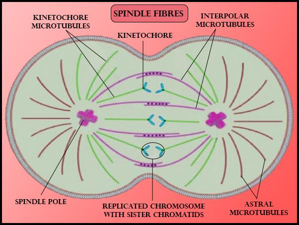

<p><strong>Bundles of microtubules</strong> that form the mitotic or meiotic spindle apparatus, responsible for moving and separating chromosomes during cell division</p><ul><li><p>microscopic protein structures made of <strong>microtubules</strong> (composed of alpha- and beta-tubulin dimers) that organize and move chromosomes during eukaryotic cell division (<strong>mitosis</strong> and <strong>meiosis)</strong></p></li></ul><p></p>
77
New cards

Microtublues

The largest component of the eukaryotic cytoskeleton, defined as hollow, rigid protein tubes made of polymerized dimers of a-tubulin and b-tubulin

  • Radiate from centrosomes to form the spindle apparatus, attaching to chromosomes at the kinetochore to separate sister chromatids during anaphase


<p><span>The largest component of the eukaryotic cytoskeleton, defined as </span>hollow, rigid protein tubes made of polymerized dimers of a-tubulin and b-tubulin</p><ul><li><p>Radiate from centrosomes to form the <strong>spindle apparatus</strong>, attaching to chromosomes at the <strong>kinetochore</strong> to separate sister chromatids during anaphase</p></li></ul><p></p>
78
New cards

Kinetochore microtubules

Specialized spindle fibers that bind directly to a chromosome's kinetochore—a protein structure built on the centromere region of a sister chromatids

  • Spindle microtubules that physically attach to the kinetochore protein complex on chromosomes

  • They shorten during anaphase to pull sister chromatids apart toward opposite poles of the cell


<p>Specialized spindle fibers that bind directly to a chromosome's kinetochore—a protein structure built on the centromere region of a sister chromatids</p><ul><li><p>Spindle microtubules that physically attach to the kinetochore protein complex on chromosomes</p></li><li><p>They shorten during anaphase to pull sister chromatids apart toward opposite poles of the cell</p></li></ul><p></p>
79
New cards

Chromosomes

A thread-like, gene-carrying structure found in the nucleus of eukaryotic cells, consisting of a single long molecule of DNA tightly wound and coiled around proteins called histones

  • a cellular structure composed of a single, long double-stranded DNA molecule tightly coiled and wrapped around histone proteins


<p>A thread-like, gene-carrying structure found in the nucleus of eukaryotic cells, consisting of a single long molecule of DNA tightly wound and coiled around proteins called <strong>histones</strong></p><ul><li><p>a cellular structure composed of a single, long double-stranded DNA molecule tightly coiled and wrapped around <strong>histone proteins</strong></p></li></ul><p></p>
80
New cards

Homologous chromosomes

A matched pair of chromosomes—one inherited from the mother and one from the father—that share the same length, centromere position, and gene sequence, though they may carry different alleles

  • a matching pair of chromosomes (one inherited from each parent) that carry the same genes in the same order. They pair up during prophase I of meiosis, swap segments through crossing over, and then separate in meiosis I so each gamete gets one of the pair


<p>A matched pair of chromosomes—one inherited from the mother and one from the father—that share the same length, centromere position, and gene sequence, though they may carry different alleles</p><ul><li><p><span>a matching pair of chromosomes (one inherited from each parent) that carry the same genes in the same order. They pair up during prophase I of meiosis, swap segments through crossing over, and then separate in meiosis I so each gamete gets one of the pair</span></p></li></ul><p></p>
81
New cards

Chromatids

One half of a duplicated chromosome connected to its identical copy at a region called the centromere

  • one of two identical halves of a replicated chromosome joined together by a region called the centromere


<p>One half of a duplicated chromosome connected to its identical copy at a region called the <strong>centromere</strong></p><ul><li><p>one of two identical halves of a replicated chromosome joined together by a region called the centromere</p></li></ul><p></p>
82
New cards

Sister chromatids

Two identical copies of a single replicated chromosome. They are joined together at a specialized region called the centromere and share the exact same DNA sequence and alleles

  • the two identical copies of a single chromosome that are created during the S phase of interphase (DNA replication). They contain the exact same alleles and are held together by proteins called cohesins until they separate


<p>Two identical copies of a single replicated chromosome. They are joined together at a specialized region called the <strong>centromere</strong> and share the exact same DNA sequence and alleles</p><ul><li><p>the two identical copies of a single chromosome that are created during the S phase of interphase (DNA replication). They contain the exact same alleles and are held together by proteins called cohesins until they separate</p></li></ul><p></p>
83
New cards

Packaging of DNA

  • DNA wraps around histone proteins (like a string of beads)

  • The histone-DNA complex is called nucleosome

  • This structure then coils to form a chromatin fiber

  • The chromatin further packs into a chromosome


<ul><li><p><span>DNA wraps around histone proteins (like a string of beads)</span></p></li><li><p><span>The histone-DNA complex is called nucleosome</span></p></li><li><p><span>This structure then coils to form a chromatin fiber</span></p></li><li><p><span>The chromatin further packs into a chromosome</span></p></li></ul><p></p>
84
New cards

End product of meiosis

The end product of meiosis is four genetically unique haploid (n) daughter cells

  • Four daughter cells are produced from a single starting diploid (2n) cell

  • Haploid (n), meaning they contain half the chromosome number of the original parent cell

  • genetically distinct from one another and from the parent cell due to crossing over (prophase I) and indepdendent assortment (metaphase I)


<p><span>The end product of meiosis is </span>four genetically unique haploid (n) daughter cells</p><ul><li><p><span>Four daughter cells are produced from a single starting diploid (2n) cell</span></p></li><li><p>Haploid (n), meaning they contain half the chromosome number of the original parent cell</p></li><li><p><strong>genetically distinct</strong> from one another and from the parent cell due to crossing over (prophase I) and indepdendent assortment (metaphase I)</p></li></ul><p></p>