chapter 5 biol-k101
 Call Kai
 Learn
Learn Practice Test
 Spaced Repetition
 Match
Match Flashcards
 Knowt Play
1/43

There's no tags or description
Looks like no tags are added yet.
Name | Mastery | Learn | Test | Matching | Spaced | Call with Kai | Chat |
|---|
No analytics yet
Send a link to your students to track their progress
44 Terms
5.1 Macromolecules are XX, built from XX
5.2 Carbohydrates = XX and XX material
Monosaccharides: XX,XX, XX…
Disaccharides: XX, XX, XX
Polysaccharides: XX, XX
XX Bond
5.3 Lipids = a diverse group of XX molecules
Triacylglycerides: XX and XX
Diacylglycerides: XX
Non-glycerol lipids: XX
XX Bond
5.4 Proteins = many structures, wide range of functions
XX and XX XX
Protein XX- 4 levels of structure
XX XX
5.5 Nucleic acids = XX and tXX hereditary information
XX A, C, G, T
2 polymers: XX and XX!
XX Bond
5.1 Macromolecules are polymers, built from monomers
5.2 Carbohydrates = fuel and building material
Monosaccharides: Glucose, Frucose, Galactose…
Disaccharides: Maltose, Sucrose, Lactose
Polysaccharides: Starch, Cellulose
Glycosidic Bond
5.3 Lipids = a diverse group of hydrophobic molecules
Triacylglycerides: Fats and oils
Diacylglycerides: Phospholipids
Non-glycerol lipids: Steroids
Ester Bond
5.4 Proteins = many structures, wide range of functions
Amino Acids and Peptide Bonds
Protein Folding - 4 levels of structure
Peptide Bond
5.5 Nucleic acids = store and transmit hereditary information
Nucleotides A, C, G, T
2 polymers: DNA and RNA!
Phosphodiester Bond
How are macromolecules synthesized and disassembled?
Monomers are XX, individual building blocks that can be joined together to form XX molecules. When two monomers are joined together, they form a XX. When many monomers are joined together into a larger chain, they form a XX.
Condensation / Dehydration Synthesis — Building Up
Condensation, also called XX XX, is the process used to join XX together to build larger molecules.
Two XX are positioned next to each other.
An XX helps carry out the reaction.
One monomer contributes an -XX and the other contributes an –X.
The XX+ X combine to form XX.
That molecule of water is XX.
Removing the water allows a new XX to form between the two XX.
Monomer + Monomer → XX + XX
Every time another monomer is added, one additional H₂O molecule is removed.
2 monomers joined → 1 H₂O removed
3 monomers joined → 2 H₂O removed
4 monomers joined → 3 H₂O removed
So, dehydration synthesis = XX XX by XX XX.
This process is constantly happening in the body. For example, amino acids can be joined together to make proteins, including proteins such as hemoglobin and insulin.
Hydrolysis — Breaking Down
Hydrolysis is the opposite process. It is used to break XX down into their smaller components or monomers.
The word helps explain what happens:
Hydro = XX
Lysis = XX
So hydrolysis means “splitting by water.”
A polymer has a XX connecting its XX.
An XX helps carry out the XX eaction.
XX is added to the bond.
The water separates into X and XX.
The bond between the monomers is XX.
The XX molecule is separated back into XX molecules/monomers.
Dimer+ H₂O → XX+ XXr
This is important when we digest food. Large molecules from food have to be broken into smaller components so the body can absorb and reuse them.
So, hydrolysis = BREAK DOWN by ADDING WATER.
How are macromolecules synthesized and disassembled?
Monomers are small, individual building blocks that can be joined together to form larger molecules. When two monomers are joined together, they form a dimer. When many monomers are joined together into a larger chain, they form a polymer.
Condensation / Dehydration Synthesis — Building Up
Condensation, also called dehydration synthesis, is the process used to join monomers together to build larger molecules.
Two monomers are positioned next to each other.
An enzyme helps carry out the reaction.
One monomer contributes an –OH and the other contributes an –H.
The OH + H combine to form H₂O.
That molecule of water is removed.
Removing the water allows a new bond to form between the two monomers.
Monomer + Monomer → Dimer + H₂O
Every time another monomer is added, one additional H₂O molecule is removed.
2 monomers joined → 1 H₂O removed
3 monomers joined → 2 H₂O removed
4 monomers joined → 3 H₂O removed
So, dehydration synthesis = BUILD UP by REMOVING WATER.
This process is constantly happening in the body. For example, amino acids can be joined together to make proteins, including proteins such as hemoglobin and insulin.
Hydrolysis — Breaking Down
Hydrolysis is the opposite process. It is used to break polymers down into their smaller components or monomers.
The word helps explain what happens:
Hydro = water
Lysis = splitting
So hydrolysis means “splitting by water.”
A polymer has a bond connecting its monomers.
An enzyme helps carry out the hydrolysis reaction.
H₂O is added to the bond.
The water separates into H and OH.
The bond between the monomers is broken.
The larger molecule is separated back into smaller molecules/monomers.
Dimer + H₂O → Monomer + Monomer
This is important when we digest food. Large molecules from food have to be broken into smaller components so the body can absorb and reuse them.
So, hydrolysis = BREAK DOWN by ADDING WATER.
Types of Macromolecules
Simple sugars ⇄ XX
Condensation: Simple sugars are joined together to form XX → water is removed.
Hydrolysis: XX are broken apart into simple sugars → water is added.
Fatty acids + glycerol ⇄ XX
Condensation: Fatty acids and glycerol are joined together to form XX → water is removed.
Hydrolysis: XX are broken apart into fatty acids and glycerol → water is added.
XX are not true polymers, but these reactions are still used to build and break them down.
Amino acids ⇄ XX
Condensation: Amino acids are joined together to form XX → water is removed.
Hydrolysis: XX are broken apart into amino acids → water is added.
Nucleotides ⇄ XX XX
Condensation: Nucleotides are joined together to form XX or XX → water is removed.
Hydrolysis: XX or XX can be broken apart into nucleotides → water is added.
Easy way to remember
Condensation = XX → XX H₂O
Hydrolysis = XX → add H2O
Types of Macromolecules
Simple sugars ⇄ Polysaccharides
Condensation: Simple sugars are joined together to form polysaccharides → water is removed.
Hydrolysis: Polysaccharides are broken apart into simple sugars → water is added.
Fatty acids + glycerol ⇄ Lipids
Condensation: Fatty acids and glycerol are joined together to form lipids → water is removed.
Hydrolysis: Lipids are broken apart into fatty acids and glycerol → water is added.
Lipids are not true polymers, but these reactions are still used to build and break them down.
Amino acids ⇄ Proteins
Condensation: Amino acids are joined together to form proteins → water is removed.
Hydrolysis: Proteins are broken apart into amino acids → water is added.
Nucleotides ⇄ Nucleic acids
Condensation: Nucleotides are joined together to form DNA or RNA → water is removed.
Hydrolysis: DNA or RNA can be broken apart into nucleotides → water is added.
Easy way to remember
Condensation = BUILD → remove H₂O
Hydrolysis = BREAK → add H₂O
What are Carbohydrates?
Carbohydrates are XX. They have a general formula of CH₂O, meaning that for every carbon, there are usually two XX and one XX. Often, if something is a carbohydrate, its name ends in “XX,” such as glucose, galactose, and ribose, although this is not always the case. Monosaccharides also have a functional group in common called the XX group (C=O), along with several hydroxyl (–OH) groups.
One way that sugars are named is by their XX, or the number of carbons they contain. A sugar with three carbons is called a XX, a sugar with five carbons is called a XX, and a sugar with six carbons is called a XX.
A second way sugars are classified is by the XX of the XX group. If the carbonyl group is at the end of the molecule, it is an XX, so the sugar is called an aldose sugar. If the carbonyl group is within the sugar molecule, it is a XX, so the sugar is called a ketose sugar. The position of the carbonyl group is important because it is involved in determining the shape of the sugar when it forms a ring.
A third way sugars can differ is by the position of their –XX functional groups. Sugars have many hydroxyl (–OH) groups, and the position of these groups can distinguish one sugar from another. For example, glucose and galactose are extremely similar, but the thing that distinguishes them is the position of the –OH group on carbon four.
What are Carbohydrates?
Carbohydrates are sugars. They have a general formula of CH₂O, meaning that for every carbon, there are usually two hydrogens and one oxygen. Often, if something is a carbohydrate, its name ends in “-ose,” such as glucose, galactose, and ribose, although this is not always the case. Monosaccharides also have a functional group in common called the carbonyl group (C=O), along with several hydroxyl (–OH) groups.
One way that sugars are named is by their length, or the number of carbons they contain. A sugar with three carbons is called a triose, a sugar with five carbons is called a pentose, and a sugar with six carbons is called a hexose.
A second way sugars are classified is by the position of the carbonyl group. If the carbonyl group is at the end of the molecule, it is an aldehyde, so the sugar is called an aldose sugar. If the carbonyl group is within the sugar molecule, it is a ketone, so the sugar is called a ketose sugar. The position of the carbonyl group is important because it is involved in determining the shape of the sugar when it forms a ring.
A third way sugars can differ is by the position of their –OH functional groups. Sugars have many hydroxyl (–OH) groups, and the position of these groups can distinguish one sugar from another. For example, glucose and galactose are extremely similar, but the thing that distinguishes them is the position of the –OH group on carbon four.
Linear and Ring Forms of Glucose
Glucose can be shown as a straight line or as a ring, but both forms are still the same glucose XX (C₆H₁₂O₆).
In the linear form, glucose has 6 carbons, numbered 1–6. Carbon 1 has the carbonyl group (C=O).
To make the ring form, the glucose molecule basically bends around and connects to itself. The OH on carbon 5 connects with carbon 1, causing the ring to close.
When the ring forms:
Carbons 1–5 are part of the ring.
An oxygen (O) is also part of the ring.
Carbon 6 stays outside the ring as CH₂OH.
So the ring has 5 carbons + 1 oxygen.
Why do some drawings look like they're missing carbons?
They aren't actually missing!
In the abbreviated ring structure, we don't have to write C at every carbon. Every corner of the ring represents a carbon unless another atom, like O, is written there.
So:
Linear glucose → bends → C1 and the OH on C5 interact → ring closes
And all the pictures on the slide are just different ways of drawing the SAME glucose molecule.
Main thing to remember:
Glucose has 6 carbons total. Carbons 1–5 are in the ring, and carbon 6 (CH₂OH) sticks outside the ring.
Linear and Ring Forms of Glucose
Glucose can be shown as a straight line or as a ring, but both forms are still the same glucose molecule (C₆H₁₂O₆).
In the linear form, glucose has 6 carbons, numbered 1–6. Carbon 1 has the carbonyl group (C=O).
To make the ring form, the glucose molecule basically bends around and connects to itself. The OH on carbon 5 connects with carbon 1, causing the ring to close.
When the ring forms:
Carbons 1–5 are part of the ring.
An oxygen (O) is also part of the ring.
Carbon 6 stays outside the ring as CH₂OH.
So the ring has 5 carbons + 1 oxygen.
Why do some drawings look like they're missing carbons?
They aren't actually missing!
In the abbreviated ring structure, we don't have to write C at every carbon. Every corner of the ring represents a carbon unless another atom, like O, is written there.
So:
Linear glucose → bends → C1 and the OH on C5 interact → ring closes
And all the pictures on the slide are just different ways of drawing the SAME glucose molecule.
Main thing to remember:
Glucose has 6 carbons total. Carbons 1–5 are in the ring, and carbon 6 (CH₂OH) sticks outside the ring.
Monosaccharides: C6H12O6 in solution.
XX-> disaccharides
Monosaccharides and Disaccharides
Glucose, galactose, and fructose are XX XX. This means they all have the same exact chemical formula, C₆H₁₂O₆, but they have different structures. Because they all have the same formula, you cannot look at C₆H₁₂O₆ and know whether it is glucose, galactose, or fructose. You have to look at how the atoms are arranged. Glucose and galactose look very similar but differ in the position of an XX group, while fructose has a different structure and commonly forms the 5-membered ring shown in the picture.
These monosaccharides can be joined together through a condensation/dehydration reaction to form XX. Di means two, so a disaccharide is made by joining two XX. When the two sugars are joined, one molecule of XX (XX) is removed, and a new XX forms between the sugars. The bond that joins two sugars together is called a XX XX.
If you put two glucose molecules together (Glu + Glu), a molecule of water is XX and a XX bond forms between them. The resulting disaccharide is called XX.
Glucose + Glucose → Maltose + H₂O
If you put one glucose and one fructose together (Glu + Fru), water is XX and a XX bond forms. This produces XX, which is table sugar. So when you eat table sugar, the sucrose is made from one glucose and one fructose joined together.
Glucose + Fructose → XX + XX
The third disaccharide shown is made by putting one glucose and one galactose together (Glu + Gal). Again, a molecule of water is XX and a XX bond forms between the sugars. This produces XX, which is commonly called XX XX.
Glucose + Galactose → XX + XX
What to recognize on the test
The three monosaccharides:
XX= C₆H₁₂O₆
XX = C₆H₁₂O₆
XX = C₆H₁₂O₆
For the disaccharides, memorize:
Glu + Glu = XX
Glu + Fru = XX (XX XX)
Glu + Gal = XX (XX XX)
And remember: two sugars join → XX XX → XX bond forms → XX
Monosaccharides: C6H12O6 in solution.
Condensation-> disaccharides
Monosaccharides and Disaccharides
Glucose, galactose, and fructose are structural isomers. This means they all have the same exact chemical formula, C₆H₁₂O₆, but they have different structures. Because they all have the same formula, you cannot look at C₆H₁₂O₆ and know whether it is glucose, galactose, or fructose. You have to look at how the atoms are arranged. Glucose and galactose look very similar but differ in the position of an OH group, while fructose has a different structure and commonly forms the 5-membered ring shown in the picture.
These monosaccharides can be joined together through a condensation/dehydration reaction to form disaccharides. Di means two, so a disaccharide is made by joining two monosaccharides. When the two sugars are joined, one molecule of water (H₂O) is removed, and a new bond forms between the sugars. The bond that joins two sugars together is called a glycosidic bond.
If you put two glucose molecules together (Glu + Glu), a molecule of water is removed and a glycosidic bond forms between them. The resulting disaccharide is called maltose.
Glucose + Glucose → Maltose + H₂O
If you put one glucose and one fructose together (Glu + Fru), water is removed and a glycosidic bond forms. This produces sucrose, which is table sugar. So when you eat table sugar, the sucrose is made from one glucose and one fructose joined together.
Glucose + Fructose → Sucrose + H₂O
The third disaccharide shown is made by putting one glucose and one galactose together (Glu + Gal). Again, a molecule of water is removed and a glycosidic bond forms between the sugars. This produces lactose, which is commonly called milk sugar.
Glucose + Galactose → Lactose + H₂O
What to recognize on the test
The three monosaccharides:
Glucose = C₆H₁₂O₆
Galactose = C₆H₁₂O₆
Fructose = C₆H₁₂O₆
Same formula + different structures = structural isomers
For the disaccharides, memorize:
Glu + Glu = Maltose
Glu + Fru = Sucrose (table sugar)
Glu + Gal = Lactose (milk sugar)
And remember: two sugars join → H₂O removed → glycosidic bond forms → disaccharide
Disaccharides are highly XX (Can you name the enzymes?)
Sucrose Glu + FruMaltose Glu + Glu Lactose Glu + Gal
A special mention – many mammals around the world lose this enzyme
as they age.
Maltose can easily be broken down into the glu, glu by the enzyme XX. Sucrose can easily be broken down to its component parts by XX. Lactose is broken down to glu gal by the enzyme XX.
Disaccharides are highly digestible (Can you name the enzymes?)
Sucrose Glu + FruMaltose Glu + Glu Lactose Glu + Gal
A special mention – many mammals around the world lose this enzyme
as they age.
Maltose can easily be broken down into the glu, glu by the enzyme maltase. Sucrose can easily be broken down to its component parts by sucrase. Lactose is broken down to glu gal by the enzyme lactase.
Polysaccharides: Starch vs. cellulose:
What’s the Difference?
The form where the OH is in the down position is called XX XX. The form where its in the up position is called XX XX.
Starch: (1-4 linkage of a -glucose):
a storage XX in XX (XX)
-XX, XX, XX, etc.
-XX, XX, etc.
-Glycogen: in XX XX
Cellulose: (1-4 linkage of B -glucose):
a structural polysaccharide (XX XX).Made out of XX
-XX by plants and animals
-XX and XX are two other
structural polysaccharides
Polysaccharides: Starch vs. cellulose:
What’s the Difference?
The form where the OH is in the down position is called alpha glucose. The form where its in the up position is called beta glucose.
Starch: (1-4 linkage of -glucose):
a storage polysaccharide in plants (energy)
-Cornstarch, pasta, crackers, etc.
-Amylose, amylopectin, etc.
-Glycogen: in animal cells
Cellulose: (1-4 linkage of -glucose):
a structural polysaccharide (cell walls).Made out of wood.
-Indigestible by plants and animals
-Chitin and Cartilage are two other
structural polysaccharides
Different ways you can put glucose together:
If you are a plant you are going to make a lot of XX and you are going to store that as XX or XX. We can take excess sugars in our diet and make XX, which our storage polysaccharide. You make a certain amount of it in your mitochondria. Chitin, is a structural polysaccharide that is XX, so chitin is glucose with an amino and other goodies on it. Chitin can be put together by insects to make their crunch exoskeleton. If you step on a bug that crunch you hear is chitin. You can make chitin into long flexible threads, stitches.
XX is our body’s preferred form of energy
Different ways you can put glucose together:
If you are a plant you are going to make a lot of glucose and you are going to store that as starch or amylose. We can take excess sugars in our diet and make glycogen, which our storage polysaccharide. You make a certain amount of it in your mitochondria. Chitin, is a structural polysaccharide that is digestible, so chitin is glucose with an amino and other goodies on it. Chitin can be put together by insects to make their crunch exoskeleton. If you step on a bug that crunch you hear is chitin. You can make chitin into long flexible threads, stitches.
Glucose is our body’s preferred form of energy
Macromolecule II: What are Lipids?
Triacylglycerols: (Fats and Oils) XX + X fatty acids
(a) Overall: 1 XX + 3 XX XX tails via a XX reaction
When you link an XX from an alcohol to a XX from a carboxylic acid, that is called an XX XX. A fat molecule is a great form of XX XX. Ester linkage: The —O—C(=O)— bond that connects a fatty acid to glycerol. It forms through dehydration/condensation, which removes H₂O. A triglyceride contains 3 ester linkages connecting glycerol to 3 fatty acids.
Examples of saturated and unsaturated fats & fatty acids:
XX XX give each fat or oil
its properties.
1. Saturated fats: (no XX XX); XX at room temp)
2. Unsaturated fats: XX at
room temperature (XX)
• Monounsaturated:
1 XX XX
• Polyunsaturated:
XX or more double bonds
3. Trans fats: XX arrangement
of XX adjacent to the
XX XX
Macromolecule II: What are Lipids?
Triacylglycerols: (Fats and Oils) Glycerol + 3 fatty acids
(a) Overall: 1 glycerol + 3 fatty acid tails via a dehydration reaction
-When you link an OH and an alchohol to a COOH, a carboxylic acid thats called an ester linkage. Fat molecule is a great energy storage. Ester linkage: The —O—C(=O)— bond that connects a fatty acid to glycerol. It forms through dehydration/condensation, which removes H₂O. A triglyceride contains 3 ester linkages connecting glycerol to 3 fatty acids.
Examples of saturated and unsaturated fats & fatty acids:
Double bonds give each fat or oil
its properties.
1. Saturated fats: (no double
bonds); solid at room temp)
2. Unsaturated fats: liquid at
room temperature (oils)
• Monounsaturated:
1 double bond
• Polyunsaturated:
2 or more double bonds
3. Trans fats: trans arrangement
of carbon atoms adjacent to the
double bonds.
What are Phospholipids/diacylglycerols?
XX + X fatty acids. Functional group = XX + XX or many other
“head groups”.
Creates an amphipathic molecule - XX XX, XX XX.
Spontaneously assembles in water to form a XX XX
What are Phospholipids/diacylglycerols?
Glycerol + 2 fatty acids. Functional group = phosphate + choline or many other
“head groups”.
Creates an amphipathic molecule - hydrophobic tails, hydrophilic head.
Spontaneously assembles in water to form a phospholipid bilayer
What are Steroids? XX-XX Lipids
Cholesterol is pretty XX except at the ho end, estradiol or estrogen and testosterone are steroid hormones that are called sex hormones.
What are Steroids? Non-Glycerol Lipids
Cholesterol is pretty hydrophobic except at the ho end, estradiol or estrogen and testosterone are steroid hormones that are called sex hormones.
Every dietary fat/oil will have differing amounts of XX &
XX fats.
Every dietary fat/oil will have differing amounts of saturated &
unsaturated fats.
What are Proteins and their general functions?
1. Enzymes (-ase): lactase, amylase, pancreatic lipase, lysozyme, Rubisco.
2. Structural Proteins: Collagen (skin), keratin (hair), dystrophin (muscle)
3. Storage / Nutritive proteins: Albumin (egg white), Zein (corn kernels),
Casein (milk), Glutein (wheat), Soya (soy protein), Globulin (peanuts)
4. Transport proteins: Aquaporins, hemoglobin, electron transport
5. Hormonal / Regulatory proteins: Insulin, growth hormone, prolactin
6. Motile proteins: Actin and myosin: muscle
7. Protective proteins: Antibodies.
Proteins are ~50% or more
of a cell’s dry weight.
The instructions for making
proteins come from genes.
What are Proteins and their general functions?
1. Enzymes (-ase): lactase, amylase, pancreatic lipase, lysozyme, Rubisco.
2. Structural Proteins: Collagen (skin), keratin (hair), dystrophin (muscle)
3. Storage / Nutritive proteins: Albumin (egg white), Zein (corn kernels),
Casein (milk), Glutein (wheat), Soya (soy protein), Globulin (peanuts)
4. Transport proteins: Aquaporins, hemoglobin, electron transport
5. Hormonal / Regulatory proteins: Insulin, growth hormone, prolactin
6. Motile proteins: Actin and myosin: muscle
7. Protective proteins: Antibodies.
Proteins are ~50% or more
of a cell’s dry weight.
The instructions for making
proteins come from genes.
How to spot an Amino Acids: the monomer.
ALL amino acids have 5 main parts:
1. XX XX
2. XX XX
3. XX XX
4. XX
5. X group: there
Are 20 different
R groups that can
be grouped
chemically:
Polar = XX
Charged = Usually
Have a charged
XX or XX
group within R group
How to spot an Amino Acids: the monomer.
ALL amino acids have 5 main parts:
1. Alpha Carbon
2. Carboxyl end
3. Amino end
4. Hydrogen
5. R group: there
Are 20 different
R groups that can
be grouped
chemically:
Polar = Hydrophilic
Charged = Usually
Have a charged
carboxyl or amino
group within R group
Now can I use my sharp intellectual skills to identify the
Nonpolar R-groups (side chains)? XX
-XX R groups all have in common the absence of XX and very few XX.
Now can I use my sharp intellectual skills to identify the
Nonpolar R-groups (side chains)? Hydrophobic
-Hydrophobic R groups all have in common the absence of oxygen and very few nitrogen.
And likewise, how can I quickly spot the Polar side chains?
XXPolar
-They all include XX, except cysteine. They have a XX letter code. They love XX and they are XX
And likewise, how can I quickly spot the Polar side chains?
Hydrophilic/Polar
-They all include oxygen, except cysteine. They have a three letter code. They love water and they are polar.
The last group is both Polar and Electrically charged Hydrophilic and either XX or XX. They also have a name that indicates if they are negatively or positively charged:aspartic acid, glutamic acid→ XX charged
The last group is both Polar and Electrically charged Hydrophilic and either acidic or basic. They also have a name that indicates if they are negatively or positively charged:aspartic acid, glutamic acid.
Proteins need to XX properly to function
properly: Four Levels of Protein Structure
1° Primary structure of a protein = unique
sequence of XX XX (XX XX)
2° Secondary structure =X-XX that coils and
folds the polypeptide chain (XX XX)
3° Tertiary structure = determined by interactions
among various side chains (X XX)
4° Quaternary structure = protein functions as
multiple XX chains (X XX)
Proteins need to fold properly to function
properly: Four Levels of Protein Structure
1° Primary structure of a protein = unique
sequence of amino acids (Peptide bonds)
2° Secondary structure = H-bonding that coils and
folds the polypeptide chain (H-bonds)
3° Tertiary structure = determined by interactions
among various side chains (R groups)
4° Quaternary structure = protein functions as
multiple polypeptide chains (2+ subunits)
The Peptide Bond: Making a polymer / polypeptide chain
(amino end) N-C-C-N-C-C (carboxyl end)
The Peptide Bond: Making a Polymer / Polypeptide Chain
The Peptide Bond: Making a polymer / polypeptide chain
(amino end) N-C-C-N-C-C (carboxyl end)
The Peptide Bond: Making a Polymer / Polypeptide Chain
What happens when you put two amino acids together?
How you make a peptide bond is when you begin to put amino acids together.
When you bring glycine next to another amino acid, the OH group of one and the H group of another—the amino part—will come together.
A covalent bond is formed that is called a XX XX.
That molecule of water is XX XX.
Every time you make a peptide bond, you remove one molecule of XX.
What happens when you put two amino acids together?
How you make a peptide bond is when you begin to put amino acids together.
When you bring glycine next to another amino acid, the OH group of one and the H group of another—the amino part—will come together.
A covalent bond is formed that is called a peptide bond.
That molecule of water is looped out.
Every time you make a peptide bond, you remove one molecule of water.
How do you know what the peptide bond looks like?
Look for this pattern:
X–X–X–X–X–X
XX = two amino acids together.
How do you know what the peptide bond looks like?
Look for this pattern:
N–C–C–N–C–C
Dipeptide = two amino acids together.
Exploring Levels of Protein Structure
Where are peptide bonds made?
Peptide bonds are formed at the XX during protein synthesis.
Primary (1°) Structure
The primary structure of a protein is the specific XX sequence/order of amino acids joined together by peptide bonds.
Instructions from your XX (XX) determine the order of the amino acids.
XX →XX→ XX
Amino acids are connected by XX XX to create a XX XX.
The chain has two ends:
XX XX (N-terminus) → beginning of the chain
XX XX (C-terminus) → end of the chain
The exact order of amino acids matters because it ultimately affects how the protein folds and functions.
Example: Glucagon
Glucagon has 29 amino acids in a specific order.
29 amino acids =XX peptide bonds
Easy rule:
# of peptide bonds = # of amino acids − 1
Exploring Levels of Protein Structure
Where are peptide bonds made?
Peptide bonds are formed at the ribosome during protein synthesis.
Primary (1°) Structure
The primary structure of a protein is the specific linear sequence/order of amino acids joined together by peptide bonds.
Instructions from your genes (DNA) determine the order of the amino acids.
DNA → RNA → Protein
Amino acids are connected by peptide bonds to create a polypeptide chain.
The chain has two ends:
Amino end (N-terminus) → beginning of the chain
Carboxyl end (C-terminus) → end of the chain
The exact order of amino acids matters because it ultimately affects how the protein folds and functions.
Example: Glucagon
Glucagon has 29 amino acids in a specific order.
29 amino acids = 28 peptide bonds
Easy rule:
# of peptide bonds = # of amino acids − 1
Secondary (2°) Structure
Immediately when the protein enters the cytoplasm, it forms what’s called the secondary structure.Secondary structure results from XX XX between different parts of the amino acid backbone. The backbone has the repeating pattern: X–X–X–X–X–X.
These hydrogen bonds cause parts of the backbone to form:
α-helix
XX
Examples: XX, XX
β-pleated sheet
XX and XX
Example: XX
Secondary (2°) Structure
Immediately when the protein enters the cytoplasm, it forms what’s called the secondary structure.Secondary structure results from hydrogen bonds between different parts of the amino acid backbone. The backbone has the repeating pattern: N–C–C–N–C–C.
These hydrogen bonds cause parts of the backbone to form:
α-helix
Elastic
Examples: wool, keratin
β-pleated sheet
Strong and flexible
Example: silk
Tertiary (3°) Structure
The tertiary structure is not talking about the XX XX backbone, but how the X-groups interact.
It results from all types of bonds between the different X-XXX of the amino acids.
These XX-XX interactions “rivet” different parts of the same protein together.
This causes the protein to XX Into its final shape needed for its function.
Tertiary (3°) Structure
The tertiary structure is not talking about the amino acid backbone, but how the R-groups interact.
It results from all types of bonds between the different R-groups of the amino acids.
These R-group interactions “rivet” different parts of the same protein together.
This causes the protein to fold into its final shape needed for its function.
Quaternary (4°) Structure
Some proteins will take it one level further to form the quaternary structure.
This is when a protein has different protein XX (XX XX).
These subunits do not function as a XX XX on their own; they have to come together with other XX to form the XX XX.
Quaternary structure = interactions between X or XX protein subunits.
Quaternary (4°) Structure
Some proteins will take it one level further to form the quaternary structure.
This is when a protein has different protein subunits (polypeptide chains).
These subunits do not function as a single protein on their own; they have to come together with other subunits to form the functional protein.
Quaternary structure = interactions between 2 or more protein subunits.
How are the subunits held together?
There is not one specific type of bond that holds them together.
They can be held together by:
XX XX
XX XX
Occasionally, XX XX
Example: Hemoglobin
Hemoglobin has X subunits:
X alpha chains
X beta chains
The alpha and beta chains are coordinated with heme, which binds oxygen.
Since hemoglobin has 4 subunits, it functions as what we call a tetramer.
Another example: Collagen
Collagen is a protein found in your skin.
What happens when 3D shape is altered?
A single nucleotide change Amino acid substitution in hemoglobin
causes Sickle-cell disease
How are the subunits held together?
There is not one specific type of bond that holds them together.
They can be held together by:
Ionic interactions
Hydrophobic interactions
Occasionally, disulfide bridges
Example: Hemoglobin
Hemoglobin has 4 subunits:
2 alpha chains
2 beta chains
The alpha and beta chains are coordinated with heme, which binds oxygen.
Since hemoglobin has 4 subunits, it functions as what we call a tetramer.
Another example: Collagen
Collagen is a protein found in your skin.
What happens when 3D shape is altered?
A single nucleotide change Amino acid substitution in hemoglobin
causes Sickle-cell disease
Protein Shape and Sickle Cell Anemia
For proteins, shape is everything. The three-dimensional shape of proteins is really important.
There are many possibilities for errors or changes in health status if you alter your genetics in a way that changes the shape of one of your proteins.
Normal Hemoglobin
In normal hemoglobin, the 6th amino acid of the beta subunit is glutamic acid.
This forms a normal beta subunit.
2 alpha subunits + 2 beta subunits come together to form the hemoglobin tetramer.
These form little plump hemoglobin tetramers that fill the red blood cell and help give it its normal plump, biconcave shape.
Sickle Cell
Some people have a mutation in one or both copies of their hemoglobin gene.
This mutation causes valine to be placed in the 6th position instead of glutamic acid.
Valine is hydrophobic.
This one amino acid change causes the beta subunit to behave differently, which affects the hemoglobin tetramer.
Instead of forming the normal plump tetramers, the hemoglobin can form long, stiff rods.
When these are inside a red blood cell, they alter the shape of the red blood cell, causing it to become sickle-shaped.
These sickled red blood cells do not carry oxygen as effectively and contribute to sickle cell anemia
Protein Shape and Sickle Cell Anemia
For proteins, shape is everything. The three-dimensional shape of proteins is really important.
There are many possibilities for errors or changes in health status if you alter your genetics in a way that changes the shape of one of your proteins.
Normal Hemoglobin
In normal hemoglobin, the 6th amino acid of the beta subunit is glutamic acid.
This forms a normal beta subunit.
2 alpha subunits + 2 beta subunits come together to form the hemoglobin tetramer.
These form little plump hemoglobin tetramers that fill the red blood cell and help give it its normal plump, biconcave shape.
Sickle Cell
Some people have a mutation in one or both copies of their hemoglobin gene.
This mutation causes valine to be placed in the 6th position instead of glutamic acid.
Valine is hydrophobic.
This one amino acid change causes the beta subunit to behave differently, which affects the hemoglobin tetramer.
Instead of forming the normal plump tetramers, the hemoglobin can form long, stiff rods.
When these are inside a red blood cell, they alter the shape of the red blood cell, causing it to become sickle-shaped.
These sickled red blood cells do not carry oxygen as effectively and contribute to sickle cell anemia
Denaturation and Renaturation of a Protein
For proteins to function properly, they have to have the correct XX-dimensional shape.When proteins are subjected to things like really XX XX or XX, proteins that should be functioning normally can start to misfold and become XX. During denaturation, the protein can start XX and losing its normal structure.The quaternary structure may be disrupted, followed by the tertiary structure, and even the secondary structure can be altered.When the protein loses its proper shape, it may no longer function properly.
Denaturation can be caused by:
XX XX
XX
XX
XX
XX XX
Etc.
Denaturation = protein loses its normal XX XX → protein may lose its XX
Denaturation and Renaturation of a Protein
For proteins to function properly, they have to have the correct three-dimensional shape.When proteins are subjected to things like really high heat or alcohol, proteins that should be functioning normally can start to misfold and become denatured. During denaturation, the protein can start separating and losing its normal structure.The quaternary structure may be disrupted, followed by the tertiary structure, and even the secondary structure can be altered.When the protein loses its proper shape, it may no longer function properly.
Denaturation can be caused by:
High heat
Acids
Bases
Alcohols
High fever
Etc.
Denaturation = protein loses its normal 3D shape → protein may lose its function.
Chaperone Proteins
Some proteins, even in their natural setting, are so big and complicated that there is a lot of folding that has to be done, so they need some help.There is specific machinery in most cells called chaperones that helps proteins XX properly.When proteins are being formed, they leave the XX and enter the XX.The unfolded protein is packaged into the chaperone structure.The XX protein enters → the cap XX → a XX Environment is created → the protein folds XX.The result is a properly folded protein that can function normally.
Chaperone Proteins
Some proteins, even in their natural setting, are so big and complicated that there is a lot of folding that has to be done, so they need some help.There is specific machinery in most cells called chaperones that helps proteins fold properly.When proteins are being formed, they leave the ribosome and enter the cytoplasm.The unfolded protein is packaged into the chaperone structure.The unfolded protein enters → the cap closes → a hydrophilic environment is created → the protein folds properly.The result is a properly folded protein that can function normally.
What are nucleic acids?
Monomer: Nucleotide
1. A X-XX sugar
(Ribose = XX,
Deoxyribose =XX)
2. A XX base
(X, X, X, X and X)
3. A XX group
What are nucleic acids?
Monomer: Nucleotide
1. A 5-carbon sugar
(Ribose = RNA,
Deoxyribose = DNA)
2. A Nitrogenous base
(A, C, G, T and U)
3. A phosphate group
How to tell if its ribose or deoxyribose:
Its very important to know if you are looking at DNA or RNA.
They both have an OH group on the three carbon, so thats not a good way to tell them apart. If there is an OH on the two carbon that is XX. If theres just an H it is XX. XX will form dna, XX will form rna.
What are the nitrogenous bases, the ones that have just a single nitrogenous base, so the pyrimidines have one ring X, X, and X, X. and X are used in DNA, X is only used in RNA. The XX are the other type they have two rings.
DNA: X,X, X, X
RNA: X, X, X, X
So the key difference is:
T (thymine) → XX only
U (uracil) →xx only
X, X, and X → BOTH
How to tell if its ribose or deoxyribose:
Its very important to know if you are looking at DNA or RNA.
They both have an OH group on the three carbon, so thats not a good way to tell them apart. If there is an OH on the two carbon that is RNA. If theres just an H it is deoxyribose. Dexoyribose will form dna, ribose will form rna.
What are the nitrogenous bases, the ones that have just a single nitrogenous base, so the pyrimidines have one ring C, U, and T. C and T are used in DNA, U is only used in RNA. The purines are the other type they have two rings.
DNA: A, T, C, G
RNA: A, U, C, G
So the key difference is:
T (thymine) → DNA only
U (uracil) → RNA only
A, C, and G → BOTH
A closer look at the 5 nitrogenous bases:
(a) Pyrimidines: XX XX
X-X-X
b) Purines: XX XX
X and X
During DNA or RNA synthesis,
XX and XX always
base-pair in specific ways:
C-G, A-T (or A-U)
A closer look at the 5 nitrogenous bases:
(a) Pyrimidines: single ring
C-U-T
b) Purines: double ring:
A and G
During DNA or RNA synthesis,
purines and pyrimidines always
base-pair in specific ways:
C-G, A-T (or A-U)
Base Pairing in DNA
This is the way that DNA base pairs, or how the nitrogenous bases help DNA base pair.
Down the DNA double helix, the two strands are held together by XX XX.
A–T Base Pair:
Adenine (A) always base pairs with XX (X).
This is called an A–T base pair.
An A–T base pair has X hydrogen bonds.
G–C Base Pair:
Guanine (G) always base pairs with XX (X).
This is called a G–C base pair.
A G–C base pair has X hydrogen bonds.
Purines and Pyrimidines:
You always bond XX purine to XX pyrimidine.
Purines: XX (X) and XX (X)
Pyrimidines: XX (X) and XX (X)
Base Pairing in DNA
This is the way that DNA base pairs, or how the nitrogenous bases help DNA base pair.
Down the DNA double helix, the two strands are held together by hydrogen bonds.
A–T Base Pair:
Adenine (A) always base pairs with thymine (T).
This is called an A–T base pair.
An A–T base pair has 2 hydrogen bonds.
G–C Base Pair:
Guanine (G) always base pairs with cytosine (C).
This is called a G–C base pair.
A G–C base pair has 3 hydrogen bonds.
Purines and Pyrimidines:
You always bond one purine to one pyrimidine.
Purines: Adenine (A) and Guanine (G)
Pyrimidines: Thymine (T) and Cytosine (C)

Identify
single ring:pyrimidine
one carbonyl group
no methyl group
cytosine
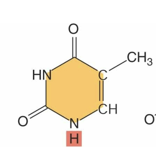
identify
single ring:pyrimidine
methyl group
two carbonyl group
thymine
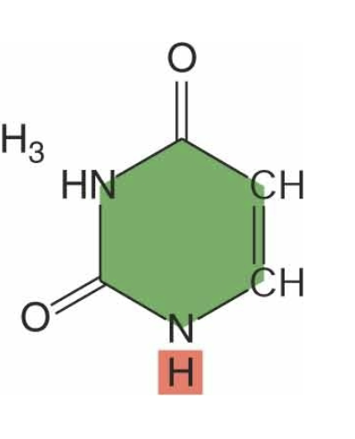
identify
single ring:pyrimidine
two carbonyl groups
no methyl present
uracil

identify
double ring: purine
no carbonyl group
adenine

identify
double ring->purine
carbonyl group
guanine
What is the DNA double helix? (the XX)
Monomers are connected by condensation:
The 5 P + 3’ OH creates a XX bond.
What is the DNA double helix? (the polymer)
Monomers are connected by condensation:
The 5 P + 3’ OH creates a phosphodiester bond.
Making a DNA/RNA Polymer: Phosphodiester Bonds
Let’s take a look at how the polymer happens.
You have one nucleotide, and then you put another nucleotide next to it.
In the image, one nucleotide has a pyrimidine, and the incoming one has a purine.
If the sugar is ribose, the pyrimidine could be X or X, and the purine could be X or X.
There is a phosphate group with an XX.
When you put the nucleotides next to each other, you loop out a molecule of XX and create a XX XX.
This new bond is called a XX bond.
Phosphodiester bonds link XX monomers to XX monomers and XX monomers to XX monomers.
What gets linked?
You link the X phosphate of one nucleotide with the X OH of another nucleotide:
X phosphate + X OH → XX bond
Enzymes
The enzyme that makes RNA is called XX XX
The enzyme that makes more DNA is called XX XX
Main idea:
5′ phosphate + 3′ OH → XX XX → XX XX
Making a DNA/RNA Polymer: Phosphodiester Bonds
Let’s take a look at how the polymer happens.
You have one nucleotide, and then you put another nucleotide next to it.
In the image, one nucleotide has a pyrimidine, and the incoming one has a purine.
If the sugar is ribose, the pyrimidine could be C or U, and the purine could be A or G.
There is a phosphate group with an OH.
When you put the nucleotides next to each other, you loop out a molecule of water and create a new bond.
This new bond is called a phosphodiester bond.
Phosphodiester bonds link RNA monomers to RNA monomers and DNA monomers to DNA monomers.
What gets linked?
You link the 5′ phosphate of one nucleotide with the 3′ OH of another nucleotide:
5′ phosphate + 3′ OH → phosphodiester bond
Enzymes
The enzyme that makes RNA is called RNA polymerase.
The enzyme that makes more DNA is called DNA polymerase.
Main idea:
5′ phosphate + 3′ OH → phosphodiester bond → nucleotide polymer
The way that RNA polymerase, or in this case DNA polymerase knows what to put in is it looks for what’s called complimentary base pairing if theres an a, it’s going to pull in a X. If there is a C, its going to pull in a X. If there is a g its going to pull in a X. So by complimentary base pairing, the original strand of DNA is used as a template for the new strand of DNA. DNA is perfectly suited for its own replication because as the two old strands separate, the new strands are built off of the information on the old strands by complimentary base pairing. A always pairs with X, C always base pairs with X.
The way that RNA polymerase, or in this case DNA polymerase knows what to put in is it looks for what’s called complimentary base pairing if theres an a, it’s going to pull in a T. If there is a C, its going to pull in a G. If there is a g its going to pull in a C. So by complimentary base pairing, the original strand of DNA is used as a template for the new strand of DNA. DNA is perfectly suited for its own replication because as the two old strands separate, the new strands are built off of the information on the old strands by complimentary base pairing. A always pairs with T, C always base pairs with G.
The more closely related two proteins are to each other or two gene sequences are to each other so to are the organisms from which those sequences came. Two sequences that have the identical DNA or amino acid sequence might be from extremely related organisms and ones that have many changes if you look at them are related but only distantly through evolutionary time. The organisms that tend to have fewer changes are more closely related in an evolutionary sense.
The more closely related two proteins are to each other or two gene sequences are to each other so to are the organisms from which those sequences came. Two sequences that have the identical DNA or amino acid sequence might be from extremely related organisms and ones that have many changes if you look at them are related but only distantly through evolutionary time. The organisms that tend to have fewer changes are more closely related in an evolutionary sense.
1. Carbohydrates: Look for XX rings = XX monomers, connected by XX
A- llinkage = oxygen projected XX in relation to XX (XX)
b -linkage = oxygen projected XX in relation to XX (XX)
2. Lipids: Look for XX
and either X fatty acids (tri-acylgycerol)
or X fatty acids and a XX (di-acylgycerol)
Remember, steroids are XX-XX lipids
3. Amino Acids: Look for the XX XX
surrounded by 4 friends: an XX, a XX,
a X and an X group
4. Proteins: Look for the X-X-X backbone.
Can you spot the peptide bond?
5. Nucleic acids: Look for 3 components:
-A XX XX
-A XX sugar
-A XX
Ribose: 2C has an XX Deoxyribose: 2C has an X (‘de-oxy’!)
Pyrimidines CUT (XX XX), Purines AG (XX XX)
1. Carbohydrates: Look for 6C rings = sugar monomers, connected by O’s
A- llinkage = oxygen projected downwards in relation to 6C (starch)
b -linkage = oxygen projected upwards in relation to 6C (cellulose)
2. Lipids: Look for glycerol
and either 3 fatty acids (tri-acylgycerol)
or 2 fatty acids and a phosphate (di-acylgycerol)
Remember, steroids are NON-glycerol lipids
3. Amino Acids: Look for the Central Carbon
surrounded by 4 friends: an amino, a carboxyl,
a H and an R group
4. Proteins: Look for the N-C-C backbone.
Can you spot the peptide bond?
5. Nucleic acids: Look for 3 components:
-A Nitrogenous Base
-A 5C sugar
-A phosphate
Ribose: 2C has an OH Deoxyribose: 2C has an H (‘de-oxy’!)
Pyrimidines CUT (single ring), Purines AG (Two rings)