Bone Practical Lab
 Call Kai
 Learn
 Practice Test
 Spaced Repetition
 Match
 Flashcards
 Knowt Play
1/189

There's no tags or description
Looks like no tags are added yet.
190 Terms
Cranium
encloses the brain

Frontal bone
anterior skull; forms the forehead, roof of orbits and nasal cavity; contains the frontal paranasal sinuses and develops as 2 bones which then fuse

Parietal bone
posterior to the frontal bone; one on each side of the skull

Temporal bone
inferior to parietal bones & posterior to sphenoid; forms parts of the sides and base of the
cranium; articulates with the mandible; houses inner and middle ear; contains numerous foramina

External auditory meatus
tube-like passageway to the ear

Mastoid process
posteroinferior to external auditory meatus; the bump posterior to your ear lobe; for
attachment of neck muscles; contains air spaces which may become infected
(mastoiditis)

Styloid process
long pointed process anteroinferior to external auditory meatus; attachment point for
muscles and ligaments of the pharynx and tongue

Occipital bone
forms the inferior portion of posterior skull and most of the base of the skull

Foramen magnum
large opening at base; connection point of brainstem & spinal cord

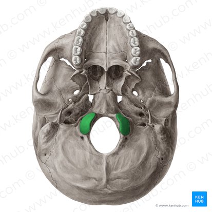
Occipital condyles
lateral to foramen magnum; articulates with cervical vertebra #1 (C1 or atlas) and allows you to nod "yes"
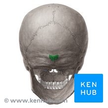
External occipital protuberance
at the midline posterior to the foramen magnum
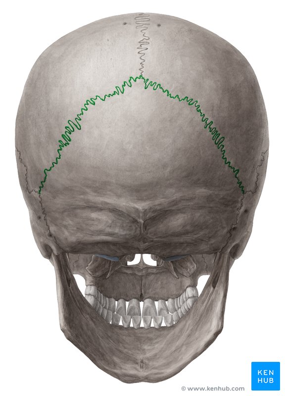
Sutures
an immovable (synarthrotic) joint where irregular edges of bones interlock or overlap

lambdoidal suture
separates the occipital & parietal bones

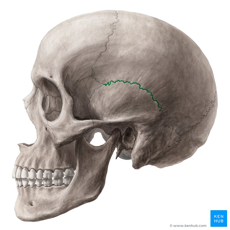
squamosal suture
separates the temporal & parietal bones
sagittal suture
separates the L & R parietal bones

Coronal suture
separates the frontal & parietal bones

Sutural (Wormian) bones
small bones located within sutures; variable number (not occiptial)
Sphenoid bone
an irregular bone anterior to the temporal bone;forms the middle floor of the cranial cavity and
part of the lateral orbits; contains numerous foramina for some cranial nerves and blood vessels;

Sphenoidal sinuses
has 2 paranasal sinuses, in the sphenoid bone

Sella turcica
on cranial floor; houses/protects the pituitary gland

Greater wing (Sphenoid)
large lateral masses of the sphenoid; like a butterfly

Lesser wing (Sphenoid)
smaller and more anterior portion of the sphenoid

Ethmoid bone
an irregular bone anterior to the sphenoid; forms part of orbits, roof of nasal cavity, upper nasal septum

Ethmoid sinuses
has 2 paranasal sinuses, in ethmoid bone

Crista galli
superior vertical projection which serves as attachment point for the outer membrane of the brain

Cribriform plate
on either side of the crista galli;

Olfactory foramina
the passageways for olfactory (smell) nerves

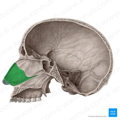
Perpendicular plate (Ethmoid)
inferior bony plate which forms the superior

Nasal septum
bone & cartilage which separates the nasal cavity into R&L compartments
Superior nasal conchae (and Medial nasal conchae)
delicate scroll-like bones which facilitate air flow and support the
nasal mucous membranes note

Inferior nasal conchae
separate bones

fontanels
(fetal skull) areas of embryonic dense CT ("soft spots") which allow for skull compression during parturition
and for brain development; fully ossified by age 2
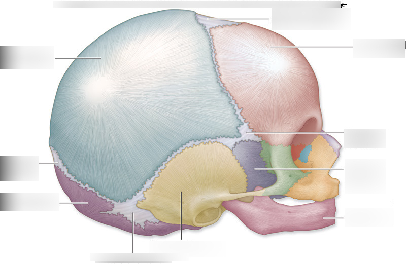
Frontal fontanels (anterior fontanels)
at the juncture of the frontal and parietal bones; the largest

Occipital fontanels (posterior fontanels)
at the juncture of the occipital and parietal bones

Mastoid fontanels
near the mastoid process

Sphenoid fontanels
near the sphenoid bone

Facial bones
form the face and jaw

Maxillae
the upper jaw; forms the floor of the orbits, sides and floor of the nasal cavity and the anterior hard palate
(roof of mouth); alveolar processes hold the upper teeth;

Maxillary sinus
sizing of sinus is large and it is in the maxillae bone

Infraorbital foramen
nerve and blood vessel passageway below the orbit

Palatine process
forms the anterior hard palate

Palatines
irregular bone which forms the posterior hard palate & part of the orbit & nasal cavity

Zygomatic bone
cheekbones; forms cheek and parts of the orbit

Zygomatic arch
forms from the zygomatic temporal process and the zygomatic process of the temporal bone
(i.e. is 2 bones); palpable anterior to your ear; for attachment of chewing muscles

Vomer
forms the lower part of nasal septum (along with the ethmoid/perpendicular plate

Lacrimal
small bones in medial orbit wall between ethmoid and maxilla; houses duct which carries tears (lacrimal =
tear) from the eye to the nasal cavity

Nasal bone
form the bridge of the nose

Mandible
the lower jaw bone, the only movable skull bone
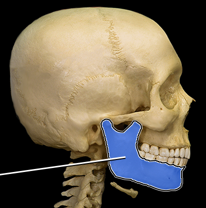
Body (Mandible)
horizontal horse-shoe shaped portion which forms the chin

Ramus
vertical extension extending from the body (ramus = bar)

Mandibular condyle
articulates with the temporal bone

Temporomandibular joint
the mandibular condyle articulate the temporal bone to form temporomandicular joint

Coronoid process (Mandible)
anterior process, for attachment of a chewing muscle

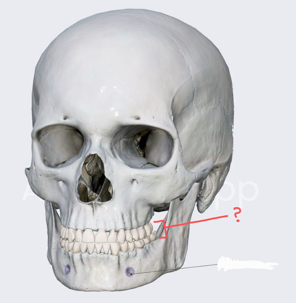
Mental foramen
In middle of the body of the mandible; for blood vessels and innervation of teeth

Alveolar processes
sockets which hold lower teeth in place (same in maxilla)
Hyoid
located in the neck (not part of skull) between the mandible and larynx; attachment point for tongue and
swallowing muscles;
the only bone that does not articulate

Thoracic cage
encloses and protects the thoracic viscera

Sternum
the "breast bone" which forms the anterior thoracic cage

Manubrium
superior portion, like the knot of a tie; articulates with the clavicle & rib 1

Body (sternum)
large middle portion; articulates with ribs

Xiphoid process
small inferior portion; attachment point for some abdominal muscles

Ribs
(12 pairs) are flat bones

True ribs
1-7 which articulate directly with sternum (via costal cartilage)
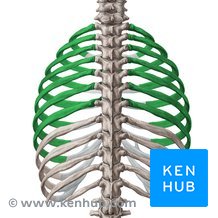
Costal cartilage

False ribs (5 pairs)
ribs 8-12 which do not articulate directly with the sternum

False ribs (ribs 8-10)
articulate with the sternum via the costal cartilage of rib 7

False ribs (ribs 11-12)
do not articulate with the sternum at all (are called floating ribs)

Head (Ribs)
articulate with the body of a vertebrae

Tubercle (Ribs)
articulates with a transverse process of a vertebrae

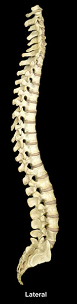
Vertebral column
the "backbone"
(33; including sacrum & coccyx); most C, T & L are separated by intervertebral disks
Cervical
form the neck and are very flexible; have transverse foramina; C1-C6 have forked transverse processes

Vertebral prominens
Part of cervical bone
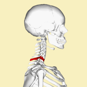
Atlas
C1; has no spine or body; large facet for articulation with occipital condyles

Axis
C2; has an odontoid process (dens), no intervertebral disk between C1 & C2

Odontoid process (dens)
which forms a pivot joint with the atlas;
allows lateral rotation of head

Thoracic
have facets on transverse processes & body for articulation with ribs; relatively inflexible

Lumbar
have large strong bodies which support the weight of the body; more flexible than T vertebrae; area where most disk herniations occur;

Intervertebral foramina
openings on the lateral side of the vertebral column between vertebrae; passage for spinal nerves

Body (Vertebrae)
thick anterior portion; contacts intervertebral disks

Vertebral foramen
passageway for spinal cord; bounded by body, pedicle and lamina

Pedicle
forms sides of the vertebral foramen, between body & transverse proces

Lamina
plate-like area between pedicle and spinous process
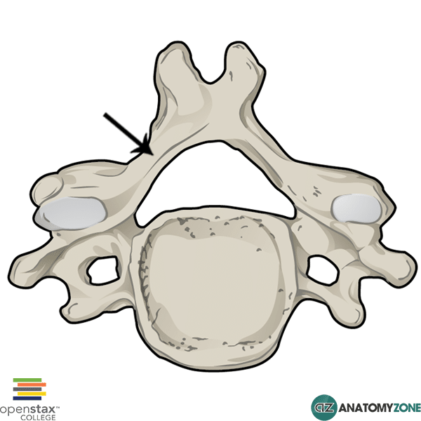
Transverse process
projects laterally and posteriorly; for muscle attachment
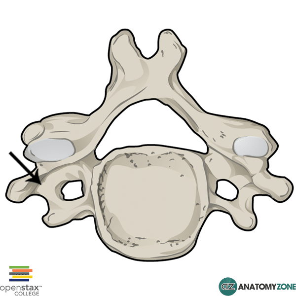
Spinous process
projects posteriorly from the medial portion; for muscle attachment

Superior articular process
paired projections lateral to the vertebral foramen on the superior aspect of the
vertebra; articulates with the inferior articular process of the adjacent superior vertebrae (is a synovial joint)

Inferior articular process
similar to the above but on the inferior aspect of vertebrae

Transverse foramen (Vertebrae)
(C only) passageway for vertebral artery & vein (goes to the brain)

Facet (rib tubercle)
(T only) on transverse process; articulates with rib tubercle

Facet (head of rib)
(T only) two on each side of a vertebra (facet or demifacet); articulates with head of rib facets are on 1, 10-12; demifacets (articulate w/ 2 ribs) are on 2-9

Sacrum
5 fused vertebrae at the base of the vertebral column; articulates with pelvic girdle/ilium and thus connects
to appendicular skeleton

Sacral foramina
four pairs; for passage of nerves and blood vessels

Sacral canal
central hollow area which is an extension of the vertebral foramina

Sacral promontory
anterior border of S1, an anatomical landmark

Coccyx
4 fused vertebrae; the "tailbone"

Pectoral girdle
The shoulder girdle; supports and connects arms to the thoracic cage

Clavicle
The "collar bone"; extends from the acromion of the scapula to the sternum

Sternal end
medial round & flat end

Acromial end
lateral end which articulates with acromion of the scapula

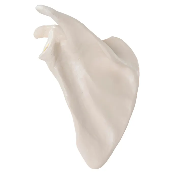
Scapula
there is two right and left pairs

Right scapula