Quiz 6: Cell Junctions, Cell Adhesion and the Extracellular Matrix
 Call Kai
 Learn
 Practice Test
 Spaced Repetition
 Match
 Flashcards
 Knowt Play
1/77

There's no tags or description
Looks like no tags are added yet.
78 Terms
two main ways animal cells held together
epithelial tissue: exs are the lining of the gut or epidermis of the skin
cells are tightly bound together into sheets
the cytoskeletons of the cells are linked from cell to cell anchoring junctions
cell-matrix attachments bond epithelial tissue to the connective tissue beneath it
connective tissue: exs are bone or tendons
formed from an ECM produced by cells that are distributed sparsely in the matrix
it is the matrix, rather than the cells, that bears most of the mechanical stress to which the issue is subjected
direct attachments between one cell and another are relatively rare, but cells have important attachments to the matrix

four functional classes of cell junctions in animal tissues
Epithelial sheets are central to the construction of multicellular animals
signal relaying junctions - allow signals to be relayed from cell to cell across their plasma membranes at sites of cell-to-cell contact; complex structures typically involving anchorage proteins alongside proteins mediating signal transduction
Chemical synapses
Immunological synapses
Transmembrane ligand-receptor pairs for cell-cell communication
Relative positions of junctions are the same in all epithelial cells:
Anchoring junctions - include cell-cell adhesions at adherens junctions or desmosomes (transmembrane cadherins) and cell-matrix adhesions at hemidesmosomes (transmembrane integrins); transmit stresses and are tethered to cytoskeletal filaments inside the cell.
Occluding (tight) junctions - seal the gaps between cells in epithelia to make a cell sheet into an impermeable or selectively permeable barrier; involve claudin proteins
Channel forming junctions - create passageways linking the cytoplasms of adjacent cells for passage of small molecules and ions; composed of connexin or innexin proteins
What do transmembrane adhesion proteins do?
link the cytoskeleton to extracellular structures
cell-to-cell anchorage. typically emdiated by ___ and they must be ___ and ___
cadherins
Cell-cell junctions must be dynamic and adaptable so they can be altered or rearranged when tissues are remodeled or repaired
cell-to-matrix anchorage, typically mediated by ___
integrin receptors
Internal linkage of transmembrane proteins to cytoskeleton is usually indirect, via intracellular anchor proteins
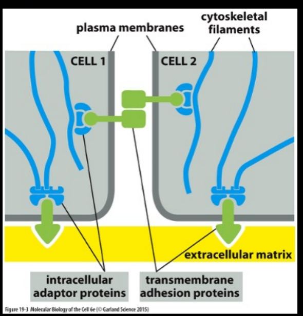
anchoring junction types
cell-cell:
adherens junction
desmosome
cell-matrix
actin-linked cell-matrix junction
hemidesmosomes
adherens junction transmembrane adhesion prot, extracellular ligand, intracellular cytoskeleton attachment, and intracellular adaptor prots
transmembrane adhesion prot: classical cadherins
extracellular ligand: classical cadherin on neighboring cell
intracellular cytoskeleton attachment: actin filaments
intracellular adaptor prots: alpha-Catenin, beta-caenin, plakoglobin (y-catenin), p120-catenin, vinculin
desmosome transmembrane adhesion prot, extracellular ligand, intracellular cytoskeleton attachment, and intracellular adaptor prots
transmembrane adhesion prot: nonclassical cadherins (desmoglein, desmocollin)
extracellular ligand: desmoglein and desmocollin on neighboring cell
intracellular cytoskeleton attachment: intermediate filaments
intracellular adaptor prots: Plakoglobin (y-catenin), plakophilin, desmoplakin
actin-linked cell-matrix junction transmembrane adhesion prot, extracellular ligand, intracellular cytoskeleton attachment, and intracellular adaptor prots
transmembrane adhesion prot: integrin
extracellular ligand: extracellular matrix prot
intracellular cytoskeleton attachment: actin filaments
intracellular adaptor prots: talin, kindlin, vinculin, paxillin, focal adhesion kinase (FAK), numerous others
hemidesmosome transmembrane adhesion prot, extracellular ligand, intracellular cytoskeleton attachment, and intracellular adaptor prots
transmembrane adhesion prot: alpha6beta4 integrin, type XVII collagen
extracellular ligand: ECM prots
intracellular cytoskeleton attachment: intermediate filaments
intracellular adaptor prots: Plectin, BP230
cadherin superfamily and integrin superfamily both exhibit
specialization based on the superfamily member expressed and the cytoskeleton filament with which it associates
types of cell-cell adhesion
homophilic binding, heterophilic binding
Large variety of plasma membrane proteins mediate cell-cell adhesion
Homophilic adhesion: an adhesion molecule expressed on the surface of one cell binds with an identical adhesion molecule on the surface of an adjacent cell
Cadherin molecules and many other types of cell adhesion molecules (CAMs) interact through homophilic adhesion
Other CAMs interact through heterophilic binding (similar to a ligand-receptor interaction), in which the interacting adhesion molecules are structurally unrelated

cadherin superfamily:
• All are transmembrane proteins except GPI-linked T-cadherin
• Multiple copies of cadherin domain motif in extracellular portions
• Intracellular portions are varied, and interact with a wide variety of intracellular ligands (signaling molecules and proteins that anchor the cadherin to the cytoskeleton)
classical cadherins:
• originally named based on tissues in which they were isolated, but also expressed in other tissues.
• closely related in sequence throughout their extracellular and intracellular domains
• These cadherins typically mediate homophilic adhesion
non-classical cadherins:
more distantly related in sequence; include proteins with known adhesive functions (desmocollin, 11 • desmoglein, protocadherins) and those primarily involved in signaling (T-cadherin, Fat, Flamingo)
• These molecules often mediate heterophilic adhesion
classical madherins mediate ___-dependent ___ adhesion
Ca2+ and homophilic
binding for homophilic adhesion occurs at the … Also descrive calcium depedent adhesion
N-terminal tips of the cadherin molecules on adjacent cells
Each of these N-terminal domains forms a knob and a nearby pocket involved in the binding interaction
Calcium dependent adhesion; calcium ions bind to the hinge region and prevent it from flexing • Spacing between the cell membranes at an anchoring junction is precisely defined and depends on the structure of the participating cadherin molecules.
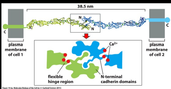
describe how classical cadheins look and how they work
Classical cadherin contains five copies of the extracellular cadherin domain
Each cadherin domain forms a somewhat rigid unit that is joined to an adjacent cadherin domain by a hinge.
Ca2+ ions bind to sites near each hinge and prevent it from flexing. When Ca2+ is removed, the hinges can flex and the structure becomes floppy, resulting in a weakened interaction with the cadherin on an adjacent cell (prone to proteolysis)

binding strength of cadherins is enhanced through ___. Describe the mechanisms
clustering at cell-cell junctions
Cadherins molecules are arrayed in parallel in the plasma membrane
Function like Velcro to hold cells together (trans interactions)
Side to side interactions between cadherins on the same cell also occur at the N-terminus (cis interactions); results in linear arrays on one cell that are perpendicular to those on the other cell
affinity
describes the strength of a single interaction (Ab to antigen; CAM on one cell interacting with CAM on adjacent cell)
CAM-CAM binding is typically a low affinity interaction
avidity
refers to the accumulated strength of multiple affinities of individual non-covalent binding interactions (i.e. functional affinity).
Similar to Velcro effect of binding; strength of this junction is much greater than that of any individual intermolecular bond
Localized clustering of CAM-CAM interactions lead to high avidity; strength of this junction is far greater than that of any individual intermolecular bond
Regulatory mechanisms can easily disassemble the junction by separating the molecules sequentially (like peeling apart Velcro)
ex of how cadherins mediate Ca2+ dependent cell-cell adhesion in all animals
Embryonic mouse cells express E-cadherin at the 8 cell stage, and become strongly and closely adherent to one another.
Mutations that inactivate E-cadherin cause embryos to fall apart and die early in development.

changing patterns of cadherin expression during construction of the vertebrate nervous system
Early chick embryo development shown: The neural tube detaches from the overlying ectoderm, and then neural crest cells detach from the neural tube for peripheral migration
As the patterns of gene expression change, the different groups of cells segregate from one another according to the cadherins they express.
Assembly of cells into an epithelium is a reversible process: Switch on expression of adhesion molecules to drive formation of an epithelium, or activate specific signaling pathways to inhibit expression of cadherins and cells become more migratory in phenotype.
epithelial-mesenchymal transitions transcription regulatory prots:
Slug, Snail, Twist
Cadherins form ___ attachments
specific (not just general adhesion)
Highly selective recognition, enables cells of a similar type to stick together and stay segregated from other types of cells
Important role during development of tissue layers (epidermis, mesoderm, and neural plate)
ex showing how cadherins form specific attachments
Experiment shown uses L cells = nonadherent fibroblast cell line that doesn’t normally express cadherins
Express cadherins in these cells in culture, and they can sort themselves out according to the type and level of cadherins they express (both qualitative and quantitative differences in the expression of cadherins have a role in organizing tissues)
Cadherins can be expressed in nonadherent L cells to illustrate homophilic adhesion (block with cadherin specific antibody)
Cadherin mediated sorting is due to highly selective recognition, not just due to glue-like adhesion of cells (reason why so many cadherins are expressed in organisms)

what interacts directly with the cytoplasmic tail of classical cadherins? what interacts with them?
p120 and beta-catenin
adherens junctions: p120 catenin regulates assembly of whole complex
linage to actin is essential for efficient cell-cell adhesion
***classical cadherins lacking their cytoplasmic tail cannot hold cells together
alpha-catenin interacts with beta-catenin and recruits other prots to provide a dynamic linkage to actin filaments
Loss of p120 catenin results in degradation of cadherin proteins, and cell-cell adhesion is lost.
Cells use changes in the level of p120 expression or its phosphorylation state as a way to regulate their strength of adhesion.

assembly of an adherens junction
Steps:membrane protrusions initate cell-cell contact —> and cadherin recruitment expands junction —> actin remodeling and myosin recruitment expands the adherens junction
Adherens junctions are enormous protein complexes containing thousands of cadherin molecules, packed into dense, regular arrays that are linked by binding at N-termini on the extracellular side
On the cytoplasmic side, a complex of catenins, actin regulators, and contractile actin bundles holds the cluster of cadherins together and links it to the actin cytoskeleton
The contractile activity generates tension (mechanotransduction) that stimulates further actin recruitment and expansion of the junction

mechanotransduction in an adherens junction
Mechanotransduction: ability to transduce a mechanical signal into a change in junctional behavior
Adherens junctions sense the forces acting on them and modify local actin and myosin behavior to balance the forces on both sides of the junction (mechanism not well understood)
Depends, in part, on proteins in the cadherin complex that alter their shape when stretched by tension (e.g. tension from non-muscle myosin II causes unfolding of alpha-catenin, that exposes a binding site for vinculin, which acts to bring additional actin filaments into the junction)

adherens junctions in epithelial cells: adhesion belt and contractile bundleof actin filaments
Adhesion belt (zonula adherens) close beneath the apical face of the epithelium, encircling each of the interacting cells in the sheet.
Contractile bundle of actin filaments, tethered to the plasma membrane by the cadherins and associated intracellular anchor proteins. This network can contract with the help of myosin motor proteins, providing the motile force for folding of epithelial cell sheets.

adhesion belts in epithelial cells
Contraction of epithelial sheets is important for folding into tubes, vesicles and other related structures.
Oriented contraction of the bundles of actin and myosin filaments running along adhesion belts causes the epithelial cells to narrow at their apex and helps the epithelial sheet to roll up into a tube
Steps: sheet of epithelial cells —> invagination of epithelial sheet caused by an organized tigthening of adhesion belts in selected regions of cell sheet —> epithelial tube pinches off from overlying sheet of cells —> epithelial tube

desmosomes form
Form spots of intercellular adhesion through non-classical cadherins (desmoglein and desmocollin)
Provide mechanical strength through linkage to intermediate filaments (keratin filaments in epithelial cells) and linkage to similar bundles in adjacent cells; creates a framework that extends throughout the tissue for tensile strength
they link the keratin IFs of one epithelial cell to those of another
Hemidesmosomes are
Epithelial cells are anchored on their basal side to the basal lamina, which lies at the interface with the underlying tissue
Alpha 6 beta 4 integrin anchors the cell to laminin in the basal lamina. Also associates with an unusual transmembrane collagen, type XVII
Intracellular attachment is to keratin (intermediate filaments), via intracellular anchor proteins plectin and BP230
keratin intermediate filament networks of adjacent cells are indirectly connected to one another through ___ and to the basal lamia through ___
desmosomes; hemidesmosomes

Epithelia are selective permeability barriers to
tight junctions
they seal gaps b/w cells
function as fences for localized distribution of apical and baslateral directed prots
describe transcellular transport of nutrients across the epithelium from gut lumen to the blood:
Example: Glucose is actively transported into the cell by Na+ driven glucose symporters on apical surface (Na+ moves inward toward lower concentration in cell, transports glucose along).
Glucose diffuses out of the cell by facilitated diffusion mediated by glucose carriers in its basolateral membrane.
Na+-K+ pump in basolateral domain returns intracellular Na+ to a lower concentration (3 Na+ out for 2 K+ in; requires ATP)
a low-molecular weight tracer added to one side of an epithelium will generally ___ beyond the tight junction
not pass
epithelia are classfied as "___” or “___” depending on ___
tight; leaky; the ability of the tight junction to prevent water nad solute movement
Tight epithelia have tight junctions that prevent most movement between cells. Examples of tight epithelia include the distal convoluted tubule, the collecting duct of the nephron in the kidney, and the bile ducts in liver tissue.
Leaky epithelia do not have these tight junctions, or have less complex tight junctions. For instance, the tight junction in the kidney proximal tubule, a very leaky epithelium, has only two to three junctional strands, and these strands exhibit infrequent large slit breaks.

tight hunctions are (structure)
form a branching network of sealing strands that completely encircles the apical end of each cell in the epithelial sheet
made of transmembrane claudin and occludin prots that hold adjacent cells together
Claudins are essential for tight-junction formation and function
Occludins are a second major transmembrane protein of tight junctions, important for limiting junctional permeability, but not essential for the assembly or structure of the tight junction •
Mice that lack the Claudin-1 gene fail to make tight junctions between the cells in the epidermal layer of the skin. As a result, the baby mice lose water rapidly by evaporation through the skin and die within a day after birth.
Expression of specific claudin proteins confers selective permeability to specific ions by forming paracellular pores (e.g. tight junctions in epithelium lining the small intestine are 10,000 times more permeable to inorganic ions, such as sodium, than the tight junctions in the epithelium lining the urinary bladder).
junctional complex prots depend on each other for their positioning:
1) tight junctions
2) adherens junctions
3) desmosomal junctions
tight junctions are positioned just apical to the adherens and desmosome junctions.
gap junctions: passageways from cell to cell
continuous aqueous pores
couple the cytoplasm between cells electrically and metabolically
only molecules smaller than ~1000 daltons can pass through gap junctions (injected fluorescent tracers of various sizes into one cell and follow diffusion to the adjacent cell)
gap junctions consist of many connexon pairs in parallel; describe them and how they work
In gap junctions, the membranes between two adjacent cells are separated by a uniform narrow gap of 2-4 nm
Gap is spanned by channel forming proteins: Connexins and Innexins
6 Connexins assemble to form a hemichannel with a 1.5 nm diameter opening: Connexon
Connexins are 4 pass transmembrane proteins; 6 assemble to form a hemichannel
Aligned hemichannels form a continuous aqueous channel that connects the cell interiors • Gap junctions consist of many connexon pairs in parallel
Pore size of ~1.4 nm, allows exchange of inorganic ions and other small water-soluble molecules, but not larger proteins or nucleic acids
Dynamic structures that can be readily assembled, disassembled or remodeled
Regulated by the cell so not always found in the open state: opening and closing triggered by a variety of stimuli, including voltage differences between cells, membrane potential of each cell, pH and concentration of free calcium ions
Connexin molecules have a half-life of only a few hours, and turnover of connexons is continuous in a cell
Cells can rapidly regulate the permeability of their gap junctions in response to ___
changes in pH, Ca2+ concentration or extracellular signals (neurotransmitters)
A. A neuron in a rabbit retina (center) was injected with a dye that passes readily through gap junctions. The dye diffuses rapidly from the injected cell to label the surrounding neurons, which are connected by gap junctions.
B. Treatment of the retina with the neurotransmitter dopamine prior to dye injection decreases the permeability of the gap junctions and hampers the spread of the dye. (Courtesy of David Vaney.)
Electrical coupling through gap junctions also synchronizes the contractions of heart muscle cells, and the smooth muscle cells responsible for the peristaltic movements of the intestine
selectins and their rloles
Selectins are lectins (carbohydrate binding proteins)
Mediate transient, cell-cell adhesion interactions in the bloodstream
conserved lectin domain binds to a specific oligosaccharide on another cepp
heterophilic adhesion
requires Ca2+ for adhesive function
Main role is in inflammatory responses and in governing the traffic of white blood cells (control binding to the endothelial cells lining blood vessels, to enable white blood cells to migrate out of the bloodstream into a tissue)
how do selectins mediate transient cell-cell adhesions in bloodshtream>
L-selectin on white blood cells binds weakly to carbohydrate ligand on endothelial cells to allow rolling along the endothelium of blood vessels, propelled by the flow of blood.
When endothelial cells are activated by an inflammatory response, they switch on expression of E-selectin which binds to carbohydrates expressed on the white blood cells.
Activation of integrin LFA1 by the white blood cell allows it to adhere more tightly by binding to ICAM1 on endothelial cells, and crawl out of the blood vessel through adjacent endothelial cells (process requires another Ig superfamily member called PECAM1)
immunoglobulin (Ig)-superfamily adhesion molecules: NCAM and ICAM
Extracellular Ig-like domain that is characteristic of antibody molecules
Ca2+ independent adhesion
weaker binding that cadherins, contribute to fine tuning of adhesive interactions
NCAM (neural cell adhesion molecule) = homophilic; found as many isotypes due to alternative splicing of an RNA transcript from a single gene
ICAM (Intercellular Cell Adhesion Molecule) = heterophilic binding (ICAM expressed on endothelial cells binds to LFA1 integrin on white blood cells)
N-Cadherin knockout mouse dies early in development; knockout of NCAM results in mostly normal development except for mild abnormalities in specific tissues of the nervous system
some forms of NCAM carry an unusually large quantity of negatively charged sialic acid; these forms of NCAM serve to inhibit adhesion due to like charges repelling each other on adjacent membranes

ECM
The extracellular matrix (ECM) is made and oriented by the cells within it – primarily secreted by fibroblasts (in cartilage and bone, it is secreted by chondroblasts and osteoblasts respectively)
ECM constructed of proteoglycans, fibrous proteins (collagens and elastic fibers), and glycoproteins (laminin and fibronectin)
Epithelial cells are separated from the underlying tissue by the Basal Lamina; laminin is the primary organizer of the basal lamina
Fibroblasts in connective tissue (ECM)
Extracellular matrix (ECM) surrounding the fibroblasts here is composed largely of collagen fibrils
Hyaluronan and proteoglycans normally form a hydrated gel filling the interstices of the fibrous network
extracellular matrix of connective tissue is composed primarily of:
1. Glycosaminoglycans (GAGS), which are large and highly charged polysaccharides that are usually covalently linked to specific core proteins to form Proteoglycans
2. Fibrous proteins, Elastin and members of the collagen family
3. Non-collagen Glycoproteins, with short N-linked oligosaccharide side chains

Glycosaminoglycan (GAG) chains
unbranched polysaccharide chains composed of repeating disaccharide units attached to a core prot
GAGs are highly (-) charged
Protein is shown in green, GAG chains in red Unbranched polysaccharide chains composed of repeating disaccharide units
one of the two sugars is always an amino sugar that is often sulfated • second sugar is usually a uronic acid
how to polysaccharide chains and GAGs form a hydrated gel?
Polysaccharide chains are too stiff to fold into compact globular structures and they are highly hydrophilic
GAGs adopt highly extended conformations that occupy a huge volume relative to their mass
Form hydrated gels even at very low concentrations; expands with water to occupy a large volume
proteoglycans in the ECM form a highly hydrated, gel-like matrix in which fibrous prots are embedded
resists compressive forces on the matrix while permitting rapid diffusion of nutrients metobolites and hormones
hyaluronan is a GAG trhat consists of repeating seq of disaccharide units that are not siulfated, acts as a space filler
major compoent of cartilage
role of charge in hydrated gels:
The negative charges on the proteoglycans trap an equal number of cations to maintain electrical neutrality.
Electrostatic forces confine these charges, both the fixed negative charges on the polysaccharide and the mobile cations, to the volume occupied by the proteoglycan.
The concentration of particles in the volume of the proteoglycan is higher than in the surrounding solution; hence, the water flows in to try to equalize the concentrations inside and outside. The proteoglycans thus trap water to form a hydrated gel by drawing in water molecules by osmosis.
collagen molecule
Gly-X-Y repeating arranngement per turn (X, Y often proline and hydroyproline)
Three left-handed alpha helical strands twist to form a right-handed triple helix.
Glycine occupies the interior of the triple helix
Collagen is a major component of skin and bone; constitute 25% of total protein mass in mammals
Major proteins of the ECM – provide tensile strength
Rich in proline and glycine (X is commonly proline and Y is commonly hydroxyproline)
Human genome contains 42 distinct genes coding for different collagen alpha chains; different combinations of these genes are expressed in different tissues
steps of collagen assembly:
Individual polypeptide chains to triple-stranded collagen molecules, then to fibrils and, finally, fibers.
Covalent intramolecular and intermolecular crosslinks organize the fibrils to form structures that resist tensile forces
collagen is synthesized as a ____ molecule that has; mechanim
procollagen molecule; large unstructered peptides at either end
Diverse COPII vesicles are required for the transport of large cargo molecules
Procollagen cargo molecules (300 nm long) bind to transmembrane packaging proteins in the ER that control the assembly of COPII coat components into much larger COPII vesicles with tighter packing of inner and outer coat proteins (The packaging proteins remain in the ER)
When the procollagen is secreted, extracellular procollagen proteinases remove its terminal peptides, producing mature collagen molecules. These molecules can then self-assemble into ordered collagen fibrils (end-to-end and side-to-side in a staggered array to form thin, cylindrical fibrils)
The striping on the collagen fibril is due to the regular, staggered arrangement of the collagen molecules within the fibril
Covalent intramolecular and intermolecular crosslinks organize the fibrils to form structures that resist tensile forces
how synthesis of collagen fibrils form resistant
In mammalian skin, collagen fibrils are woven in a wickerwork pattern, alternating layers with different orientations, so they resist tensile stress in multiple directions.
In tendons, which attach muscle to bone, they are organized in parallel bundles aligned along the major axis of tension.
In mature bone and in the cornea, they are arranged in layers, with the fibrils in each layer lying parallel to one another but nearly at right angles to the fibrils in the layers on either side.
type II collagen:
Fibril forming collagen molecule, secreted into the extracellular space, where it assembles into higher order polymers called collagen fibrils.
Collagen fibrils often aggregate into larger cable-like bundles, called collagen fibers
Localized to cartilage and intervertebral discs
type IX collagen:
Fibril-associated collagen that decorates the surface of collagen fibrils
Thought to link the fibrils to one another and to other components in the ECM
Cells interact with the ECM both chemically and mechanically; fibroblasts will tug on a randomly oriented collagen meshwork, causing the gel to contract by drawing in the collagen fibers
cells shape the ECM by aligning ___ fibers
collagen fibers
Cells interact with the ECM both chemically and mechanically; fibroblasts will tug on a randomly oriented collagen meshwork, causing the gel to contract by drawing in the collagen fibers
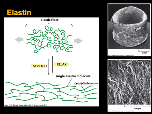
ECM molecules II: Fibrous Prot; Elastin molecules form ___
random coils that can easily stretch and contract; joined together by covalent bonds to generate a cross-linked network
Elasticity of elastin fibers derives from their lack of secondary structure
Elastic fibers in the ECM of skin, blood vessels and lungs give these tissues the resilience to recoil after transient stretch
Shown: elastic fibers in the outer layer of a blood vessel
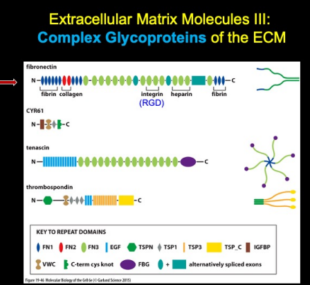
ECM Molecules III: Complex Glycoprots
Many matrix glycoproteins are large scaffold proteins containing multiple copies of specific proteininteraction domains
Each domain is folded into a discrete globular structure, with many domains arrayed along the protein like beads on a string
Roughly 200 matrix glycoproteins are known in mammals

Complex Glycoprot: Fibronectin; what do they bind to, through what, more info…
Fibronectin binds to integrins through its RGD sequence (Arg-Gly-Asp)
Large glycoprotein dimer composed of two very large subunits joined by disulfide bonds at one end. Each subunit is folded into a series of functionally distinct domains separated by regions of flexible polypeptide chain.
Domains consist of smaller modules which are serially repeated
No self assembly of fibronectin into fibrils; fibrils only form on the surface of cells that express integrin receptors. Binding of fibronectin with integrin receptors provides linkage to the actin cytoskeleton and transmits tension to the fibronectin molecules.

How do binding of fibronectin w/ integrin receptors provide linkage to the actin cytoskeleton and transmit tension to the fibronectin molecules?
(A)Actin monomers assemble on the barbed end of actin filaments at the leading edge. Transmembrane integrin proteins help form focal adhesions that link the cell membrane to the substrate.
(B) If there is no interaction between the actin filaments and focal adhesions, the actin filament is driven rearward by newly assembled actin. Myosin motors also contribute to filament movement.
(C) Interactions between actin-binding adaptor proteins and integrins link the actin cytoskeleton to the substratum. Myosin-mediated contractile forces are then transmitted through the focal adhesion to generate traction on the extracellular matrix, and new actin polymerization drives the leading edge forward in a protrusion.
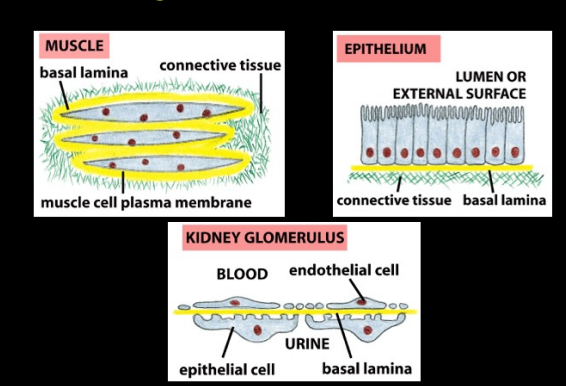
organization of basal lamia
• Basal lamina (basement membrane) is a thin, flexible layer of secreted extracellular matrix molecules with a critical structural role in the architecture of the body.
Basal lamina underlies all epithelial cells and surrounds muscle, fat and Schwann cells to separate them from the underlying or surrounding connective tissue, and forms a mechanical connection between them
function of the basal lamia
selective filter in the kidney glomerulus; proteoglycan role
selective barrier to movement of cells:
no fibroblast contact w/ epithelial cells
macrophages, lymphocytes and nerve processes can move through
tissue regeneration after injury; provides a scaffold for cell migration
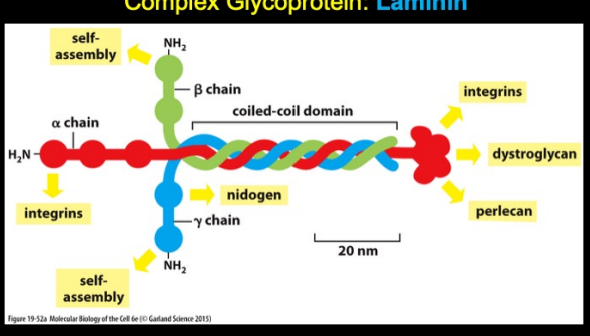
complex glycoprot: Laminin
• Laminin is the primary organizer of the sheet structure of basal lamina ; first join to each other while bound to receptors on the surface of the cells that produce them (integrins and dystroglycan)
Large, flexible protein composed of 3 polypeptide chains held together by disulfide bonds, with chains twisted together at one end, but separated portions of the chains at the other end.
The heterotrimers can self-assemble in vitro into a network, but require interaction with cells to organize the network into an orderly sheet.
Mice lacking the y-alpha-µµa-1 chain die during embryogenesis because they are unable to make basal lamina.
Type IV collagen gives the basal lamina tensile strength
Nidogen and perlecan serve as linkers to connect the laminin and type IV collagen networks once the laminin is in place

junctional basal lamia at NMJ has what?
special organization characteristics
When the nerve, but not the muscle, is allowed to regenerate, the junctional basal lamina directs the regenerating nerve to the original synaptic site
When the muscle, but not the nerve, is allowed to regenerate, the junctional basal lamina causes newly made acetylcholine receptors to accumulate at the original synaptic site
The neuromuscular junction is reestablished based on information contained in the basal lamina: Special molecules present in the basal lamina mark the site of the junction and allow it to be reconstituted exactly
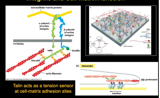
what are integrins? what does thetail bind to?what are some other relavent prots?
Integrins are the principal receptors on animal cells for binding most extracellular matrix (ECM) proteins; transmembrane heterodimers that link to theChat actin cytoskeleton; divalent cation binding domains (calcium and magnesium)
The head of an integrin molecule attaches directly to an ECM protein, and the intracellular tail binds to talin, which links the integrin to filamentous actin.
Talin as a tension sensor at cell-matrix adhesion sites: the tension between integrin and actin filaments causes talin to unfold and reveal a binding site for vinculin, which provides additional linkages to actin filaments
Kindlin is a regulatory protein that binds to the tail of the beta integrin subunit
Vinculin, alpha-actinin and filamin help to reinforce the linkage to actin
integrin structure
Integrins are a diverse family of transmembrane adhesion receptors that mediate cell-cell and cellmatrix adhesion
Each integrin contains one alpha subunit and one beta subunit
At least eighteen alpha subunits and eight beta subunits have been characterized so far and form 24 different integrins in vertebrates
The specific linkage of the extracellular domains of integrins to ECM proteins, or in some cases, to counter receptors on adjacent cells, supports cell adhesion and is crucial for embryonic development, tissue maintenance and repair, host defenses and haemostasis (coordinated effort between platelets and numerous blood clotting proteins, resulting in the formation of a blood clot and subsequent stopping of a bleed from a damaged blood vessel)

integrins switch b/w an active site and an inactive site
Allosteric regulation: as an integrin binds to or detaches from its ligands, it undergoes conformational changes that affect both the intracellular and the extracellular ends of the molecule.
Structural change at one end is coupled to structural change at the other.
“Outside-In Activation”: Binding of extracellular ligand causes an unfolding of the integrin, the intracellular domains move apart and reveal the talin binding site on the tail of the beta chain, binding of talin leads to assembly of actin filaments
“Inside-Out Activation”: Talin competes with alpha chain for binding site on beta chain. When talin binds to the beta chain, it causes the two chain molecules to spring apart, driving the extracellular portion of the integrin into its extended, active conformation. Triggered by intracellular regulatory molecules (PIP2)

integrin activation in platelets:
Integrin activation in platelets:
Thrombin activates a G protein coupled receptor à activates Rap1 GTPase, which interacts with RIAM protein à recruits talin to the plasma membrane
Talin and kindlin interact with the integrin beta chain to trigger integrin activation
Talin then interacts with adaptor proteins, such as vinculin, resulting in the formation of an actin linkage
Integrins recruit what intracellular signaling prots at sites of cell-substratum adhesion?
Focal Adhesion Kinase (FAK) is activated at sites of cell-matrix contacts
focal contacts are important in cell spreading and adhesion to ECM substrates
Integrin binding can stimulate localized changes in the cytoplasm close to the cell-matrix contact
Integrins cluster at cell-matrix contacts to form focal adhesions, where activation of the small GTPase Rho plays a part in the maturation of the adhesive complex by recruiting actin filaments
FAK is a protein tyrosine kinase that is recruited to focal adhesions when integrins cluster at cell-matrix contact, cross-phosphorylate and provide a docking site for additional intracellular signaling proteins.
integrin clustering
As adhesions mature, multiprotein complexes assemble at the cytoplasmic face of clustered, ligand-bound integrins. These complexes connect integrins to the actin cytoskeleton and transmit signals into the cell.
Integrin clustering at focal adhesions is a reversible process.
Depends on the specific integrins that are involved, presence of co-signaling receptors and the nature and mechanical properties of the extracellular matrix proteins
main prots to focus on in integrin clustering:
Focal Adhesion Kinase (FAK)
Src-family Kinases (Src)
Vinculin
Vinculin
Vinculin
Important for integrin-mediated adhesion and cell spreading, although it does not directly bind to the cytoplasmic tail of beta integrin
Src-family Kinases (Src)
Src-family Kinases (Src)
Src-family protein tyrosine kinases are rapidly activated following integrin-ligand interactions. • Src-kinase activity contributes to the reinforcement of initial integrin-mediated adhesions by activating downstream kinases and adaptors.
Can bind directly to the beta-integrin tail, which contributes to activation of kinase activity and controls cell spreading. Integrin-linked Kinase (ILK)
Major role as a signaling scaffold protein; required for correct targeting of its components to integrin-mediated adhesions and protects its components from proteasomal degradation Paxillin
Another essential scaffold protein that is recruited early to integrin adhesions
Numerous phosphorylation sites provide multiple regulated sites of protein-protein interaction. Together, they mediate the binding of kinases (FAK, Src, ILK) phosphatases (PTP-PEST), actin binding proteins (vinculin and the parvins), and regulators and effectors of the Rho family of small GTPases.
Also binds directly to cytoplasmic tail of alpha4-integrin, which aids in the ability of this integrin to promote cell migration and recruitment of leukocytes to inflammatory sites
Focal Adhesion Kinase (FAK)
Focal Adhesion Kinase (FAK)
Acts as a phosphorylation-regulated signaling scaffold and is important for adhesion turnover, Rhofamily GTPase activation, cell migration and cross-talk between growth-factor signaling and integrins.
In response to integrin clustering, the autophosphorylation of FAK generates docking sites for SH2- domain-containing proteins, including Src-kinases, which in turn become activated and phosphorylate FAK, which promotes its kinase activity and interaction with other proteins.
why cells must be able to degrade matrix for migration
Cells produce proteases (matrix metalloproteases, serine proteases) which give them the ability to cut through matrix:
Enables cells to divide while embedded in matrix
Enables cells to travel through matrix
Needed during normal branching growth of epithelial structures, such as glands
Migration of white blood cells across the basal lamina of a blood vessel into tissues
Important for the spread of cancer cells through the body, and their ability to proliferate in the tissues that they invade
Matrix proteases are tightly controlled for activation only at the right time and place:
Confined to the cell surface by specific anchoring proteins, by membrane-associated activators, and controlled through the production of specific protease inhibitors in regions where protease activity is not neede