chapter 9 -- PACS fundamentals
 Call Kai
 Learn
 Practice Test
 Spaced Repetition
 Match
 Flashcards
 Knowt Play
1/36

There's no tags or description
Looks like no tags are added yet.
37 Terms
fundamentals
MIMPS
formerly picture archiving and communication system (PACS)
PACS consists of the following:
digital acquisition (picture)
display workstations
storage devices (archiving)
components are interconnected through an intricate network (communication)
PACS is the electronic version of the radiologist’s reading room and the file room
First PACS
early 1980s
served one single modality
large research institution
most developed by scientists in those institutions
later
vendors become more involved
modality-specific proprietary systems were developed
standardization
MIMPS
medical image management and processing system
defined as a device that provides one or more capabilities for review and digital processing of medical images for interpretation by a trained practitioner for disease detection, diagnosis, or patient management
PACS
a computer system designed or digital image that can receive, store, archive, distribute, and display digital images
DICOM
first version — 1985
universally accepted standard
groundwork for future integrated PACS development
components of PACS
can be broken into three fundamental parts:
image acquisition
display workstations
archive servers
image acquisition
acquired in digital format:
ultrasound
computer tomography (CT)
magnetic resonance imaging (MRI)
digital workstation
any computer used to view a digital image
most interactive part of a PACS
used inside and outside of the radiology department
archive servers
file room of the PACS
consists of the following:
database server or image manager
short and long-term storage
workflow manager
central part of the PACS
houses all of the historic and current data
may also serve as the central hub that receives all images before release for interpretation
workflow
how a process is done step by step or how a task is completed
how one completes an exam from order entry to transcribed report
exact workflow is different in every radiology department
the technologist gets the order via an electronic worklist or a paper requisition
verification of patient ID and the patient history is still required
the order is input into the radiology information system (RIS), and the RIS sends a message to the PACS to find all historical images and put them in the short-term archive
the technologist prepares the room, retrieves the patient, and acquires the patient history
the history is recorded on the requisition or input electronically into the patient’s electronic medical record
the technologist performs the exam, and the images are processed and repeated if necessary and sent to the appropriate PACS device
images are tagged with RIS information so the historic image reports are available
if the patient’s physician does not have access to the electronic images, a CD or DVD can be made
the requisition is given to the radiologist or he or she may pull the images from an electronic worklist
the radiologist may also pull historic images and reports for comparison
the radiologist dictates a report and has it transcribed
voice recognition software may be used
review of the report and correction can be made immediately after dictation, and then it is signed, making it final
with PACS, the time from completion of the exam to final report can be hours instead of days
system architecture
can be defined as the hardware and software infrastructure of a computer system
PACS architecture consists of:
acquisition devices
storage
display workstations
image management system
three common PACS architectures
client or server based
distributed systems
web-based systems
client or server based system
images are sent to the archive server
display workstation function as a client of the archive server
accesses images based on a centralized worklist
person at the client chooses a name from the list
archive server sends the image data to the client
most systems allow basic image manipulation at the client
changes are saved on the archive server
advantages
any exam sent to the PACS is available anywhere without other interventions
disadvantages
the archive server is seen as a single point of failure
if the archive goes down, the entire system is down, and no image movement can take place
newly acquired images must remain at the modality until the archive is up and can receive the images
system is network dependent
images fly back and forth between the archive and the workstation
network can become bogged down
archive server handles many requests at once and can become bottlenecked
distributed systems
also known as distributed or stand-alone models
acquisition modalities send the images to a designated reading station and possibly to review stations
in some systems, the images are sent from the modality to the archive server, and the archive server distributes the images to the designated workstation
reading station designation may be designed on radiologist’s reading preferences
example
MRI may send to one station
CT sends to another
or all cross-sectional neurologic images may be sent to one station, whereas all body imaging may be sent to another
designation is decided after extensive workflow observation
workstations can query and retrieve images from the archive
all images are locally stored
images are then sent to the archive server after the images have been read
images remain on the local hard drive of workstation until they are deleted by the user or system
advantages
if the archive server goes down, local reading at the workstations is not interrupted
after archive comes back up, the images that have been changed and signed off are automatically forwarded to the archive to be saved
PACS data less likely to be lost because of multiple copies in various locations
system is less dependent on network for speed
user can work on one exam while the workstation is pulling and getting the next exam ready to be read
the workstation can fetch historic images based on rules set up by the user
disadvantages
personnel rely heavily on the system to perform image distribution correctly
if distribution is wrong, then prefetching of historic exams will be wrong
each workstation has a different worklist; only one person at a time can work on that list
it can be inconvenient to read additional studies
the radiologist would have to move to another workstation to read the images
users must be depend on query-and-retrieve function when nonschedule exams arrive at the workstation
it is possible for two radiologists to be reading the same exam and not knowing it until they try to start dictation
paper requisition becomes an important piece of information
web-based systems
are similar to client and server systems
the biggest differences are that images and application software are held centrally and load to the client display
only the images are held at the archive
most interactive part of PACS
consists of the following:
monitor
one of the most important elements
LCD is most popular
computer with a mouse and keyboard
hardware and software fit the user’s requirements
resolution and orientation of the monitor are factors in determining which type of monitor is to be used
most cross sectional imaging is read on a 1K square monitor
most digital projection images are read on at least a 2K portrait monitor
resolution
number of pixels in the display
more pixels = higher (more information that can be displayed)
also defined as the process or capability of distinguishing among individual adjacent parts of an image
pixels are arranged in a matrix
display workstations
medical displays are generally higher quality than displays for other applications
radiologists use the highest resolution monitors available for the modality that is being read
mammography requires 5K or 5 megapixel resolution
cross-sectional image only requires a 1K monitor
1K monitors are sufficient for referring physicians
can be categorized by primary use:
primary reading stations for radiologists
review stations for referring physicians
technologist quality control (QC) station for technologist review of images
image management station for the file room personnel
each has a main purpose and is strategically located near the end user of that designated purpose
may be made up of different hardware depending on demand and the need of the user and requirements of the software that will be used
radiologist reading stations
used by radiologist to make a primary diagnosis
will have the highest quality hardware, including the best monitor
computer hardware used depends on the needs of the PACS vendor but usually is robust with little downtime
the keyboard and mouse can be customized to the needs of department
many different styles of mice are available
access to the RIS is nearby
a dictation system is near or is connected to PACS station
many systems are integrating the RIS and dictation system within the PACS software
physician review station
step-down model of the radiologist’s reading station
may have the same level of software but may reduce some advanced functions
one of the most important features is the ability to view current and previous reports with images
many vendors are integrating the RIS function with PACS software
most referring physicians want to read the radiologist’s report along with viewing images
often the report is more important to them than the images
software may be loaded on a stand-alone station that is dedicated to viewing images
software may be delivered over a web browser on any personal computer within an office or floor
in high-volume areas such as the emergency department and intensive care unit, there are dedicated PACS workstations strictly for image viewing
these may have the higher end monitors, but many may have a lower end monitor because of the costs
one of the greatest advantages of a PACS is the ability to view the same set of images in multiple locations at one time
the referring physician can pull up the patient’s images in the office and read the radiologist’s report and then call the radiologist on the phone and consult while both parties are viewing the same set of images
continuity and speed of patient care have shown improvement with the use of PACS
technologist QC stations
used to review images after acquisition but before sending them to the radiologist
may be used to improve or adjust image-quality characteristics
may be used to verify patient demographic information
placed between the CR and DR acquisition modalities as a pass-through to ensure that the images have met the department quality standard
generally has a 1K monitor
when manipulating images, the technologist should be careful not to change the appearance too much from the original acquired image
technologists should consult frequently with the radiologist to ensure quality
QC workstation can also be used to query and retrieve historic images to check previous pathology or body characteristics
QC workstations can help with the selection of technical factors or procedural protocol
QC stations can afford same benefit as pulling the film jacket before the examination
file room workstation
before PACS, file was a large open room with endless rows or shelves full of film jackets
today, often as simple as several computers and a dry laser to make copies for outside needs
a workstation may be used to look up exams for a physician or to print copies of images for the patient to take to an outside physician
many hospitals are moving away from printing films because of the cost
hospitals are moving toward burning compact disks (CDs) with the patient’s images
less expensive
made quickly
come with easy-to-use software burned into them for viewing on any PC
patient demographics may be corrected
incorrect information may make retrieval difficult
the database is only as good as the information put into it
common functions found on a PACS workstation
can be divided into four categories:
navigation function
image manipulation and enhancement function
image management function
advanced workstation function
navigation function
used to move through images, series, studies, and patients
the customizable worklist is used to navigate through patient files

hanging protocol
defines how a set of images will be displayed on the monitor
users can customize the protocol for each modality
CT exam is selected
four images can be viewed on each monitor
CR image is selected
can be viewed as one image on each monitor
study navigation
a study in PACS is the current or previous exam being viewed
may consist of 2 or 3 single images (radiography) or several (CT, MRI)
images can be run through in stacks
stack mode of scrolling through images made is called “cine”
cine function is used most often in cross-sectional imaging
image manipulation and enhancement function
window width and window level
annotations
flip and rotation
zoom (pan) and magnify
measurements
flip and rotate
used to orient the image in the correct anatomic hanging position
tool is usually a left-to-right flip or a 90 degree clockwise and counterclockwise icon
use of lead markers is important to ensure that the radiologist is reading the correct side
digital markers may be a legal liability

zoom (pan) and magnify
used primarily by the radiologist to increase the size of an area on the image
magnify usually enlarges a square area of the image
the square can be moved around the image to quickly see various areas magnified
pan and zoom functions are usually used together
image is first zoomed up to the desired magnification level
pan icon is activated so that the zoomed image can be moved around to view the different areas of the image
measurements
most common are distance and angle
another common measurement tool used is region of interest (ROI)
distance measurement
software uses pixel size to measure structures on the image
angle measurement
can give an angle measurement between two structures
commonly used when reading spine studies
region of interest (ROI)
also called volume of interest — 3-D
determines the pixel intensity of a certain area
pixel intensity: primary information stored within pixels (grayscales)
different tissues or fluid has a different intensity reading
radiologist uses this to determine whether something is solid or fluid
image management functions
patient demographics can be modified at the technologist workstation, reading station, and file room station
patient demographics must be correct
if incorrect at the archive level, the images could be lost
many hospitals allow only certain persons the access to change demographics just to keep the errors to a minimum
query/retrieve ion
used to retrieve on demand any studies from the archive
allows user to query a study on multiple fields
patient’s name or identification, date of service, modality, diagnosis code or comment field
CD burning option
feature may only be available in the file room to control the CDs that are sent out
HIPAA compliance must also be maintained
copy and paste
used with the web-based systems to create presentations for conferences
patient information must be removed from the image before it is placed into a presentation
many hospitals gave retained the ability to print films for outside use
printing is usually only done in the file room so that control can be maintained over the printed films for HIPAA purposes and cost reasons
workstations may be connected to paper printers for quick consults and for medical records

advanced workstation functions
usually placed on specialty workstations for radiologists; some are found on the technologist QC station to further enhance the images
most common advanced functions:
multiplanar reconstruction (MPR)
maximum intensity projection and minimum intensity projection (MIP and MinIP)
volume rendering technique (VRT)
shaded surface display (SSD)
stitching
multiplanar reconstruction (MPR)
one of the most commonly used 3D techniques
usually used to produce coronal images from the axial set to reduce radiation to the patient and scan time

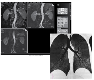
maximum intensity projection and minimum intensity projection (MIP and MinIP)
used to visualize vessels (MIP) and air-filled structures (MinIP)
commonly performed after injection of contrast on CT and MRI studies
contrasts shows areas of structures and blockages within the vessels
volume rendering technique
function similar to MIP
allows user to assign colors based on the tissue intensity
bone, contrast medium, and organs can be visualized using various colors
uses a histogram-type graph to differentiate the various structures

shaded surface display
uses a threshold of pixel intensity values
everything below the threshold is removed
everything above is assigned a color and is shown as a three-dimensional object

image postprocessing
regarded as an advanced function of the workstation
edge enhancement, smoothing, contrast enhancement
many other advanced workstation functions are available to be added to the PACS workstation
this is a growing field with advancements coming each year
specific information about how to perform these procedures can be found in the vendor’s user manual