Coronary Artery Disease and Acute Coronary Syndrome
 Call Kai
 Learn
 Practice Test
 Spaced Repetition
 Match
 Flashcards
 Knowt Play
1/43

There's no tags or description
Looks like no tags are added yet.
44 Terms
Coronary Artery Disease (CAD)
Commonly caused by atherosclerosis: Lipid-rich plaque in coronary artery walls
Endothelial injury → lipid buildup + inflammation → plaque growth
Causes narrowing and plaque rupture/erosion (leads to platelet activation + thrombus)
Risk Factors: Smoking, HTN, ↑LDL, diabetes, obesity, inactivity, age, family history
Ischemia
Myocardial O2 demand > supply → insufficient O2 to myocardium (heart)
Brief ischemia may reverse, prolonged severe ischemia → necrosis → MI
Angina, dyspnea, diaphoresis, nausea, fatigue, lightheadedness
Can impair pumping + arrhythmias; early reperfusion may prevent MI
↓Supply in Myocardial Ischemia
Atherosclerosis → narrowing
Plaque rupture → thrombus → ↓coronary flow
Vasospasm/Small vessel dysfunction → ↓flow (even w/o major blockage)
↑Demand in Myocardial Ischemia
Exercise/Stress → ↑HR, BP, contractility → ↑cardiac work
Collateral Flow
Small coronary connections may enlarge with gradual narrowing, providing alternate blood flow around a blockage
May reduce ischemia but often insufficient, especially with sudden occlusion
Ischemic Cell Injury
↓O2 = ↓ATP + anaerobic metabolism
↓Energy = impaired relaxation/contraction + disturbed ion balance → arrhythmias
Angina
Chest pressure/tightness/discomfort from ischemia ± radiation to one/both arms, jaw, neck or back
Chest pain
Stable Angina
Narrowed coronary artery + ↑demand → predictable exertion/stress symptoms
Relieved by rest or sublingual nitroglycerin
Management: Beta blocker (↓demand) or calcium channel blocker (↑supply, vasodilator) = symptom control; NTG (relief)
Lipid lowering + risk reduction = event prevention
Acute Coronary Syndrome
New, worsening, or rest symptoms. Often due to plaque rupture/erosion → thrombus
Unstable angina
NSTEMI/STEMI = non ST/ST elevation MI
ECG + troponin test
Suspect ACS if change in usual angina
Myocardial Infarction
Plaque rupture → platelets + fibrin thrombus
Requires injury + ischemic evidence (symptoms, ECG changes or image findings)
If troponin rise/fall = acute injury
Primary, secondary, or procedure-related MI
STEMI/NSTEMI describes presentation, type of MI describes cause and guides treatment
Primary MI
Acute coronary problem, commonly atherothrombosis
Secondary MI
Acute illness → O2 supply/demand imbalance (anemia, hypoxemia, hypotension, tachyarrhythmia)
Procedure-Related MI
Cardiac procedure complication → ischemic injury
Troponin
Myocardial injury marker
MI = rise/fall above sex-specific 99th percentile + ischemic evidence
Unstable Angina (UA)
Ischemic symptoms w/o acute ischemic troponin rise/fall
ST depression/T inversion or normal ECG
Risk-based NSTE-ACS care; NO fibrinolysis
NSTEMI (non-ST elevation MI)
Troponin will likely be elevated; acute injury + ischemic evidence
No persistent ST elevation; ECG may be normal
Risk-based invasive strategy; NO fibrinolysis
STEMI (ST elevation MI)
EMERGENCY
Acute ischemic presentation; troponin may be normal initially and then rise
Diagnostic ST elevation
Immediate reperfusion, do NOT wait for troponin
CAD/ACS Diagnostic Testing
ECG (12-lead ECG ≤10min)
High-Sensitivity Troponin
Echocardiogram (assess wall motion, pumping fx, complications)
Stress Test (↑cardiac workload to check for ischemia in stable pts)
Coronary Imaging (CT angiography, cardiac catherization)
ST-Depression
Downward ST segment ≥1mm
Suggests subendocardial ischemia; can also be reciprocal change or posterior MI

T-Wave Inversion
Inverted/Negative T wave
May indicate ischemia, especially when new, deep or dynamic

ST Elevation
Upward ST segment ≥1mm
Suggests acute injury/occlusion (STEMI)
EMERGENCY

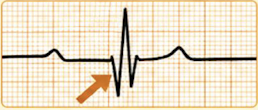
Q Waves
Wide and deep (≥0.04sec and/or ≥25% of R wave)
May indicate prior MI or evolving myocardial necrosis
STEMI
Restore blood flow immediately
PCI preferred (balloon/stent opens the artery)
Fibrinolytics if PCI will take >120min or symptoms <12h and no contraindications
Antiplatelets, anticoagulation, anti-ischemic therapy, high-intensity statin
NSTEMI/Unstable Angina (UA)
Persistent pain despite treatment or unstable BP/rhythm → immediate angiography
Stable but high-risk → angiography <24h
Antiplatelets, anticoagulation, anti-ischemic therapy, high-intensity statin
Percutaneous Coronary Intervention (PCI)
Balloon opens the narrowed artery and stent supports the artery
Rapidly restores blood flow
Preferred for STEMI pts, single-vessel disease, or suitable anatomy
Coronary Artery Bypass Grafting (CABG)
Creates a new pathway around blocked arteries using grafts
Improves blood flow to large areas of the heart
Preferred for left main disease, multivessel disease (diabetics), or complex anatomy
Use if PCI is not possible or failed
Aspirin
Antiplatelet
Cox-1 inhibition → ↓thromboxane A2 → vasoconstriction
Clopidogrel
P2Y2 Inhibitors
P2Y12 blockade → ↓ADP signaling → ↓platelet aggregation
ACS + stent thrombosis prevention, usually use w/ aspirin (DAPT)
ADRs: Bleeding/bruising, GI upset, rare TTP (thrombotic thrombocytopenic purpura)
Contraindications: Active bleeding/allergy
Avoid omeprazole/esomeprazole (↓drug activation)
DAPT (Dual Antiplatelet Therapy)
Aspirin + P2Y12 inhibitor = inhibit platelet aggregation
ACS PCI: Ticagrelol/prasugrel preferred if suitable
Fibrinolysis: Clopidogrel
Anticoagulants
Inhibit clotting factors → ↓fibrin formation → limit clot extension
Unfractionated Heparin (UFH)
↑Antithrombotic activity → inhibits thrombin + Xa → limits fibrin/clot extensionn
STEMI/NSTEMI, PCI anticoagulation
ADRs: Bleeding, HIT (heparin-induced thrombocytopenia, thrombosis despite ↓platelets)
Contraindications: Prior HIT, uncontrolled bleeding, drug/pork allergy, inability to monitor coagulation
Monitor aPTT/anti-Xa, blood count, protamine reverses UFH
Enoxaparin (Low Molecular Weight Heparin)
↑Antithrombotic activity → inhibits Xa > IIa → limits clot formation
UA/NSTEMI + selected STEMI (based on reperfusion strat)
ADRs: Bleeding, thrombocytopenia/HIT, injection reactions. Neuraxial hematoma = neurologic injury
Contraindications: Major active bleeding, immune HIT ≤100 days/HIT antibodies, allergy
↓Renal clearance → ↑exposure/bleeding. Protamine = partial reversal
Eptifibatide (GP IIb/IIIa inhibitors)
GP IIb/IIIa blockade → prevents fibrinogen linking platelets → ↓aggregation
Selected PCI (large thrombus or inadequate restored flow)
ADRs: Bleeding, acute thrombocytopenia ± hypotension/bradycardia
Contraindications: Bleeding ≤30 days, major surgery ≤6wks, stroke ≤30 days or any hemorrhagic stroke
Potent IV antiplatelet → close bleeding surveillance; select situations only
Fibrinolytics
Plasminogen → plasmin → fibrin breakdown
Not for everyone — major bleeding risk
Tenecteplase (TNKase)
Fibrinolytic
Plasminogen → plasmin → fibrin breakdown → clot dissolution
STEMI only when PCI unavailable
ADR: Major bleeding/intracranial hemorrhage (ICH), reperfusion arrhythmias
Contraindications: Active internal bleeding, bleeding diathesis, severe uncontrolled HTN, hi-risk brain lesions; brain/spinal surgery/trauma ≤2mo, prior ICH, ischemic stroke ≤3mo
Give within 12h of symptoms (PCI not possible in 120min); door-to-needle goal ≤30min
Nitrates
Venodilation → ↓preload (venous return) → ↓wall stress/O2 demand → angina relief
Nitroglycerin (NTG)
Convert to nitric oxide → venodilation → ↓preload (ventricular filling) → ↓O2 demand
Angina/ACS pain. IV: Persistent ischemica, HF, or HTN
ADRs: Headache, ↓BP, dizziness, reflex tachycardia
Contraindications: PDE-5 drugs/riociguat, shock, severe anemia, ↑intracranial pressure
Sublingual. NO PDE-5 (severe hypotension)
Statin
Atorvastatin OR rosuvastatin
HMG-CoA reductase inhibition → ↓hepatic cholesterol/LDL → ↓recurrent events
Assess muscle symptoms/interactions
Metoprolol
B1 blockade → ↓HR/contractility → ↓O2 demand
Oral start within 24h if appropriate
Avoid acute HF/shock risk, severe bradycardia/block, active bronchospasm
ACE Inhibitor/ARM
↓Afterload (ejection resistance) + ↓harmful remodeling (ventricular size/shape change)
Monitor BP, K, renal function
Pump Failure
Damaged myocardium → ↓contraction/output → HF or shock
Assess lungs, BP, urine output, peripheral perfusion
Escalate if worsening perfusion
Arrhythmias
Ischemia/Reperfusion → Ventricular tachycardia/fibrillation, bradycardia, or AV block
Assess continuous ECG + pt perfusion
Report/Treat unstable rhythms immediately
Mechanical Rupture
Papillary muscle → acute mitral regurgitation
Septum → shunt
Free wall → tamponade
New murmur + sudden deterioration → emergency escalation
Recurrent Ischemia
New pain → reassess + obtain ECG
Pain relief ≠ proof of reperfusion
Escalate recurrent ischemic findings