Third Eyelid and Cornea
 Call Kai
 Learn
 Practice Test
 Spaced Repetition
 Match
 Flashcards
 Knowt Play
1/73

There's no tags or description
Looks like no tags are added yet.
74 Terms
What is the function of the third eyelid?
protection of the cornea, distribution of tear film, nictitans gland contributes about 30-40% of the aqueous component of the tear film
What diseases can affect the third eyelid?
nictitans gland prolapse, scrolling of the third eyelid cartilage, neurological disease (prolapse)
What is nictitans gland prolapse characterized by?
loss of anchoring if gland to orbital periosteum, may be associated with inflammation
What does neurological disease in the third eyelid (prolapse) a clinical sign of?
horner’s syndrome or Haws syndrome
What causes scrolling of the cartilage of the third eyelid?
unknown ?lack of support of external eyelids ?overlong third eyelid
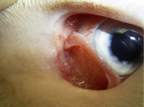
What is this image showing?
scrolling of cartilage of the third eyelid

What is this image showing?
prolapsed nictitans gland (“cherry eye“)
What does normal corenal structure contain?
Tear film (lipid, aqueous, mucin), epithelium, stroma, descemet’s membrane and endothelium

What is this histology picture of?
cornea
What is the function of the cornea?
maintains globe structure, transparency for vision
List diseases that affect the cornea (8)
Keratoconjunctivitis sicca
Infectious keratitis
Eosinophilic keratitis
Other non-ulcerative keratitis
Sequestrum
Lipid keratopathy
Primary and secondary oedema
Neoplasia
What is corneal ulceration?
loss of epithelium, ± stroma, ± descemet’s membrane (rupture)
What are the 4 phases of epithelial injuries (corneal healing)?
latent, migration, proliferation, cell substrate attachment
What occurs during the latent phase of corneal healing?
apoptosis of cells damaged by wound stimulus
increased metabolic activity and cell structural changes
fibronectin polymerases onton wound bed - provisional anchoring extracellular matrix
reduction in adhesion of basal cells to basement membrane
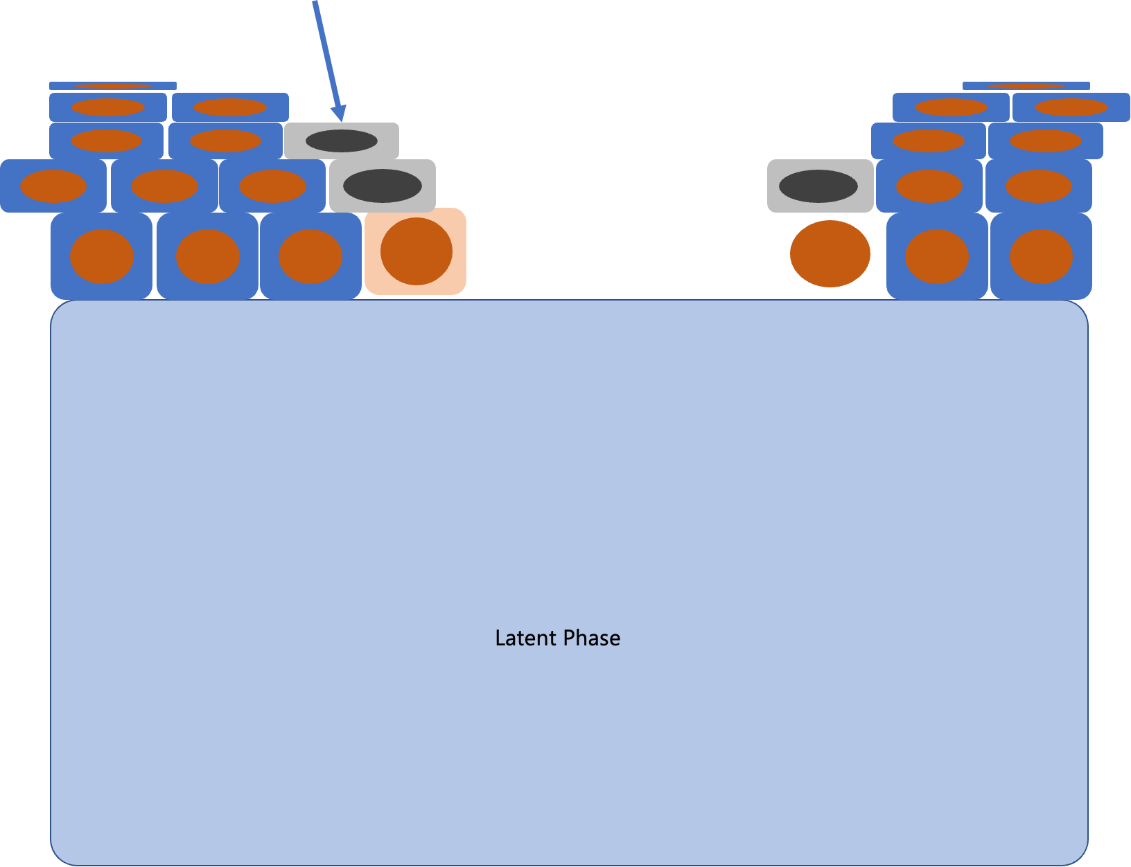
What does this arrow point to during the latent phase of corneal healing?
apoptosis of cells damaged by wound stimulus
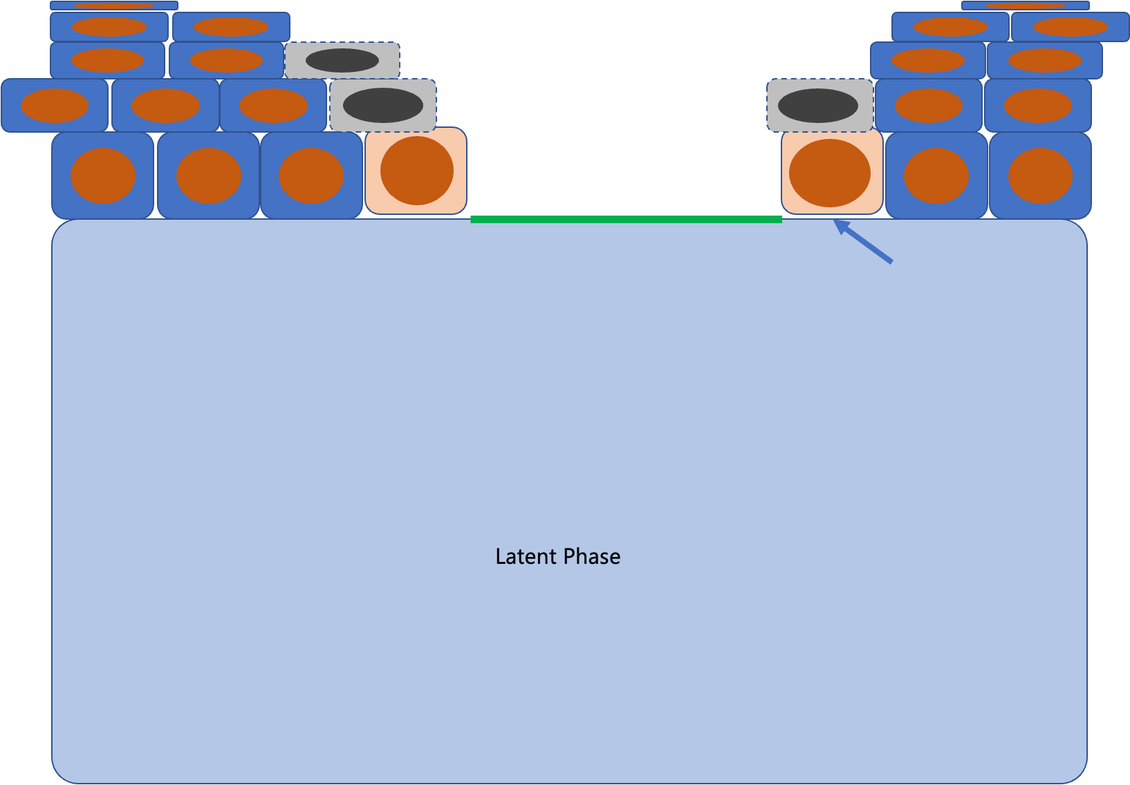
What does this arrow point to during the latent phase of corneal healing?
reduction in adhesion of basal cells to basement membrane

What does this arrow point to during the latent phase of corneal healing?
fibronectin polymerases onton wound bed - provisional anchoring extracellular matrix
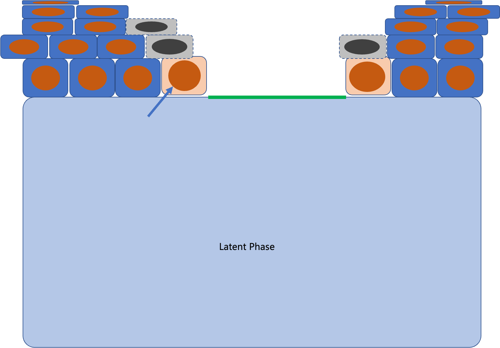
What does this arrow point to during the latent phase of corneal healing?
increased metabolic activity and cell structural changes

What does this arrow point to during the migratory phase of corneal healing?
migration of entire epithelial sheet over the wound bed

What does this arrow point to during the migratory phase of corneal healing?
leading edge cells flatten out over wound area and develop filopodia
What occurs during the migratory phase of corneal healing?
leading edge cells flatten out over wound area and develop filopodia
migration of entire epithelial sheet over the wound bed
NO cell division at this stage
What occurs during the proliferation phase of corneal healing?
Cells at the periphery of cornea undergo mitotic division while the wound bed is migrated over
transformation of basal cells into the layers of stratified epithelium and eventually squamous cells
basement membrane remodeling

What does this arrow point to during the proliferation phase of corneal healing?
transformation of basal cells into the layers of stratified epithelium and eventually squamous cells

What does this arrow point to during the proliferation phase of corneal healing?
basement membrane remodeling
How long can the attachment phase of corneal healing last?
the process can take up to a year to complete
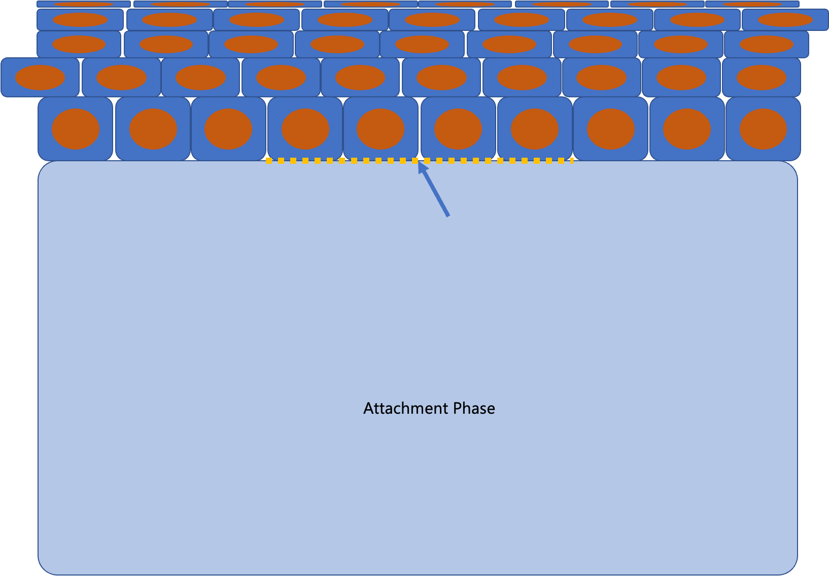
What does this arrow point to during the attachment phase of corneal healing?
new hemidesmosomes attachments formed
What occurs during the attachment phase of corneal healing?
new hemidesmosomes attachments formed, can take up to 1 year
Depending on the balance of mediators, MMPs and extent of the wound the outcome of stromal healing can be?
regeneration of normal stromal structure
scarring
stromal melting
neovascularization
What occurs during stromal healing?
epithelial compromise → swelling of stroma, apoptosis of keratocytes/fibroblasts and invasion of inflammatory cells → transformation of keratocytes within the stromal collagen into fibroblasts → remaining fibroblasts migrate to the wound → firboblasts deposit fibrotic material to fill defect
Where do the inflammatory cells come from during stromal healing?
recruited from conjunctival vessels to the site of injury
What is the function of inflammatory cells during stromal healing?
protect against infection but can also contribute to tissue damage
What happens to the fibrotic tissue deposited during stromal healing?
may contract and opacify
What are common clinical signs associated with ulcers?
pain (blepharospasm, increased blink rate, ocular discharge)
conjunctival hyperemia
corneal edema (focal)
corneal neovascularization
What are some broad causes of corneal ulceration?
TRAUMA
external irritation
tear film changes
Corneal pathology
infectious disease
What tear film changes can lead to poor corneal health?
reduced quantity and reduced quality
What can reduce quality of tear film?
lagophthalmos, reduced blink (facial vs trigeminal nerve dysfunction or trigeminal nerve cells increase would healing rates in vitro), tear film instability, macropalpebral fissure, globe enlargement/proptosis
What corneal pathologies cause corneal ulceration?
lipidosis, calcification, corneal edema (endothelial dystrophy, intraocular disease), SCCED (superficial chronic corneal epithelial deficit)
What breeds are predisposed to spontaneous chronic corneal epithelial deficits (SCCED)?
boxers, pembroke corgis, others
What occurs to the eye in spontaneous chronic corneal epithelial deficits (SCCED)?
superficial, non healing involving epithelium only deficit
What occurs during deeper ulceration leading to loss of stroma?
stromal breakdown due to increased collagenase activity (pseudomonas and streptococcus spp, and neutrophils), rapid time scale, can lead to globe rupture
What is the cytology of melting ulcers?
neutrophils, epithelial cells and bacteria

What is this image showing?
corneal sequestrum
Who is affected by corneal sequestrum?
feline, rarely equine
What occurs during corneal sequestrum?
nectoric cornea, superficial ulceration
black pigment absorbed from feline tear film
What can cause corneal sequestrum?
Underlying FHV
What is a lipid keratopathy?
central corneal lipidosis
Who is affected by lipid keratopathies?
cavaliers, boxers and huskies
What does lipid keratopathies cause?
excessive accumulation of lipid in corneal fibroblasts - cholesterol crystals
benign usually non-progessive
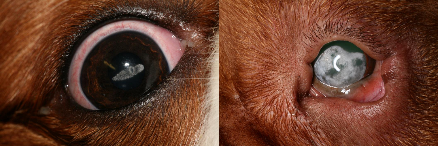
What does this image show?
lipid keratopathies
What can cause lipid keratopathies?
response to corneal insult or systemic lipid abnormalities
What can cause chronic superficial keratitis?
neovascularization, pigmentation, fibrosis or UV induced
Who is affected by eosinophilic keratitis?
feline and equine
What can causes eosinophilic keratitis?
FHV
What does the eye look like if it has eosinophilic keratitis?
eosinophils in dense white plagues or neovascularization

What does this image show?
eosinophilic keratitis
What can cause corneal edema?
endothelial cell loss, internal ocular disease (glaucoma or uveitis), ulceration, limbia disease
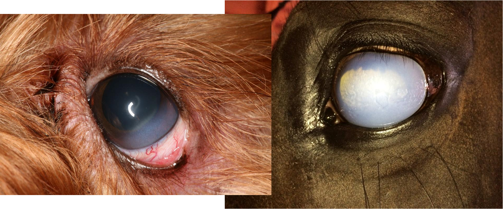
What do these image show?
corneal edema
What are inclusion cysts?
epithelial cells implanted into stroma
grow and shed - leaves creamy white fluid in center of epithelial cell lined cyst

What does this image show?
inclusion cyst

What does this image show?
neoplasia - rare
What is the function of the lacrimal system?
tear production and drainage
What are the components of tear film?
mucus - goblet cells in conjunctiva
aqueous - lacrimal gland in upper eyelid and nictitans gland in third eyelid
lipid - meibomian glands on eyelid margin
What is the function of tear film?
lubrication, immune modulation, removal of debris, nutrition to the cornea
What can cause lacrimal gland insufficiency leading to tear film deficincies?
congenital, drug induced (sulphonamides), immune mediated, neurogenic, iatrogenic, systemic disease
What happens with immune mediated keratoconjunctivitis sicca?
the body recognizes self antigens in the lacrimal gland → starts T cell response against the antigens → reduction in tear film quantity and quality
What are the clinical signs of keratoconjunctivitis sicca?
blepharospasm, mucoid/mucopurulent discharge, conjunctivitis, keratitis, ulceration
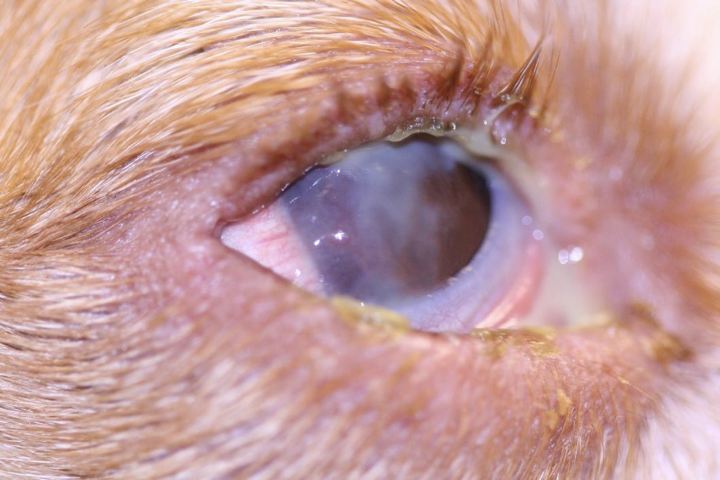
What does this image show?
keratoconjunctivitis sicca

What does this image show?
tear testing

What does this image show?
Corneal fibrosis, pigmentation and neovascularisation
What is the function of the nasolacrimal system?
remove tears from eye and LDALT + CALT = EALT
What is LDALT?
Lacrimal Drainage Associated Lymphoid Tissue
How does the nasolacrimal system vary in different species?
Usually have upper and lower puncta - only lower in rabbit
exit is variable - obvious in horses, can be nasopharyngeal in dogs
What are imperforate ducts?
part of nasolacrimal system at either end for tear overflow
What can cause external compression of the nasolacrimal system?
cysts, trauma, dental disease (rabbit), parasites (onchocerca)