Alveolar Bone
 Call Kai
 Learn
Learn Practice Test
 Spaced Repetition
 Match
Match Flashcards
 Knowt Play
1/44

There's no tags or description
Looks like no tags are added yet.
Name | Mastery | Learn | Test | Matching | Spaced | Call with Kai | Chat |
|---|
No analytics yet
Send a link to your students to track their progress
45 Terms
Gingiva
PDL
Cementum
Alveolar bone
4 principal components of the periodontium
Ectomesenchymal tissues
The alveolar process develops from the ?
cribriform plate, bundle bone, or lamina dura on radiographs
Alveolar bone proper is aka ?
Alveolar bone proper
forms inner wall of tooth socket; insertion site for Sharpey’s fibers
Alveolus (tooth socket)
bony cavity within the alveolar process that houses and supports the root of each tooth
Alveolar crest
forms the most coronal portion of the alveolar process; normally lies approx. 1-2 mm apical to the CEJ
Fenestrations and dehiscences
localized defects in the cortical alveolar bone that exposes portions of the tooth root
fenestration
an isolated “window” of missing cortical bone in which the marginal (alveolar crest) bone remains intact
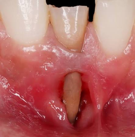
dehiscence
a more extensive defect in which the loss of cortical bone extends coronally to involve the alveolar crest

prominent root anatomy
thin cortical bone
facial tooth positioning
orthodontic tooth movement
occlusal trauma
factors associated with fenestrations and dehiscences
osteons or haversian systems
densely packed lamellae arranged into cylindrical units
bundle bone
refers to the incorporation of the terminal portions of the Sharpey’s fibers into the bone
bone lamellae
organized layers of mineralized bone matrix that contribute to the strength and structural organization of alveolar bone while minimizing weight
interstitial lamellae
remnants of older osteons left behind after bone remodeling
concentric lamellae
arranged in rings around the Haversian canal within each osteon
lamellar bone
Mature, highly organized bone characterized by parallel collagen layers and superior mechanical strength
woven bone
Immature bone that forms rapidly during growth, fracture repair, and early socket healing before gradually being replaced by lamellar bone through bone remodeling
bone marrow spaces
cavities located between the trabeculae of cancellous alveolar bone and are filled with either red or yellow marrow
canaliculi
microscopic channels that allow the diffusion of nutrients and waste products while facilitating cell-to-cell communication among osteocytes
nutrient canals
small channels within the alveolar bone that transmit blood vessels, lymphatic vessels, and nerves between the bone marrow spaces and the PDL
volkmann canals
Transverse or oblique channels in compact bone that connect adjacent Haversian canals
volkmann canals
their primary function is to provide vascular and neural communication by connecting blood vessels throughout the bone
osteoprogenitor cells
precursor cells capable of differentiating into osteoblasts during growth, repair, and regeneration
extracellular matrix
the strength and resilience of alveolar bone depend on its ?
Type I
Collagen type in the organic matrix (or osteoid)
periodontal ligament vessels
nourish the alveolar bone proper and maintain the vitality of the attachment apparatus
periosteal vessels
supply the cortical plates and contribute significantly to bone repair following injury or surgery
intraosseous vessels
nourish the cancellous bone and bone marrow spaces while supporting hematopoiesis and bone metabolism
bone metabolism
the continuous processes of bone formation and bone resorption that maintain skeletal integrity and mineral homeostasis
bone modeling
alters the overall shape and size of bone during growth and tooth eruption through independent formation and resorption
bone remodeling
a lifelong process in which osteoclast-mediated resorption is followed by osteoblast-mediated bone formation at the same site
Mechanical regulation
Functional forces stimulate bone remodeling according to the principle that bone adapts to mechanical stress
Wolff’s Law
states that bone remodels in response to the magnitude and direction of functional loading
Molecular regulation
Bone remodeling is controlled by signaling molecules produced by osteoblasts, osteocytes, immune cells, and periodontal tissues
RANK-RANKL-OPG signaling system
Principal molecular regulator of bone remodeling
RANKL
stimulates osteoclast differentiation and bone resorption by binding to RANK receptors on osteoclast precursors
Osteoprotegerin (OPG)
A decoy receptor protein that prevents bone breakdown by binding to RANKL and blocking osteoclast development
Parathyroid hormone (PTH)
increases osteoclast activity indirectly
raises blood calcium levels
promotes bone resorption
Calcitonin
inhibits osteoclast activity
reduces bone resorption
helps preserve bone mass
Vitamin D
enhances intestinal calcium absorption
supports mineralization
essential for normal bone development
Estrogen
suppresses osteoclast activity
maintains bone density
Growth hormone
stimulates osteoblast activity
promotes skeletal growth
Horizontal bone loss
Most common bone loss pattern
occurs when alveolar crest is uniformly reduced while remaining generally parallel to a line connecting the CEJ of adjacent teeth
reflects generalized destruction of periodontal support
Vertical (angular) bone loss
occurs when resorption progresses unevenly
create infrabony pockets that may be favorable for regenerative periodontal therapy because portions of the surrounding bone remain intact
1-2 mm apical to CEJ
normal alveolar crest level