Radiographic Interpretation of Dental Caries
 Call Kai
 Learn
Learn Practice Test
 Spaced Repetition
 Match
Match Flashcards
 Knowt Play
1/48

There's no tags or description
Looks like no tags are added yet.
Name | Mastery | Learn | Test | Matching | Spaced | Call with Kai | Chat |
|---|
No analytics yet
Send a link to your students to track their progress
49 Terms
multifactorial disease involving complex risk and protective factors; localized destruction of teeth by acid demineralization
dental caries

simultaneous demineralization and remineralization (achieve a balance to prevent caries)
dynamic process of enamel
- metabolic microbial organic acids decrease pH
- dissolution of Ca and PO4
demineralization
- changes in color/glossiness
- cavitation/destruction
- dentin and pulp exposure
3 effects of continuous enamel demineralization
less tissue density, less absorbed by photons (more radiolucent)
How does demineralization present on radiographs?
- occlusal (pits and fissures)
- recurrent/secondary
- facial/lingual (smooth surface)
- root/radicular
- interproximal
5 possible sites for carious lesions
- outer 1/2
- inner 1/2
2 possible progression levels of enamel caries
- outer 1/3
- middle 1/3
- inner 1/3
- directly involving pulp
4 possible progression levels of dentin caries
starts on pits and fissures, hard to detect clinically and radiographically
early stages of occlusal caries
incipient lesions (enamel only), not detected radiographically (superimposition), detected clinically
initial stages of occlusal caries
diffuse radiolucency below enamel occlusal surface (can advance into pulp)
occlusal caries that have lesions into dentin
occlusal caries (into dentin)
What is this an example of?

occlusal caries (into dentin)
What is this an example of?

occlusal caries (into dentin)
What is this an example of?

occlusal caries (into dentin)
What is this an example of?
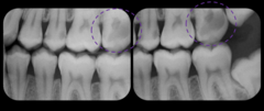
caries adjacent to margins of restorations
recurrent/secondary caries
diffuse radiolucency below restorations
How do recurrent/secondary caries present on radiographs?
recurrent/secondary caries
What is this an example of?

recurrent/secondary caries
What is this an example of?

recurrent/secondary caries
What is this an example of?
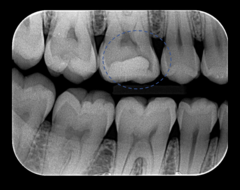
usually close to gingival margin, better seen clinically
facial/lingual (smooth surface) caries
sharply defined round or oval radiolucency
How do facial or lingual caries present on radiographs?
facial/lingual (smooth surface) caries
What is this an example of?

facial/lingual (smooth surface) caries
What is this an example of?

facial/lingual (smooth surface) caries
What is this an example of?

facial/lingual (smooth surface) caries
What is this an example of?

facial/lingual (smooth surface) caries
What is this an example of?

facial/lingual (smooth surface) caries
What is this an example of?

facial/lingual (smooth surface) caries
What is this an example of?

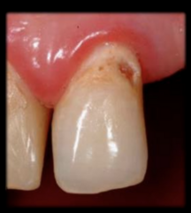
below CEJ, gingival recession
root surface/radicular caries
diffuse radiolucency below CEJ
How do root surface/radicular caries present on radiographs?
root surface/radicular caries
What is this an example of?
root surface/radicular caries
What is this an example of?

root surface/radicular caries
What is this an example of?

root surface/radicular caries
What is this an example of?

NOT carious lesions!
- collar or wedge-shaped radiolucency
- between CEJ and alveolar bone
cervical burnout
cervical burnout
What is this an example of?

cervical burnout
What is this an example of?

arise at or slightly below contact point; viewed a lot better radiographically than clinically
interproximal caries

if dentin is affected, it will appear gray and discolored on marginal ridges
How can interproximal caries be viewed clinically?

irregular radiolucency with base at proximal surface near contact point
How do enamel interproximal caries appear on radiographs?

triangular radiolucency with base at CEJ
How do dentin interproximal caries appear on radiographs?
- bitewings for posterior teeth
- PAs for anterior teeth
What is the best radiographic exam to detect carious lesions?

- controversial results (less detailed compared to intraoral)
- produce ghost images, noise, abnormal configuration of arches
- used for uncooperative patients, severe gag reflex, severe anatomic limitations
3 characteristics of extraoral bitewings
- correct horizontal and vertical angulation
- look at both premolar and molar BWX images (+ PAs for anteriors)
What are important things to remember when viewing bitewings for caries?
usually only seen when large enough to be clinically apparent
How do carious lesions appear on panoramic radiographs?
can show carious lesions at early stages (about 1/2 of proximal surface caries are not seen clinically)
What is the largest contribution for BWX when diagnosing carious lesions radiographically?
25-50% demineralization is needed before visualization (radiographs underestimate actual lesion depth)
What is important to remember about demineralization on radiographs?
- radiographs cannot determine if a lesion is active/cavitated or inactive/arrested
- superficial layers can remineralize
What is important to remember about cavitated lesion progression on radiographs?