BIOL1006 - ETest 1
 Call Kai
 Learn
 Practice Test
 Spaced Repetition
 Match
 Flashcards
 Knowt Play
1/25

There's no tags or description
Looks like no tags are added yet.
26 Terms
The anatomical organisation of the human nervous system

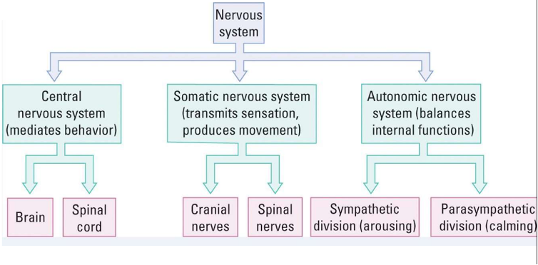
The functional organisation of the human nervous system
What is afferent information
Sensory info coming into the nervous system
What is efferent information
Info leaving the nervous system
What is white matter
· Long myelinated axons (fatty)
· When bundled together, they form white matter tracts
· Form complex connections between brain areas, spinal cord and the PNS
· Forms majority of the deep parts of brain
· Myelination (increases in white matter) occurs across the lifespan
· Associated with MS, psychiatric disorders
What is grey matter
· Comprises neuron cell bodies, dendrites, short myelinated and unmyelinated axons, glial cells, capillaries
· Neurons receive information distally through the white matter tracts and locally from nearby neurons
· Collects and modifies info collected by white matter
· Cerebral cortex, basal ganglia, hypothalamus
Grey matter loss seen in schizophrenia
The spinal cord
· Long bundles of tissue extending from the medulla oblongata in the brainstem
· Main pathway for info connecting the brain and PNS
· Produces most movements following instructions from the brain but can act independently
· Each segment receives and sends info to and from discrete parts of the body
The brain stem
· Begins where spinal cord enters the skull and extends into the lower areas of the forebrain
· Comprises of 3 regions: hindbrain, midbrain and diencephalon
· Receives afferent nerves from all senses and sends efferent nerves out to spinal cord
The forebrain
CEREBRAL CONTEX = outer layer of neural tissue of the cerebrum
Comprised of 3 main structures:
1. Neocortex (4 lobes): regulates host of cognitive activites and individuals traits
2. Basal ganglia: controls voluntary movement
3. Limbic system: regulates emotions and behaviours that create and require memory
Frontal lobe
· Largest lobes in the brain
· Frontally positioned
· Voluntary movement, expressive language, higher level executive functions, personality
· Damage = catastrophic and impact language, attention, flexibility, understanding, impulsivity, personality
Parietal Lobe
· Positioned towards back and top, above temporal lobe
· Integratrs sensory info
· Involved in visuospatial processing, spatial mapping, knowledge of numbers and their relations, object manipulation
· Damage = sensory impairment, neglect, apraxia, dyslexia
Temporal Lobe
· Located below lateral fissure and processes multiple sensory inputs
· Includes primary auditory cortex and Wernicke’s area (language)
· Role in language comprehension
· Communicates with hippocampus to contribute to formation of explicit long term memory
· Damage = aphasia, prosopagnosia, deafness
Occipital Lobe
· Smallest lobe at the very back
· Contains visual cortex
· Divided into functional visual areas, each containing a full map pf the visual world
· Receives info from retinal cells via complex optical tracts through thalamus
· Lesions cause blindness, hallucinations, epilepsy
What are Gyri
ridges on the surfaces
What are sulci
grooves on the surface
What are the 3 major cerebral arteries
anterior, middle, posterior
The 4 ventricles of the brain
1 and 2 in each cerebral hemisphere = left and right lateral. 3rd is in the median slit like cavity between the 2 thalami. 4th cavity of the hindbrain between pons and cerebellum.
The limbic system
· Located both sides of the thalamus, right under the cerebrum
· 2 of the major structures are the hippocampus and amygdala
· Palys a role in emotion, sexual behaviour, memory, motivation
· Highly interconnected with the nucleus accumbens – brain’s pleasure centre and plays a role in addiction
· Significant role in reward – dopaminergic projection play role in learning and reinforcement
Somatic Nervous System
· Part of the PNS associated with skeletal muscles voluntary control of the body movements
· Comprising of afferent and efferent nerves
Consists of 3 parts
1. Spinal nerves: carry sensory info into and motor commands out of the spinal cord
2. Cranial nerves: carry info into and out of the brain stem
3. Association nerves: intergrates sensory input and motor output numbering thousands
Functions of 12 cranial nerves
1. Olfactory (smell)
2. Optic (vision)
3. Oculomotor (eye movement)
4. Trochlear (eye movement)
5. Trigeminal (sensation & chewing)
6. Abducens (eye movement)
7. Facial (movement & sensation)
8. Auditory vestibular (hearing & balance)
9. Glossopharyngeal (tongue/pharynx)
10. Vagus (heart, vessels, larynx)
11. Spinal accessory (neck muscles)
12. Hypoglossal (tongue muscles)
Spinal nerves
· 31 pairs of spinal nerves, one on each side of the vertebral column
· Grouped into the corresponding cervical, thoracic, lumbar, sacral and coccygeal regions of the spine
o 8 pairs of cervical nerves (C)
o 12 pairs of thoracic nerves (T)
o 5 pairs of lumbar nerves (L)
o 5 pairs of sacral nerves (S)
o 1 pair of coccygeal nerves
Dorsal fibres
carry info from receptors
Ventral fibres
carry info out of spinal cord
Role of the autonomic nervous system
· A division of the peripheral nervous system that influences the function of internal organs
· Control system that acts largely unconsciously and regulates heart rate, digestion, respiratory rate, pupillary response, urination, sexual arousal
2 divisions of ANS
· Sympathetic nervous system is considered to be the "fight or flight" system
· Parasympathetic nervous system is considered to be the "rest and digest" system