VIBS 243 Midterm
 Call Kai
 Learn
Learn Practice Test
 Spaced Repetition
 Match
Match Flashcards
 Knowt Play
1/127

Earn XP
Name | Mastery | Learn | Test | Matching | Spaced | Call with Kai | Chat |
|---|
No analytics yet
Send a link to your students to track their progress
128 Terms
Which of the following images DO NOT/DOES NOT illustrate scanning transmission electron microscopy?
A
B
C
A and B
A, B, and C

A
Which of the following is/are considered a toluidine blue stain?
A
B
C
A and B
A, B, and C

C
Which of the following types of microscopy matches its/their description AND is/are best for viewing cellular detail?
A- transmission electron microscopy
B- toluidine blue light microscopy
C- periodic acid/Shiff light microscopy
A and B
A, B, and C

A
Which item – description(s) about magnification is(are) correct?
A. MAGNIFICATION - INCREASE IN IMAGE SIZE
B. RESOLUTION - Largest DISTANCE BETWEEN TWO POINTS THAT CAN BE SEEN (DISTINGUISHED)
C. MAGNIFICATION - Smallest DISTANCE BETWEEN TWO POINTS THAT CAN BE SEEN (DISTINGUISHED
D. A and B
E. A, B, and C
A
Which of the following statements about this image is/are correct?

A. This is an H&E (hematoxylin and eosin) stain
B. In this stain nucleic acids will stain more blue
C. In this stain proteins will stain more red
D. A and B
E. A, B, and C
E
The Golgi apparatus matches which icon of the cell?

A- Gate keeper
B- Construction team
C- Packaging department
A and B
A, B, C
C
Which structure(s) match(es) the definition(s)?

A- Smallest unit of protoplasm
B- Groups of cells with same general function and texture
C- Two or more types of tissue in a larger working unit
D- A and B
E- A, B, and C
E
Which major class of cytoplasmic structures (component groups within cells) best match(es) descriptive feature(s)?
A. Inclusions – expendables not needed for the life of the cell but important for cell function
B. Membranous organelles - common structures in cells for metabolic functions of cell
C. Non -Membranous organelles – cytoskeletal components
D. A and B
E. A, B, and C
E
Which major class(es) of cytoplasmic structures (component groups within cells) best match(es) the example(es) of the classes?
A. Inclusions - microfilaments
B. Membranous organelles – cell membrane (plasma membrane)
C. Non -Membranous organelles – melanin granules
D. A and B
E. A, B, and C
B
The following is demonstrating an increase in image _________. Fill in the blank

A. Magnification
B. Carification
C. Resolution
D. A and B
E. A, B, and C
A
The benefit(s) to transmission electron microscopy in comparison to light microscopy is/are:

A. more cellular detail
B. Better organelle detail
C. Great for surface details
D. A and B
E. A, B, and C
D
Muscle cells that need a lot of energy to contract will have an abundant amount of ______________. Choose the best answer.
A. Mitochondria
B. Lysosomes
C. Cilia
D. Microvilli
A
Why is it important to identify cells in mitosis?
A. To identify if regulation of the cell cycle is abnormal
B. A decrease in cells in mitosis could indicate cancer
C. A & B
D. None of the above
A
Which micrograph(s) illustrate(s) conventional electron microscopy?

A
B
C
A and B
A, B, and C
A&B
Which major class(es) of cytoplasmic structures (component groups within cells) best match(es) the examples of the class(es)?
A. Inclusions - lipid
B. Membranous organelles - RER
C. Non -Membranous organelles – free ribosomes
D. A and B
E. A, b, and c
E
Which of the following images is an example of electron microscopy?

A
B
C
A and B
A, B, and C
A, B, and C
Which structure(s) match(es) the definition(s)?

a- Groups of cells with same general function(s)
b- Smallest unit of protoplasm
c- Several organs working in a common function
d- A and B
e- A, B, and C
C
Leydig cells which produce a lot of steroid hormones will have an abundant amount of_____________. Choose the best answer
a) Smooth ER
b) Rough ER
c) Nuclei
d) Lysosomes
e) a, b, and c
A
Why is it important to identify cells in mitosis?
A) To identify if regulation of the cell cycle is abnormal
B) An increase in cells in mitosis could indicate cancer
C) A & B
D) None of the above
C
The cell nucleus matches which icon?

a- Gate keeper
b- Computer
c- Construction team
A and B
A, B, and C
B
The following image describes which of the following EM types?

A) Scanning EM
B) Conventional EM
C) Carbon replica EM
A and B
A, B, and C
C
Which of the following images illustrate(s) carbon replica transmission electron microscopy?

A
B
C
A and B
A, B, and C
A
Which is/are examples of simple squamous epithelium cells?

A
B
C
A and B
A, B, and C
A, B, and C
What type of epithelium is in the picture below?

A. Simple columnar
B. Simple cuboidal
C. Pseudostratified columnar
D. Transitional
A
Stratified squamous epithelium is classified as keratinized or non-keratinized bases on the presence of nuclei in cells of which layer?

A
B
C
A and B
A, B, and C
A
Where can you find this type of epithelium in the picture below?

a. Respiratory system
b. Urinary System
c. Skin
d. Digestive System
a
What type of epithelium is in the picture below?

a. pseudostratified columnar
b. Transitional
c. Simple columnar
d. Stratified columnar
B
What is the apical modification that the arrow is pointing to in the picture?

a. Microvilli
b. Cilia
c. Stereocilia
d. Goblet cell
B
What type of epithelium is in the picture below?

Simple cuboidal
Stratified cuboidal
Simple squamous
Pseudostratified columnar
1
Which statement(s) is/are correct about epithelium?
Epithelium lines the walls of all passages leading to the exterior of the body
Epithelium (endothelium) lines blood vessels
Epithelium (mesothelium) lines the outside of organs
A and B
A, B, and C
5
Which cellular junction helps to seal off the external environment from deeper tissues?
Desmosomes
Gap Junction
Tight Junction
Zonula Adherens
A, B, and C
3
Which statement(s) is/are true about the structure the arrow is pointing to in the image?

The structure functions in absorbing
The structure functions in secreting mucous
The structure functions in holding the cells onto underlying tissue
A and B
A, B, and C
B and C
3
What type of epithelium is in the picture below?


Simple columnar with goblet cells
Pseudostratified Columnar with goblet cells
Transitional
Stratified Columnar with goblet cells
1
What type of epithelium is pictured below?


nonkeratinized stratified squamous
keratinized stratified squamous
transitional
pseudostratified columnar
2
Which type(s) of epithelium match(es) the image(s) in skin?
A- simple squamous
B- stratified cuboidal
C- stratified squamous

Stratified cuboidal of sweat ducts
Simple squamous endothelial
Stratified squamous epithelium
A and B
A, B, and C
3
Epithelium is classified based on
Shape of cells on its surface
Shape of cells on its base
Number of nuclei in each cell
A and B
A, B, and C
1
Which cellular structure(s)/cell(s) of epithelium match(es) the image(s)?

a-microvilli
b-Goblet cells
c-microvilli
A and B
A, B, and C
1
Which of the following is true in regards to epithelium?
A)Epithelium is one of the four main tissue types that make up our bodies
B) Epithelium is a lining- it lines our body cavities, organs, and blood vessels for example
C) Epithelium is classified by the shape of the non-superficial cells as well as how thick each layer is
D)Answers A and B are true
E)Answers B and C are true
D
Which cellular structure(s) of epithelium match(es) the image(s)?

a- brush boarder
b-goblet cell secretion
c- basement membrane
d- A and B
e- A, B, and C
e
What type of epithelium is in the picture below?

Pseudostratified Columnar
Transitional
Simple Columnar
Stratified Columnar
1
What type of epithelium is in the picture below?

Non-Keratinized Stratified Squamous
Keratinized Stratified Squamous
Transitional
Pseudostratified Columnar
1
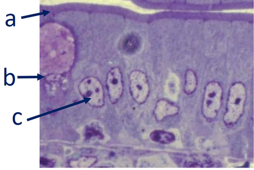
Which cellular structure(s)/cell(s) of epithelium do not/does not match the image(s) in these cells?
a- Basement membrane
b- Goblet cell
c- Nucleus of cell
A and B
A, B, and C
a
What is the function of the structure the arrow is pointing to in the image?

moving debris
secreting mucous
Absorbing
attaching cells to underlying tissue
1
Which type(s) of epithelium match(es) the image(s) in skin?
A) Shape of cells on its surface
B) Shape of cells on its base
C) Number of nuclei in each cell
D) A and B
E) A, B, and C
A
Where can you find this type of epithelium?

Respiratory System
Urinary System
Skin
Digestive System
2
Which one(s) of these flattened nuclei belong(s) to connective tissue:

A
B
C
A and B
A, B, and C
1
The collagenous connective tissue supporting an epithelium is called_____________.
A. Lamina propria
B. Basement membrane
C. Apical surface
D. Basal lamina
A
Identify the type of tissue in the image

Multilocular Adipose Tissue
Unilocular Adipose Tissue
Collagenous Connective Tissue
Cartilage
2
Connective tissue:
Provides mechanical support and metabolic exchange for epithelia
Provides site of battle for blood-borne immune cells against pathogens
Stores energy via adipose tissue
A and B
A, B, and C
5
The yellow arrow in the image is pointing to which structure?

Fibroblast cytoplasm
Collagen fibers
Nucleus of fibroblast
Ground substance
2
The yellow star is in which structure?

Adipose cells
Cytoplasm of fibroblasts
Ground substance
Nucleus of fibroblast
None of the above
3
Which structure(s) do not/does not match(es) the image(s) in connective tissue?

A. Collagen fibers (bundles)
B. Smooth muscle cells
C. Ground substance
D. A and B
E. A, B, and C
B
Which of the following are classified under connective tissues?
Adipose Tissue
Bone
Cartilage
A and B
B and C
A, B, and C
6
Classification of connective tissue proper is based on:
A. Ratio of cells to extracellular matrix
B. Arrangement of fibers
C. The closeness to epithelium
D. A and B
E. A, B, and C
d
Poco Bueno passed down a hereditary disease called Hereditary Equine Regional Dermal Asthenia which causes abnormal formation of which of the following?
Collagen fibers
Adipose tissue
Ground substance
Fibroblast cells
1
Which of the following type(s) of cartilage are labeled correctly in the image below?

A) Hyaline Cartilage
B) Elastic Cartilage
C) Fibrocartilage
B and C
All of the above
5
Which of the following is labeled correctly in the image below?

Chondroblast
Osteoblast
Periosteum
All of the above
None of the above
5
What is NOT true of bones?
Functions for support and protection
Acts as a reservoir for calcium and phosphate
Receives nutrients via diffusion from the perichondrium
Encloses the sides of developing blood cells
3
What cell type is indicated by the arrow below?

Osteocyte
Osteoblast
Osteoclast
Chondrocyte
1
Which of the following is true about endochondral ossification?
It is the deposition of bone matrix onto preexisting cartilage matrix
A growth plate of fibrocartilage will be replaced by bone
It allows for the extension of compact bone in width
All of the above
1
The yellow star is located in which structure?

Bundles of collagen fibers
Ground substance
Adipose cells
Nuclei of fibroblasts
Cytoplasm of fibroblasts
1
true or false: Adipose tissue is resting, and its only function is storage of energy.
false
Which of the following is NOT true of cartilage?
It acts as a cushion for joints
It is a provisional model for bone
It contains an extensive blood supply through Haversion systems
It is lined by perichondrium
3
What is true about the type of cartilage pictured below?

It is the least common type of cartilage
It acts as the provisional model for most bones
It is elastic cartilage
None of the above
2
What statement is FALSE about the cell indicated by the arrow below?

It is an osteoblast
It synthesizes the organic bone matrix
It will become an osteoclast when it is entrapped in a lacunae
It expands bone by appositional growth
3
Which statement below is FALSE of the organization of bone?
They are arranged in concentric lamellae
They are given nutrients through Haversion systems
They are constantly being rebuilt by chondrocytes
They are composed of cells and mineralized extracellular matrix
3
Which of the following is incorrect regarding connective tissue?
A. Connective tissue usually has widely separated cells with an abundant extracellular matrix
B. Connective tissue is often avascular (lacks blood vessels)
C. Connective tissue acts as the histological glue that holds things together
D. More than one of the above are incorrect
B
The yellow arrows are pointing to which structure?

Bundles of collagen fibers
Ground substance
Adipose cells
Nuclei of fibroblasts
Cytoplasm of fibroblasts
4
What is true about the structure indicated by the arrows below?

A) It is a capsule of dense irregular connective tissue
B) It holds a vascular supply
C) It is the periosteum
D) A and B
E) All of the above
D
Classification of connective tissue proper is based on:
A. ratio of cells to extracellular matrix
B. arrangement of fibers
C. shape of surface/apical cells
D. a & b
E. a, b & c
D
Identify the type of tissue in the image below....

Multilocular Adipose Tissue
Unilocular Adipose Tissue
Bone
collagenous connective tissue
1
What do humans and guinea pigs have in common?
A. They can both get a vitamin C deficiency
B. They can both get scurvy
C. They can't produce their own vitamin C
D. A and C
E. B and C
F. A, B, and C
F
Connective tissue:
A. provides mechanical support and metabolic exchange for epithelia
B. provides site of battle for blood borne immune cells against pathogens
C. stores energy via adipose tissue
D. a & b
E. a, b & c
E
What cell type is indicated by the arrow in the image below?

A. Chondrocyte
B. Osteocyte
C. Osteoblast
D. Osteoclast
D
The black arrow in the image is pointing to a light band. Which of the following is true regarding this structure?

This band contains myosin filaments
This band is also viewed in smooth muscle
This band is viewed in cardiac muscle
A and B
A, B, and C
3
Which of the following is true regarding the tissue in the image?

You can voluntarily control this tissue
The acidophilic staining in the image corresponds to extracellular fibers
Striations are a histological trait of this tissue
A and C
A, B, and C
None of the above
6
Cells of which type(s) of muscle match(es) both the image(s) and the stated characteristic(s)?

Smooth muscle - Involuntary contraction of muscle
Skeletal - Voluntary contraction of muscle
Cardiac - Involuntary contraction of muscle
A and B
A, B, and C
5
Which of the following is true about the structure indicated by the arrow in the image.

A. This is a nucleus of the skeletal muscle
B. This represents the neuromuscular junction
C. This is the motor end plate
D. A and C
E. B and C
F. A, B, and C
E
What is/are the function(s) of muscle?
Enable the heart to beat
Locomotion of multicellular animals
Movement of their internal organs
A and B
A, B, and C
5
Which of the following is true regarding the structure indicated by the blue arrow?

This represents where to Schwann (Neurolemmocytes) cells meet
There is less myelin in this area
This is the Schmidt-Lantermann cleft
A and B
A and C
A, B, and C
4
Which structure(s) of the nerve cell/associated cells best match(es) the image(s)?

A. Nuclei
B. Satellite cells
C. Nissl bodies
D. A and B
E. A, B, and C
A
Which connective tissue structure(s) of the nervous system best match(es) the image(s)?

Epineurium
Perineurium
Endomysium
A and B
A, B, and C
4
Which structure(s) of the dorsal root ganglion and attached nerve best match(es) the image(s)?

Cell bodies of ganglion neurons
Cell body of ganglion neuron
Myelinated axon
A and B
A, B, and C
5
Which structure(s) of these two nerves best match(es) the image(s)?

A. Fascicles in nerves
B. Myelinated axons
C. Nerve terminal/motor end plate
D. A and B
E. A, B, and C
E
. Which of the following is true regarding the type of tissue in the image?

Nuclei are located in the periphery
There is only one nucleus per cell
The term fiber means cell
A and B are correct
A and C are correct
A, B, and C are correct
5
What type(s) of muscle do not/does not match both the image(s) and the stated characteristic?

Cardiac – branched
Skeletal - striated
Smooth - non-striated
A and B
A, B, and C
1
Which characteristic(s) of muscle match(es) both of the paired image(s) in these muscles?

Voluntary
Involuntary
Involuntary
A and B
A, B, and C
3
The dog, Rocky, in the case study had an issue at which structure?
Neuromuscular junction/motor end plate
Axon
Myelin sheath
Bone
Muscle
1
Which structure(s) of the nervous system best match(es) the image(s)?

Dendrites of neurons
Myelin sheath of axons
Epineurium
A and B
A, B, and C
2
Which structure(s) of the nerve cell best match(es) the image(s)?

a- Neuronal cell body
b- Nerve terminals
c- Dendrites
d- A and B
e- A, B, and C
C
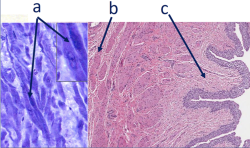
Which image(s) is not/are not composed of long, spindle-shaped smooth muscle cells?
A
B
C
A and B
A, B, and C
C
True or False: A reflex involves a sensory pathway to the spinal cord and a motor pathway back out to the muscle.
true
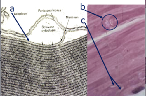
Which structure(s) match(es) both the image(s) and characteristic(s)?
A) Cell membrane of Schwann cells – form the myelin sheath around axons
B) Node of Ranvier – Where two fibroblasts join around the same axon
C) Schmidt-Lantermann cleft – complete compaction of myelin around an axon
A and B
A, B, and C
A
Which structure(s) of these neurons best match(es) the image(s)?

a- Node of Ranvier
b- Node of Ranvier
c- Node of Ranvier
d- A and B
e- A, B, and C
e
Which of the following is true about the tissue in the image?

A) Involuntary contraction
B) Intercalated discs close off cells to decrease communication
C) These cells would have abundant mitochondria
d) A and B
e) A and C
f) A, B and C
e
Identify the structure indicated by the blue arrow.

A. Schwann cell nucleus
B. Schwann cell plasma membrane
C. Node of Ranvier
D. Axon
D
True or False: A signal needs to make it to the brain in order for pain to be perceived.
True
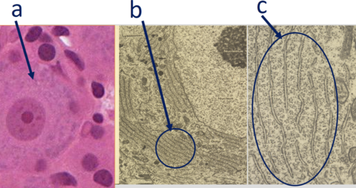
Which structure(s) of the nerve cell best match(es) both the definition(s)/location and the image(s)?
a- Nissl bodies - cytoplasm for coarse aggregates of basophilic substance
b- Nissl bodies - rough endoplasmic reticulum and polyribosomes
c- mitochondria in cells that produce ATP
d- a and b
e- A, B, and C
d
Identify the structure indicated by the blue arrow.

Schwann cell nucleus
Schwann cell plasma membrane
Axon
Node of Ranvier
1
Function of blood is transportation of cells and fluid. Which function(s) of blood cells/structures/fluids best match(es) the image(s)?

A. Immune defense for the body
B. Transportation of oxygen and carbon dioxide
C. Prevent loss of transportation of blood
D. A and B
E. A, B, and C
C