M3: Urinary/Renal System
 Call Kai
 Learn
 Practice Test
 Spaced Repetition
 Match
 Flashcards
 Knowt Play
1/14

There's no tags or description
Looks like no tags are added yet.
15 Terms
Urinary System: Function
The urinary system maintains internal body equilibrium and eliminates metabolic waste products.
Waste Elimination: Filters and removes nitrogenous wastes (e.g. urea, creatinine) generated by cellular metabolism to prevent toxic build-up.
Fluid & Electrolyte Balance: Regulates body water volume and essential electrolytes (Na+, K+, Ca2+) vital for nerve signal conduction and muscle contraction.
Renal Hormones
Erythropoietin: Stimulates red blood cell production within the bone marrow.
Renin: Enzyme secreted by the kidneys that plays a central role in controlling blood pressure.
Active Vitamin D (Calcitriol): Activated by the kidneys to enable intestinal calcium absorption, supporting bone health and cellular processes.
Urinary System Anatomy
Composed of four main structures: paired kidneys, paired ureters, a single urinary bladder, and a single urethra.
Kidney Location: Paired bean-shaped organs positioned retroperitoneally (behind the abdominal peritoneal lining) on either side of the spine below the diaphragm.
Asymmetry: The right kidney sits slightly lower than the left to accommodate the space occupied by the liver.
Protective Layers: Enclosed by three distinct tissue layers: innermost renal capsule, middle adipose capsule, and outermost renal fascia.
Kidney Structure + Urine Flow
Outer Renal Cortex: Structural zone extending from the outer renal capsule to the bases of the renal pyramids.
Inner Renal Medulla: Contains cone-shaped renal pyramids separated by intervening renal columns.
Urine Transport Pathway: Collecting ducts in renal pyramids -> Minor calyx -> Major calyx -> Renal pelvis -> Ureter -> Urinary bladder -> Urethra.
Ureters, Bladder & Urethra
Ureters: Muscular tubes connecting each kidney to the bladder; move urine via peristaltic contractions to prevent backflow into the kidneys.
Urinary Bladder: Hollow pelvic organ storing urine; stretch activation triggers the micturition reflex to initiate urination.
Urethra: Terminal duct for urine excretion; shorter in females (solely urinary function) and longer in males (serves both urinary and reproductive functions).
Glomerular Filtration
Filtering blood plasma from glomerular capillaries into the glomerular capsule
Tubular Reabsorption:
Moving essential water and solutes from tubular filtrate back into blood.
Tubular Secretion:
Transferring waste products and excess ions from blood into tubular filtrate for excretion.
Nephron: Structure
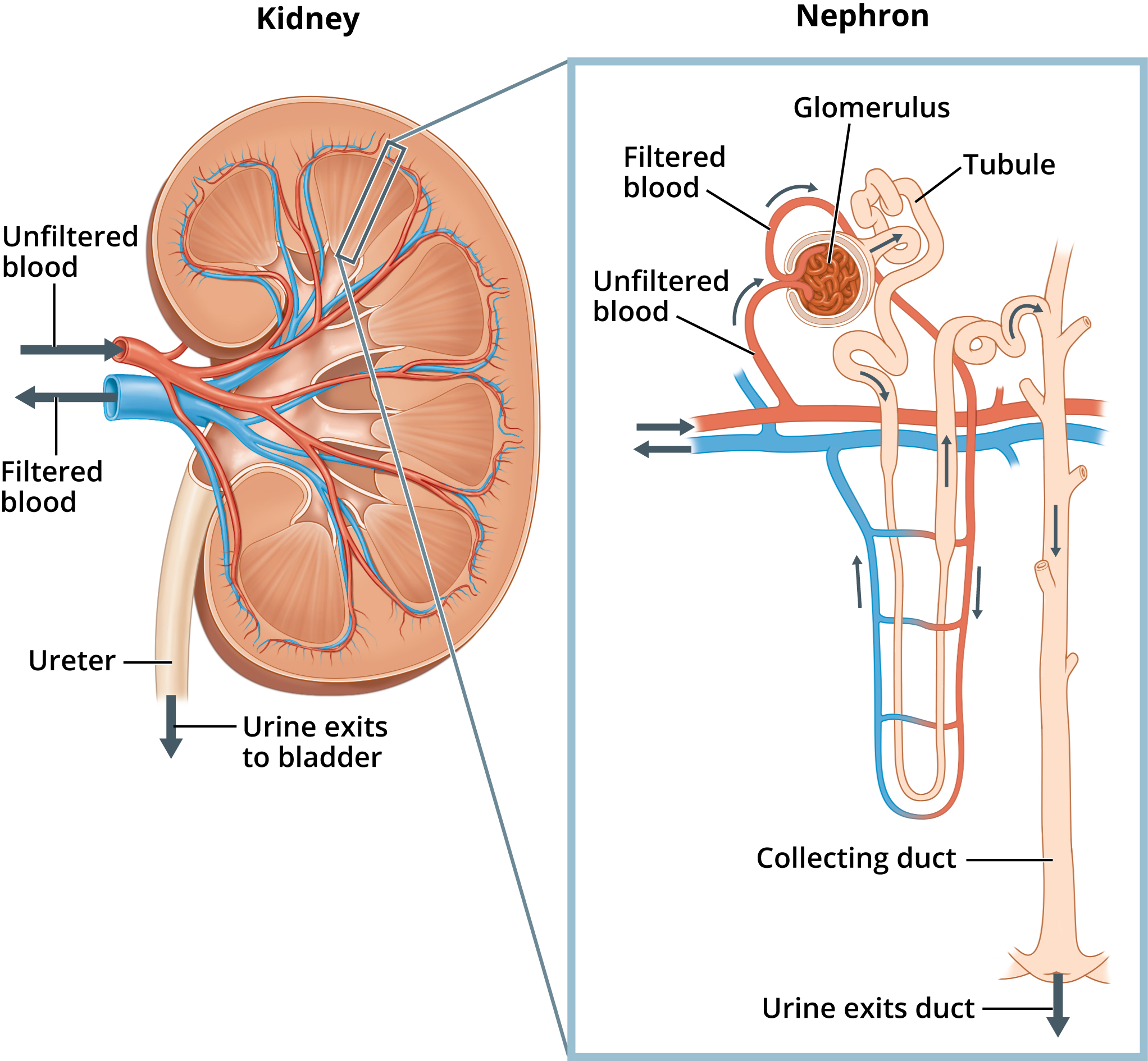
Nephron: Microscopic functional unit of the kidney responsible for filtering blood and forming urine.
Histology of Nephron Components
Renal Corpuscle: Consists of the glomerulus (capillary network) surrounded by Bowman's capsule (double-sided cup) to filter blood plasma into glomerular filtrate.
Proximal Convoluted Tubule (PCT): Lined with simple cuboidal epithelium featuring a prominent apical microvilli brush border.
Loop of Henle:
Juxtaglomerular Apparatus (JGA): Specialized region where the final portion of the thick ascending limb contacts the afferent arteriole.
Distal Convoluted Tubule (DCT):
Renin-Angiotensin-Aldosterone System (RAAS)
Renin Enzyme: Synthesised and secreted by specialised juxtaglomerular (JG) cells within the juxtaglomerular apparatus of the kidneys.
Secretion Triggers: Released into the bloodstream when the body senses a drop in blood pressure or reduced renal blood flow.
RAAS Cascade: Renin acts on liver-derived angiotensinogen -> converts it into Angiotensin I -> Angiotensin-Converting Enzyme (ACE) converts Angiotensin I into Angiotensin II.
RAAS: Renal Actions of Angiotensin II
Vasoconstriction: Acts as a potent systemic vasoconstrictor -> causes blood vessels to constrict -> increases peripheral resistance -> raises blood pressure.
GFR Reduction: Constricts afferent arterioles -> reduces Glomerular Filtration Rate (GFR) -> conserves water and salts.
Aldosterone Secretion: Triggers the release of aldosterone from the adrenal cortex.
RAAS: Aldosterone
Secretory Triggers & Cellular Action
Triggers: Secreted by the adrenal cortex in response to Angiotensin II or elevated plasma potassium (K+) concentrations.
Mechanism: Stimulates insertion of sodium (Na+) channels and Na+/K+ pumps into cells of the distal convoluted tubules (DCT) and collecting ducts.
Physiological Actions
Sodium & Water Retention: Increases active reabsorption of Na+ -> drives passive reabsorption of water -> expands blood volume and supports blood pressure.
Potassium Excretion: Promotes secretion of excess K+ into collecting ducts for urinary excretion.
RAAS: Erythropoietin (EPO) + RBC Production
Synthesis & Triggers
Production Site: Synthesised by peritubular interstitial cells located in the kidneys.
Secretion Trigger: Released into the bloodstream in response to decreased blood oxygen levels (e.g. high altitude or anaemia).
Actions on Erythropoiesis
Target Site: Stimulates red blood cell (erythrocyte) production within the bone marrow.
Physiological Effect: Increases the blood's oxygen-carrying capacity -> ensures adequate oxygen delivery to sustain cellular function.
RAAS: Vit D (Calcitriol)
Activation Pathway
The kidneys activate vitamin D by converting inactive calcidiol into its active form, calcitriol.
Physiological Roles
Intestinal Reabsorption: Enhances intestinal calcium reabsorption into the blood by promoting calcium-binding protein synthesis.
Mineral Regulation & Bone Health: Regulates plasma calcium and phosphate levels for bone tissue mineralization, working alongside parathyroid hormone (PTH).
Urethra: Male vs. Female
Urethra: Terminal duct connecting the urinary bladder to the exterior of the body for urine discharge.
Female Urethra:
Shorter (~4 cm long), positioned posterior to the pubic symphysis.
Serves an exclusively urinary function.
Mucosal lining transitions from transitional epithelium (near bladder) -> non-keratinised stratified squamous epithelium (near external orifice).
Male Urethra:
Longer (~20 cm long), serving dual urinary and reproductive functions (discharges urine and semen).
Divided into 3 anatomical regions: prostatic urethra (passes through prostate), membranous urethra (passes through deep perineal muscles), and spongy urethra (extends through penis).