KINS 4500 chapter 9 respiration
 Call Kai
 Learn
 Practice Test
 Spaced Repetition
 Match
 Flashcards
 Knowt Play
1/106

There's no tags or description
Looks like no tags are added yet.
107 Terms
you can hold your breath longer after hyperventilating. What makes you breathe again?
An increase in carbon dioxide (CO₂) levels in the blood—and the resulting drop in blood pH (acidification)—stimulates the medulla chemoreceptors in the brainstem to force you to breathe again
respiration is four processes, and only the first one is breathing
the four processes of respiration, each with the condition it falls in, and transport drawn as a step that is not one of the four. Acid base regulation is the fifth role and is the last slide of the second meeting
pulmonary ventilation
external respiration
internal respiration
cellular respiration

pulmonary ventilation
atmospheric air nose and mo9th
alveoli of lungs
falls in asthma
external respiration
alveoli of lungs
pulmonary capillary blood
falls in pulmonary edema
transport
not one of the four
falls in anaemia
internal respiration
systemic capillary blood
muscle cells
falls in carbon monoxide
cellular respiration
muscle cells
aerobic metabolism
yields ATP
falls in cyanide
one path from the nose to the alveolus and every branch is narrower
air travels a single branching path from the nose and mouth to the alveolar sacs, and the structures along that path are what the next slide divides into two functional zones
Nose and mouth, then pharynx, larynx, and trachea
The trachea divides into the left and primary bronchi
Bronchi branch to secondary and tertiary bronchi, then to bronchioles
Bronchioles at the terminal bronchioles
past them: respiratory bronchioles, alveolar ducts, alveolar sacs
each generation is narrower, and there are far more of them
maximize surface area by branching

path of air
nose > mouth > pharynx > larynx > trachea > bronchi > bronchioles > alveolar ducts > alveolar sacs
conductive zone
nose and mouth to the terminal bronchioles
moves air, warms it, humidifies it, filters it
No gas exchange, anatomical dead space
trachea
respiratory zone
respiratory bronchioles
alveolar ducts and sacs
every gas exchange here
the conductive zone moves air and exchanges nothing
everything from nose to terminal bronchioles is plumbing, and the volume it holds is air you moved but could not use
Conductive: nose and mouth to terminal bronchioles (brings air in)
Transport air, warms it to 37 degrees C, humidifies it to 99.5% saturated, filters it
No gas exchange, so it is also called anatomical dead space
Respiratory zone: respiratory bronchioles, alveolar ducts, alveolar sacs
Every gas exchange in the lungs happens here

Dead space is a fixed pipe, so it costs most when you breathe small
anatomical dead space scales with body size rather than with fat, and because the pipe does not change, the share of each breath it wastes falls as tidal volume rises
About 1 mL per pound of LEAN body mass. A 150 lb adult: about 150 mL
Fowler measured it by single-breath N₂ washout: 156 plus or minus 28 mL in 45 men That is 26% of their tidal volume, measured not assumed
It is airway volume, so it scales with frame. That is why one rule fits a child and an adult
At rest it wastes about a third of every breath In exercise it wastes about a fifth, because tidal volume rises and the airway does not
Alveolar dead space: alveoli ventilated but not perfused. Minimal in healthy lungs
Anatomical plus alveolar dead space is physiological dead space
Physiological dead space needs an arterial sample: the Bohr equation
Goes down with exercise
inspiration always costs muscle while
expiration is free until it is not
inspiration
always needs muscle
diaphragm contracts and flattens, ribs lift outward
thoracic volume increases
intrapulmonary pressure falls below atmospheric pressure air flows in
expiration
passive at rest
at rest: diaphragm relaxes, ribs fall, lungs recoils elastically. No muscle work
thoracic volume decreases
intrapulmonary pressure rises above atmospheric
air flows out
in exercise it turns ACTIVE: abdominals and internal intercostals drive it
airflow is a pressure gradient divided by a resistance
V= delta P/ R
V is airflow, in liters per second
Delta P is the pressure gradient, P1-P2, in mmHg
R is resistance, in mmHg per liter per second
A bigger gradient gives more flow. More resistance gives less flow
Resistance rises steeply as airway radius falls
Breathing a little top of mouth, helps open nose open up and nose breathing
A 130 lb woman at her ideal weight gains 20 lb. What happens to her anatomical dead space?
it stays at about 130 mL, because airway volume has not changed
the whole breath happens between 758 and 762 mmHg
Boyle’s law > P x V= constant
chest height is volume from Boyle’s law, inverse in pressure, with the deviation from rest exaggerated 60 times. The real swing from full inspiration to full expiration is 0.53 percent of resting volume

lung pressure crosses atmospheric twice on every breath
the same breath as a pressure trace, so the size of the swing is visible. Four mmHg moves a half of liter of air
harder breathe more pressure gradient

the lung has two blood supplies and they do opposite jobs
pulmonary circulation and bronchial circulation
pulmonary circulation
Right ventricle to pulmonary artery to alveolar capillaries
Serves EXTERNAL respiration: takes up O2, unloads CO2
Low pressure: about 25/10 mm Hg arterial, about 7 mmHg venous
Carries deoxygenated blood in an artery
bronchial circulation
Thoracic aorta to lung tissue, drains to the pulmonary veins
Serves INTERNAL respiration of the lung tissue itself
Part of the systemic circulation
So not all pulmonary venous blood is fully oxygenated
ventilation and perfusion are matched, but not evenly
V/Q is the ratio of ventilation to perfusion. Ideal is about 1.0
Apex: relatively under perfused, so V/Q is above 1.0
Base: relatively over perfused, so V/Q is below 1.0
The best perfused region is also the best ventilated region
Light exercise improves matching. Heavy exercise reintroduces inequality
Lungs direct blood flow
apex
relatively under perfused, so V/Q is above 1.0
base
relatively over perfused, so V/Q is below 1.0
Example talked about he asked RPE during VO2 max and guy got distracted and flow ended
During exercise > take more deeper breathes, because less dead space
Want breaths to be matched, functional rate
Deeper breaths more ventilation and surface area for gas exchange and blood exchange, there is a limit
there are only four volumes, and every capacity is a sum of them

inspiratory reserve volume IRV
the greatest amount of air that can be inspired at the end of a normal respiration
tidal volme
the amount of air that is inspired or expired in a normal breath
expiratory reserve volume ERV
the greatest amount of air that can be expired at the end of normal respiration
residual volume RV
the amount of air that is left in the lungs following a maximal exhalation
inspiratory capacity IC
the greatest amount of air that can be inspired from a resting expiratory level
IC= IRV + Vt
vital capacity VC
the greatest amount of air that can be exhaled following a maximal inhalation
VC= IRV + VT +ERV
functional residual capacity FRC
the amount of air left in the lungs at the end of a normal expiration
FRC= ERV + RV
total lung capacity TLC
the greatest amount of air that can be contained in the lungs
TLC= VC + RV
IC + FRC
IRV + VT + ERV + RV
a spirometer cannot see residual volume, and that matters twice
the shaded is the air in a spirometer never sees. Each capacity is drawn as the sum of the volumes beneath it

How much air you can movie one second predicts how long you live
FEV1 as a percentage of predicted is a long term predictor of all-cause mortality in the general population, not just a lung disease measure
Buffalo Health Study, 29-year follow-up of a general population sample
Low FEV1 percent-predicted predicted death from ALL causes, not only respiratory
Aronson 2006 adds an inverse association between pulmonary function and CRP
So lung function tracks something systemic, plausibly inflammation
This is why a spirometry number is worth taking in an apparently healthy person
All cause mortality > looking at causes of death, risks of dying
notation is a system, not a vocabulary list
Capital letter is the variable: V volume, P pressure, S saturation, F fraction
Capital subscript is the gas phase: A alveolar, I inspired, E expired, T tidal, D dead space
Lowercase subscript is the blood phase: a arterial, v venous, c capillary
A volume per unit time is a flow, so VO2max is a flow rate in mL . Min-1
A bar means a mean, so bar v is MIXED venous, the average of all venous return
A dot above means per unit time. A bar above means a mean value
Lowercase f standing alone is frequency, breaths per minute, not the F above
So PAO2 is alveolar and PaO2 is arterial. One letter apart
capital letter
variable
capital subscript
gas phase
lowercase subscript
blood phase
a volume per unit time is a
flow
so VO2max is a flow rate in mL . Min-1
bar
meanso bar v is MIXED venous, the average of all venous return
dot
above means per unit time. A bar above means a mean value
lowercase f
standing alone is frequency, breaths per minute, not the F above
minute ventilation is what moved whereas
alveolar is what you used
minute ventilation
VE= VT x f
At rest: 12 breaths/min x 0.5 L- 6.0 L.min-1
Counts every liter that moved, including the wasted ones
alveolar ventilation
VA= (VT-VD) x f
At rest: 12 (x 0.5-0.15)L- 4.2 L.min-1
Counts only the liters that reached an alveolus
same 6 liters per minute. Short and Shallow, or slow and deep
Person 1 (Short and shallow)
Math: (30 times (0.20- 0.15)
Result: = 1.5 L/min
Person 2 (Slow and deep))
Math: 6 times (1.00 - 0.15)
Math: (6 times 0.85)
Result: = 5.1 L/min
Conclusion
Slow and deep breathing provides a much higher alveolar ventilation rate (5.1 L/min vs. 1.5 L/min), because short and shallow breathing wastes 75% of each breath just filling the dead space.
two people each move 6.0 L of air per minute with 0.15 L of dead space. One breathes 30 times at 0.20 L, the other 6 times at 1.00 L. Which one does more for their alveoli?
fast and shallower breather
RR >30 breaths/min
VT > 0.2 L
AV = 30 x (0.2-0.15) > 1.5 L/min
slow and deep breather
RR > 6 breaths/min
VT > 1.00 L
AV = 6 x (1.00-0.15) > 5.1 L/min
Person 2 delivers significantly more fresh air to their alveoli (5.1 L/min vs. 1.5 L/min). Because Person 1 has a very low tidal volume, a massive portion of each breath (75%) is wasted entirely on clearing the anatomical dead space.
one breath, three volumes, depending on where you measure it
three gas laws, three changes, every one of them staring from the same 5 liters on one shared axis
pressure falls > Boyle’s Law
temperature falls > Charles’ law
water vapor removed

pressure falls > Boyle’s Law
760 to 600 mmHg, held at 0 degrees Celsius and dry
temperature falls > Charles’ Law
37 to 0 degrees Celsius, held at 760 mmHg and dry
water vapor removed
saturated and dry, held at 37 degrees Celsius and 760 mmHg
breathing rhythm is generated in the medulla and smoothed by the pons
the control of breathing as a left to right flow. The limb from the medulla out to the muscles is added from the chapter text
Opigenetics > gene activation. Light activation

high brain centers (stimulate or inhibit)
cerebral cortex, voluntary control
hypothalamus strong emotion and pain
DRG
chemoreceptors (stimulate)
mental, in the medulla, rising PCD, falling pH
peripheral, carotid and aortic, also falling PO2, rising K+
DRG
lung and airway receptors (inhibit)
stretch, the Harling-Breur reflex, on overinflation
irritant, to fumes, mucus and particulates
DRG
muscle and joint receptors
proprioceptors, sensitive to movement
DRG
inspiratory muscles
diaphragm, external intercostals
VRG
expiratory muscles
internal intercostals, abdominals, only when breathing is forceful
ventilation is
12-15 breaths per minute
dorsal respiratory group integrates whereas
ventral respiratory group generates the rhythm
three classes of input reach the respiratory centers
higher brain centers, mechanical receptors and chemoreceptors all modulate breathing, and they do not matter equally
higher brain centers
Hypothalamus: sympathetic drive, pain, strong emotion, cold water immersion
Cerebral cortex: conscious control for speech, singing, lifting, swimming
mechanic receptors
Lung stretch: the Hering- Breur reflex, which cuts off an over deep breath
Irritant: cough and bronchoconstriction
Muscle and joint: movement signals the centers
chemoreceptors
Central, in the medulla: responds to CO2 by way of H+
Peripheral, carotid, and aortic bodies: O2, CO2, H+ and K+
Arterial PO2 falls from 100 to 80 mmHg while arterial PCO2 rises from 40 to 45 mmHg. Which change drives ventilation harder?
The 5 mmHg rise in PCO2, which nearly doubles ventilation
The rise in PCO2 drives ventilation harder because central and peripheral chemoreceptors are acutely sensitive to even tiny increases in carbon dioxide and the resulting drop in pH, triggering an immediate surge in breathing to blow it off. Conversely, the drop in PO2 to 80 mmHg has almost no effect because oxygen levels must fall below a critical hypoxic threshold of roughly 60 mmHg before the hypoxic ventilatory drive is strongly activated.
over the same 35 mmHg, CO2 does everything and O2 does nothing
Both panels span 35 mmHg of their own gas. Carbon dioxide crosses that span from 4 to 39 liters a minute. Oxygen crosses it without responding at all
normal arterial PO2 is 95 mmHg and normal arterial PCO2 is
40 mmHg
makes cost you comfort, not performance, and UCONN ran one of the studies
three controlled trials found no performance, saturation or core-temperature cost to masked exercise; the real signals are retained CO2 under an N95 and breathing discomfort
Shaw 2020: randomized crossover, n=14, cloth vs surgical vs none, maximal cycling
No difference in time to exhaustion, peak power, SaO₂, tissue oxygenation, RPE or HR
Epstein 2020: ramp protocol, n=16, none vs surgical vs N95
No difference in HR, respiratory rate, BP, saturation or time to exhaustion
BUT significantly higher end-tidal CO₂ with the N95, rising with intensity
Yoshihara 2021, OUR DEPARTMENT: n=12, 60 min in 32 °C heat, 4 masks vs none
No difference in rectal temperature or heart rate. Breathing discomfort DID rise
All three studied healthy young adults only
only some of those inputs actually drive exercise hypernea
the inputs that dominate at rest are not the ones that drive exercise hyperpneas and the mechanism is still not fully explained
what drives exercise hypernea
pH, by way of CO2 and of lactate
Potassium, released from working muscle
Muscle and joint mechanoreceptors
Central command, feeding forward
what does not drive exercise hypernea
PaO2, flat as a stimulus above 60 mmHg
PaCO2, tightly regulated, barely rises
Hering-Breuer reflex, minimal even in exercise
what is not explained about exercise hypernea
Nothing explains how closely VE tracks metabolic rate
No single stimulus accounts for the match, after seventy years
shallow water blackout: it kills in guarded pools, and that is the point
hyperventilation extends breath hold time by lowering Co2, not by adding oxygen, so it removes the warning without adding the reverse
The name for it is shallow-water blackout, or hypoxic blackout
The urge to breathe comes from rising CO₂, not from falling O₂
Hyperventilation blows off CO₂, so the urge arrives late or not at all
Oxygen keeps falling on the same schedule. Nothing has been added
Craig 1976: 58 cases of loss of consciousness in underwater swimming and diving
23 of the 58, 40%, were fatal. Most occurred in GUARDED pools
Swimming continues for a short time after blackout, so it is hard to spot
Recommendation: limit underwater swimming to one length of a 25 yd or 25 m pool
Fick’s Law of diffusion (this chapter)
Vgas= (A x D X delta P) divided by T
A area, D diffusion coefficient, which rises with solubility
Delta P the pressure gradient, T the thickness of the barrier
Governs how fast a gas crosses the alveolar membrane
Answers: why does oxygen get into the blood at all
Fick equation chapter 11
VO2= Q x a-vO2 diff
Relates oxygen uptake to cardiac output and extraction
Introduced in session 7 as equations 11.3 and 11.13
Why does hyperventilating before an underwater swim make blackout more likely?
hyperventilating lowers CO2, delaying the urge to breathe past safe oxygen
four things that se how fast a gas crosses the membrane
diffusion rate rises with the pressure gradient, the surface area and the gas’ solubility and falls as the barrier thickens
The pressure gradient across the barrier, in ,mmHg
The surface area available for diffusion
The thickness of the barrier between two locations
The solubility of the gas in the barrier liquid

altitude does not change the percentage of oxygen, only the pressure
percent times barometric pressure equals partial pressure, there times over. the oxygen percentage is identical at sea level and at altitude, only the pressure it multiples has changed
partial pressure changes

oxygen crosses downhill at both ends of the circuit
arrow size is the LOCAL gradient, so it falls nothing as the blood equilibrates
both panels share one 0 to 110 mmHg axis
deep breaths better

carbon dioxide crosses downhill at both end of the circuit
Arrow size the LOCAL gradient, so it falls to nothing as the blood equilibrates. Both panels share 0 to 110 mmHg axis
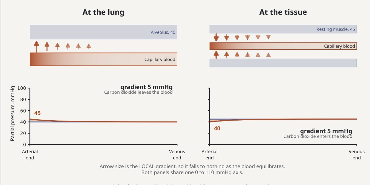
working muscle holds a gradient 1.7 times the resting one, and never lets the blood catch up
exercising muscle PO2 of 3.8 mmHg is MEASURED, by myoglobin desaturation and is invariant beyond 60 percent of maximal work rate

without hemoglobin, blood would
carry 1/70th of the oxygen
dissolved in plasma, 1.5 to 3% blood
Solubility constant 0.00304 mL.dL-1 mmHg-1 at 37 degrees C
At PaO2, 95 mmHg: 95 x 0.00304=0.29 mL.dL-1
Tiny, and it is what SETS the partial pressure of oxygen
Only dissolved gas exerts pressure, so only is drives diffusion
bound to hemoglobin, 97 to 98.5% blood
four heme groups per hemoglobin, one O2 each
binding capacity is constant 1.34 mL O2 per g Hb
fully saturated means all four hemes occupied
SbO2% is how much of that capacity is in use
it multiples the blood’s oxygen capacity about seventyfold
hemoglobin is four subunits, and its shape is the point
binding one oxygen makes the next easier to bind, which gives the dissociation curve its S shape
Four globin chains, each wrapped around one iron containing heme
One O2 binds per heme, so four per hemoglobin
The four do not bind at the same time
Binding the first changes the shape, making the second easier
This cooperativity is why the curve is sigmoid, not straight
RBC count: 4.3 to 5.2 million per cubic female, 5.1 to 5.8 male

arterial blood carries 19.79 mL of oxygen per deciliter
oxygen content is hemoglobin times its binding constant times saturation, plus dissolved fraction and the dissolved fraction is not optimal in the arithmetic
Take Hb = 15 g·dL−1, SaO₂ = 97%, PaO₂ = 95 mmHg
1.34 is hemoglobin's oxygen-binding capacity, in mL O₂ per g Hb
Bound: 15 × 1.34 × 0.97 = 19.50 mL·dL−1
Dissolved: 95 × 0.00304 = 0.29 mL·dL−1
Total: 19.50 + 0.29 = 19.79 mL·dL−1
Venous, at 75% and PvJO₂ 40 mmHg: 15.08 + 0.12 = 15.20 mL·dL−1
Looking at stuff as a whole
Hydrogen drives change
the a-vO2 difference is what the tissues actually took
arterial content minus venous content is the oxygen extracted in one pass, and at rest that is only about a quarter of what was carried
A-vO2 diff= arterial O2 content- venous O2 content
At rest: 19.79-15.20=4.59 mL.dL-1
Only about 25% of the oxygen carried, extracted in one pass
The other 75% returns to the lungs unused
It is the shaded band at the top of the figure

exercise double extraction without changing the arterial side
At rest
Arterial O2 content: 19.79 mL.dL-1
Venous O2 content 15.20 mL.dL-1
A-vO2diff= 19.79-15.20=4.59
4.59 mL.dL-1, about 25% extracted
During heavy exercise
Arterial O2 content: 19.79 mL.dL-1, essentially unchanged
Venous O2 content: about 10.00 mL.dL-1
A-vO2diff=19.79-10.00=
9.79 mL.d-1. more than double
the S shape is a safety margin on top, a delivery zone below
the curve is flat above about 60 mmHg and steep below it, so loading is protected and unloading is sensitive
Flat above about 60 mmHg: big falls in PO2 cost little saturation
That is the loading safety margin, at altitude and in lung disease
Steep below about 40 mmHg: small falls in PO2 release a lot of oxygen
That is the unloading zone, at the working tissue
Arterial sits at about 97%, on the flat part
Venous sits at about 75%, on the shoulder
Loading easy, unloading hard

three things shift the curve right and they live in chapter 10
decreased pH
increased temperature
increased 2, 3 DPG
decreased pH
Lowers Hb-O2 affinity, shifts the curve right
Favours offloading O2 to the tissues
This the bohr effect
increased temperatures
Lowers Hb-O2 affinity, shifts the curve right
A working muscle is warm, so it unloads more
increased 2,3 DPG
A byproduct of glycolysis in the red cell
May shift the curve right
Rises at ALTITUDE. Not a major cause during exercise
Inflamed good > muscle will get remodeled
inside the muscle cells the carrier is myoglobin
myoglobin has a higher affinity for oxygen than hemoglobin, so it accepts oxygen at low PO2 inside the cell and shuttles it to the mitochondria
Myoglobin, Mb, sits inside the muscle fibre
Shuttles O₂ from the cell membrane to the mitochondria
Higher O₂ affinity than hemoglobin, so O₂ moves Hb to Mb, not the reverse
Also a small O₂ store for the onset of exercise
One heme per molecule, not four: no cooperativity, no S curve
carbon dioxide travels three ways, mostly as bicarbonate
about 70% of Co2 moves as bicarbonate which is why CO2 transport and acid base balance are the same mechanism
can also be dissolved or carbamino
dissolved
5-10% of CO2
Dissolved in plasma
Set the partial pressure of CO2
So it is what the chemoreceptors read