Comprehensive Review of Helminthic Infections, Breast Histology, and Surgical Breast Anatomy
 Call Kai
 Learn
 Practice Test
 Spaced Repetition
 Match
 Flashcards
 Knowt Play
1/104

Earn XP
Description and Tags
Vocabulary practice flashcards integrating clinical parasitology (intestinal and tissue helminths), histological features of the mammary gland across physiological states, and surgical breast and axillary anatomy.
105 Terms
Nematodes
Non-segmented, cylindrical roundworms possessing a complete digestive tract.
Cestodes
Segmented tapeworms featuring an anterior attachment organ (scolex) with suckers (and sometimes hooks) that completely lack a digestive tract.
Trematodes
Unsegmented, leaf-like flukes that lack hooks and possess an incomplete digestive tract.
Eosinophilia
The cardinal laboratory and hematologic hallmark of a tissue infection caused by parasitic worms.
Echinococcus granulosus
The primary exception among parasitic helminths that undergoes asexual multiplication within the human host inside hydatid cysts.

Trichinella spiralis
The only intracellular helminth of humans; its infective larvae encyst within active skeletal muscle cells.

Enterobius vermicularis
The human pinworm; an intestinal nematode living in the cecum and colon, diagnosed by recovering eggs from perianal folds using the Scotch tape test, and treated with pyrantel pamoate or mebendazole.

Trichuris trichiura
The whipworm; an intestinal nematode acquired by ingesting embryonated eggs from contaminated soil or food, capable of causing rectal prolapse, tenesmus, and bloody diarrhea in heavy infections.
Löeffler syndrome
A pulmonary inflammatory response consisting of cough, marked eosinophilia, and transient pulmonary infiltrates caused by larvae of Ascaris lumbricoides migrating through lung tissue.

Ascaris lumbricoides (Adult)
The giant human roundworm; its anterior end characteristically displays three sensory 'lips' surrounding the mouth opening.
Ground itch
An intensely pruritic, erythematous maculopapular cutaneous reaction at the site of skin penetration by infective hookworm filariform larvae.

Ancylostoma duodenale
Old World hookworm; an intestinal nematode whose anterior buccal capsule contains sharp cutting teeth to anchor into the small intestinal mucosa.

Necator americanus
New World hookworm; an intestinal nematode whose anterior buccal capsule possesses pairs of semilunar cutting plates rather than teeth.
Parthenogenesis (Strongyloides stercoralis)
The specialized reproductive mechanism allowing parasitic adult female threadworms to reproduce in the human small intestine without requiring fertilization by males.
Larva currens
A rapidly advancing, highly pruritic, serpiginous cutaneous track caused by autoinfective filariform larvae of Strongyloides stercoralis migrating through the dermis.

Fasciolopsis buski
The giant intestinal fluke; a trematode acquired by ingesting metacercariae encysted on aquatic plants, which colonizes the small intestine and responds to praziquantel.

Scolex of Taenia solium
The anterior attachment structure of the pork tapeworm, distinguished by four large muscular suckers and a rostellum bearing two rows of chitinous hooks.
Scolex of Taenia saginata
The attachment organ of the beef tapeworm, bearing four prominent suckers but completely devoid of an armed rostellum or hooks.
Cysticercosis
A systemic infection resulting from the ingestion of Taenia solium eggs via the fecal-oral route, causing encysted larval stages to develop in the central nervous system, striated muscles, subcutaneous tissues, and eyes.

Diphyllobothrium latum
The broad fish tapeworm; acquired through ingestion of larvae in undercooked raw fish, characteristically absorbing dietary vitamin B12 and causing pernicious anemia.

Hymenolepis nana
The dwarf tapeworm; an intestinal cestode capable of completing its full life cycle without an obligatory intermediate host, displaying craspedote (overlapping) proglottids.

Dipylidium caninum
The dog tapeworm; an intestinal cestode transmitted via the ingestion of infected dog fleas, identified microscopically by egg packets containing multiple eggs.
Schistosoma haematobium
A trematode whose adults reside within the venous vessels of the urinary bladder; diagnosed by microscopic identification of eggs with a prominent terminal spine in urine.
Schistosoma mansoni
A blood fluke whose adults reside in mesenteric veins of the large intestine and liver, shedding diagnostic eggs with a prominent lateral spine in the feces.

Mammary crest (Milk line)
Paired bilateral ectodermal thickenings extending from the axillae to the groin along which mammary glands develop embryologically.
Compound branched tubuloalveolar gland
The histological classification of the mammary gland, which represents a highly specialized modification of apocrine sweat glands embedded in dense stroma and adipose tissue.
Merocrine secretion (Mammary gland)
The cellular secretory mechanism wherein the protein components of breast milk are packaged in Golgi vesicles and released into the alveolar lumen via exocytosis without cellular membrane loss.
Apocrine secretion (Mammary gland)
The cellular secretory mechanism wherein milk lipid droplets coalesce and are released from alveolar cells along with a surrounding envelope of the apical cell membrane and cytoplasm.
Suspensory ligaments of Cooper
Fibrous connective tissue bands spanning from the dermis of the skin through the breast parenchymal stroma to the deep pectoral fascia; tumor-induced retraction causes characteristic skin dimpling.
Terminal Duct Lobular Unit (TDLU)
The basic functional and histological secretory unit of the adult female breast, consisting of a terminal ductule and its cluster of secretory alveoli (acini).

Lactiferous duct epithelium
The lining of the main mammary excretory ducts, transitioning from stratified squamous epithelium near the nipple pore to a two-layered cuboidal epithelium overlying myoepithelial cells deeper in the gland.
Myoepithelial cells (Mammary)
Spindle-shaped contractile cells located between alveolar secretory cells and the basal lamina that contract under the influence of oxytocin to promote milk ejection.

Alveolar switch
The marked histological transformation occurring during pregnancy where breast stroma diminishes and ductules proliferate into expanded, hyperplastic secretory lobules and alveoli.

Prolactin vs. Oxytocin
Prolactin, released by the anterior pituitary, drives milk production and synthesis in alveolar cells, whereas oxytocin, released by the posterior pituitary, stimulates myoepithelial contraction for milk let-down.
Colostrum
A thick, protein- and antibody-rich fluid produced by the glandular alveoli during late pregnancy and the initial days of lactation before mature milk production ensues.

Axillary tail of Spence
A lateral extension of parenchymal tissue from the upper outer quadrant of the breast that passes through an aperture in the deep axillary fascia.
Foramen of Langer
The specific opening in the deep fascia of the axilla through which the axillary tail of Spence extends into the axillary fossa.

Wuringer's horizontal septum
A horizontal ligamentous fascial septum that divides the breast into superior and inferior anatomical compartments while carrying key neurovascular branches to the nipple-areola complex.
Rotter's interpectoral nodes
Lymph nodes situated between the pectoralis major and pectoralis minor muscles that provide an alternative lymphatic drainage pathway from the mammary gland.
Axillary lymph node levels
Surgical lymphatic staging regions classified by their relationship to the pectoralis minor muscle: Level I is lateral and inferior, Level II is directly posterior (deep), and Level III is medial and superior.

Sentinel lymph node biopsy
A surgical mapping procedure identifying the first draining axillary lymph node(s) from a breast tumor using injected radiotracer or blue dye.
Long thoracic nerve of Bell
The motor nerve supplying the serratus anterior muscle; inadvertent surgical injury during axillary dissection leads to winged scapula.
Thoracodorsal nerve
The motor nerve innervating the latissimus dorsi muscle; iatrogenic transection causes loss of shoulder adduction and inability to push up from a seated position.
Intercostobrachial nerve
A sensory nerve crossing the axilla originating from the second intercostal nerve; injury causes numbness or paresthesia along the medial skin of the upper arm.
Batson's vertebral plexus
A valveless network of epidural and vertebral veins connecting with posterior intercostal veins, serving as a route for breast carcinoma metastasis to the spine and central nervous system.

Boundaries of the axilla
A pyramidal anatomical space defined by an apex (cervicoaxillary canal), a base (skin and axillary fascia), an anterior wall (pectoralis major and minor), a posterior wall (subscapularis, teres major, latissimus dorsi), a medial wall (ribs 1 to 4 and serratus anterior), and a lateral wall.

Lateral wall of the axilla
The narrow anatomical boundary of the axilla formed by the intertubercular sulcus (bicipital groove) of the humerus.
Enterobius vermicularis
pinworm
Trichuris trichiura
whipworm
Ascaris lumbricoides
human roundworms
Strongyloides stercoralis
human threadworm
Fasciolopsis buski
giant intestinal fluke
Taenia saginata
beef tapeworm
Taenia solium
pork tapeworm
Diphyllobothrium latum
broad fish tapeworm
Hymenolepis nana
dwarf tapeworm
Dipylidium caninum
dog tapeworm
hookworms, human threadworms, schistosomes
larvae penetrate the skin
filarids
vectoborn
trematode
pathology is generally associated with the adult stage
Echinococcus granulosus
only exception which multiplies asexually within the hydatid cysts
Trichinella
only intracellular helminth
embryonated egg
infective stage of enterobius vermicularis (pinworm)
eggs on perianal folds (Scotch tape test)
diagnostic stage of enterobius vermicularis (pinworm)
ingestion of eggs
mechanism of infection of enterobius vermicularis (pinworm)
lumen of cecum, colon
site of infection of enterobius vermicularis (pinworm)
perianal pruritus
main symptoms of enterobius vermicularis (pinworm)
Pyrantel pamoate, Mebendazole
treatment of enterobius vermicularis (pinworm)
eggs of enterobius vermicularis in a cellulose-tape preparation

eggs of enterobius vermicularis in a wet mount

male and female of enterobius vermicularis in mounted specimens
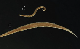
trichuris trichiuria
whipworm-intestinal nematode
embryonated egg
infective stage of trichuris trichiura
unembryonated eggs in the feces
diagnostic stage of trichuris trichiura
ingestion of eggs from fecally contaminated soil/food
mechanism of infection of trichuris trichiura
cecum, colon
site of infection of trichuris trichiura
asymptomatic, lower abdominal pain, distention, diarrhea, profuse bloody diarrhea, cramps, rectal prolapse, may migrate to appendix
main symptoms of trichuris trichiura
mebendazole, albendazole
treatment stage of trichuris trichiura
egg of trichuris trichiura in an iodine-stained wet mount
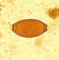
adult if t. trichiura removed during a colonoscopy

anterior end of trichuris trichiura

ascaris lumbrico
roundworm