Ankle & Foot, arthro, gait, ligaments
 Call Kai
 Learn
 Practice Test
 Spaced Repetition
 Match
 Flashcards
 Knowt Play
1/105

There's no tags or description
Looks like no tags are added yet.
106 Terms
During initial contact, both ankle and MTP are
in neutral
During loading response, the ankle and MTPs dof?
Ankle 5 degrees PF, MTP in neutral
In midstance the ankle and MTPs dof?
Ankle in 5 degrees of dorsiflexion, MTP in neutral
During terminal stance, MTPs and Ankle dof?
Ankle 10 degrees of DF
MTPs 30 degrees of extension
During pre-swing ankle and MTPs dof?
ankle 15 degrees of plantarflexion
MTPs 60 degrees of extension
Initial swing Ankle and MTP dof?
ankle 5 degrees plantarflexion
MTPs are neutral
during midswing ankle and MTPs are
in neutral
During terminal swing ankle and MTPs are
in neutral
The interosseous membrane of the leg is a
tough fibrous sheet of connective tissue
The interosseous membrane of the leg spans the distance between
interosseous borders of tibial and fibular shafts
The subtalar is stabilized by the
interosseous talcocalcaneal ligament
within dorsiflexion of the talocrural joint, the talus rolls ____________ and the leg and simulatneously slides ______________
rolls anteriorly and slides posteriorly
the deltoid ligament is 2 layers of strong connective tissue that connects the tibia to...
navicular, calcaneus, talus
posterior slide allows the talus to rotate...
forward with only limited anterior translation
The subtalar joint is stabilized by...
interosseous talocalcaneal ligament
What ligament of the talocrural joint becomes taut in repose to posterior sliding of the talus
The calcaneofibular ligament
Any collateral ligament that becomes increasingly taut from posterior translation of the talus also becomes increasingly taut during
dorsiflexion
Dorsiflexion of the talocrural ligament elongates
the posterior talofibular ligament,
posterior tibiotalar fibers of the deltoid ligament
the dome of the talus is wider _____________ than ______________
wider anteriorly than posteriorly
As dorsiflexion occurs in a closed chain, the talus gets wedged between...
medial and lateral malleoli
The wedging of the talus causes the distal tibia and fibula to
spread apart slightly
This spread of the tibia and fibula is resisted by tension in the
distal tibiofibular ligaments and the interosseous membrane
during plantar flexion in the talocrural joint the talus rolls ______________ as the bone simultaneously slides _____________
rolls posteriorly as bone simultaneously slides anteriorly
Any collateral ligament that becomes increasingly taut from anterior translation of the talus also becomes increasingly taut during
plantarflexion
What ligament is stretched in full plantar flexion in the talocrural joint
anterior talofibular ligament
plantar flexion of the talocrural joint also stretches...
dorsiflexor muscles and the anterior capsule of the joint
deltoid ligament
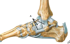
The subtalar joint (STJ) arthrokinematics involve a sliding motion among
the three sets of facets
Axis of rotation is typically described as a line that pierces the lateral-posterior heel and courses through the subtalar joint in
anterior, medial and superior direction
supination/inversion of the subtalar joint involves and __________ roll and __________ glide
medial roll, lateral glide
pronation/eversion of the subtalar joint involves and __________ roll and __________ glide
lateral roll and medial glide
Inversion exceeds eversion by nearly
double
inversion DOF
22.6
eversion DOF
12.5
What two structures naturally limit eversion
lateral malleolus, deltoid ligament
During weight-bearing activities in the stance phase of walking, pronation and supination occur as the leg and talus rotate over...
a relatively stationary calcaneus
Subtalar motion allows the foot to assume positions that are independent of...
The orientation of the superimposed ankle and leg
The talocalcaneonavicular joint (TCN) is a complex synovial joint where the
The head of the talus articulates with the calcaneus and the plantar calcaneonavicular ligament inferiorly
talocalcaneonavicular joint (TCN)

The talocalcaneonavicular joint (TCN) is a complex synovial joint where the head of the talus articulates with the calcaneus and the plantar calcaneonavicular ligament inferiorly and the head of the talus
articulates with the navicular anteriorly
The TCN joint is reinforced by
interosseous talocalcaneal ligament
talonavicular ligament
plantar calcaneonavicular ligament
interosseous talocalcaneal ligament
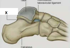
The interosseous talocalcaneal ligament reinforces the TCN and capsule
posteriorly
The talonavicular ligament reinforces the TCN and capsule
superiorly
The plantar calcaneonavicular ligament reinforces the TCN and capsule
inferiorly
The calcaneocuboid joint is a synovial joint between
facet of the anterior surface of the calcaneus and the facet on the posterior surface of the cuboid
calcaneocuboid joint

Facet on the anterior surface of the calcaneus is (shape)
convex
Facet on the posterior surface of the cuboid is (shape)
concave
The plantar calcaneocuboid ligament connects the __________ tubercle to _________ surface of cuboid
calcaneal tubercle to inferior surface of cuboid
plantar calcaneocuboid ligament

The long plantar ligament

the longest ligament in the sole of the foot
long plantar ligament
the long plantar ligament is ___________(____________) to plantar calcaneocuboid ligament
inferior (superficial)
cuneonavicular joint

The cuneonavicular joint is supported by
Plantar cuneonavicular ligament
Dorsal cuneonavicular ligament1
The cuneonavicular joint is between
navicular and cuneiforms
Dorsal cuneonavicular ligament

Plantar cuneonavicular ligament
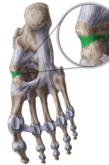
The forefoot contains what three sets of joints, those being
tarsometatarsal joints (TMT)
metatarsophalangeal joints
interphalangeal joints
tarsometatarsal joints

metatarsophalangeal joints

interphalangeal joints

the first MT joint is an articulation between
medial cuneiform with the first metatarsal
The second and third TMT joints are articulations of
intermediate (2) and lateral (3) cuneiforms with 2nd and 3rd Metatarsals
The fourth and fifth TMT joints are articulations of
cuboid with 4th and 5th Metatarsals
the metatarsophalangeal joints are numbered..
1 to 5
the metatarsophalangeal joints are articulations betwen
heads of metatarsal (convex) and bases of proximal phalanges (concave)
Metatarsophalangeal joints are located about ____ cm proximal to the "web spaces" of the toes
2.5 cm
what cartilage covers the distal end of each metatarsal head
articular cartilage
with the metatarsophalangeal joints flexed, the prominant heads of the ________________ are easily palpable on the dorsum of the foot
metatarsals
the metatarsophalangeal joints have _____ DOF
two DOF
The three metatarsophalangeal joint ligaments include
medial collateral ligament
lateral collateral ligaments
plantar ligaments
The metatarsophalangeal joints two movements in the sagittal plane are
extension and flexion
The metatarsophalangeal joints two movements in the horizontal plane are
abduction and adduction
Toes can be passively extended about ___ degrees
65 degrees
Toes can be passively flexed about
30 to 40 degrees
Great toe typically allowsgreater extension, to near ___ degrees
85 degrees
The intermetatarsal joints are extensions of
each tarsometatarsal joint
The intermetatarsal joints are between
the bases of the metatarsals
Lisfranc joint

The Lisfranc joint is the combination of...
all intertarsal joints and tarsometatarsal joints
longitudinal arch

lateral longitudinal arch
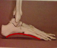
The longitudinal arch is formed between
posterior end of calcaneus and heads of metatarsals
the longitudinal arch is highest ___________ and lowest _____________
highest medially and lowest laterally
The medial longitudinal arch (MLA) is the primary....
load-bearing and shock-absorbing structure of the foot
Bones that contribute structurally to the medial longitudinal arch include..
_
_
_
_
_
-calcaneus
-talus
-navicular
-cunieforms
-three medial metatarsals
Without an arched configuration large and rapidly produced forces applied against the foot would likely
exceed the physiologic weight-bearing capacity of the bones
Additional structures that assist the arch in absorbing loads include
fat bads, sesamoid bones of the great toe, and superficial plantar fascia
The ligaments that support the medial longitudinal arch (MLA)
plantar calcaneonavicular (spring)
plantar calcaneocuboid (short plantar ligament)
long plant ligament
plantar fascia/aponeurosis
transverse arch

The medial side of the transverse arch is
head of the talus and navicular
the lateral side of the transverse arch is
end of the calcaneus and cuboid
the transverse arch flattens anteriorly at
metatarsal heads
what four extrinsic muscles can provide dynamic support for the arches:
tibialis anterior
tibialis posterior
fibularis longus
fibulris brevis
Nonmuscular structures responsible for maintaining height and general shape of the arch are
plantar fascia
spring ligament
first tarsometatarsal joint
The primary passive support to the medial longitudinal arch is provided by..
plantar fascia
plantar fascia

well standing still, the weight of the body falls through the foot near the region of the...
talonavicular joint