Anatomy Exam 2 (Module 6)
0.0(0)
Studied by 1 person Call Kai
 Learn
Learn Practice Test
 Spaced Repetition
 Match
Match Flashcards
 Knowt Play
Card Sorting
1/88

Last updated 3:58 PM on 10/11/22
Name | Mastery | Learn | Test | Matching | Spaced | Call with Kai | Chat |
|---|
No analytics yet
Send a link to your students to track their progress
89 Terms
1
New cards
Bones in the skull
- 28 bones
- most are "flat" bones formed via intramembranous ossification
- bones united by sutures (interlocking, immovable joints)
- most are "flat" bones formed via intramembranous ossification
- bones united by sutures (interlocking, immovable joints)
2
New cards
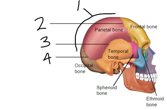
Major sutures of the skull
- coronal suture
- squamous suture
- lambdoid suture
- sagittal suture
- squamous suture
- lambdoid suture
- sagittal suture
3
New cards
1) sagittal suture
2) coronal suture
3) squamous suture
4) lambdoid suture
2) coronal suture
3) squamous suture
4) lambdoid suture
4
New cards
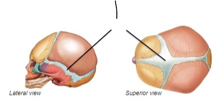
Fetal/Infant skulls
- connect by flexible CT to allow head to deform during birth & rapid brain growth
- area between bone = fontanelles (soft spots), will fuse over time
- area between bone = fontanelles (soft spots), will fuse over time
5
New cards
1) fontanelles
6
New cards
Craniosynostosis
premature fusion of the skull bones, leading to unusual cranial vault shape
7
New cards
Subdivisions of the skull
1) cranial division
2) facial division
2) facial division
8
New cards
Cranial bone functions
- protect brain
- provide attachment sites for some neck/head muscles
- provide attachment sites for some neck/head muscles
9
New cards
Facial bone functions
- form the framework of face openings for passage of air and food
- hold the teeth
- anchor muscles of the face
- hold the teeth
- anchor muscles of the face
10
New cards
Cranium subdivisions
1) vault = superior, lateral, and posterior bones of the skull (forehead included)
2) base = inferior part of the cranium
2) base = inferior part of the cranium
11
New cards
How many bones are in the cranium?
8 bones
12
New cards
Bones of the cranium
- frontal (forehead)
- occipital
- sphenoid
- ethmoid
- 2 parietals (left & right)
- 2 temporals (left & right)
- occipital
- sphenoid
- ethmoid
- 2 parietals (left & right)
- 2 temporals (left & right)
13
New cards
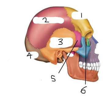
1) frontal bone
2) parietal bone
3) temporal bone
4) occipital bone
5) sphenoid bone
6) ethmoid bone
2) parietal bone
3) temporal bone
4) occipital bone
5) sphenoid bone
6) ethmoid bone
14
New cards
Parietal bones
- make up most of the superior part of the skull, extend posteriorly and laterally
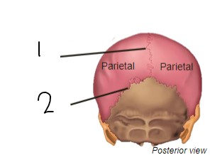
- sagittal suture between two parietal bones
- lambdoid suture between parietal and occipital bones
- sagittal suture between two parietal bones
- lambdoid suture between parietal and occipital bones
15
New cards
1) sagittal suture
2) lambdoid suture
2) lambdoid suture
16
New cards
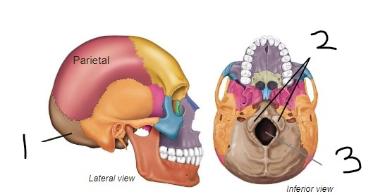
Occipital bone
- posterior part of the cranium and cranial base
- contain foramen magnum and occipital condyles
- contain foramen magnum and occipital condyles
17
New cards
Foramen magnum
large opening in the occipital bone that allows the spinal cord to pass & connect w/ the brain
18
New cards
Occipital condyles
region where the skull articulates w/ the vertebral column (atlas or C1 vertebra)
19
New cards
1) occipital
2) occipital condyles
3) foramen magnum
2) occipital condyles
3) foramen magnum
20
New cards
Temporal bones
paired bones (left & right side) that house the openings to the ear, forming the base of the cheekbone
21
New cards
Main regions of temporal bones
1) petrous
2) tympanic
3) squamous
2) tympanic
3) squamous
22
New cards
Petrous region of temporal bones
- best seen internally
- contains middle & inner ear cavities
- hard
- contains middle & inner ear cavities
- hard
23
New cards
Tympanic region of temporal bones
contains external acoustic meatus (EAM) opening, leading to the middle & inner ear
24
New cards
Squamous region of temporal bones
vertical portion (part of cranial vault)
25
New cards
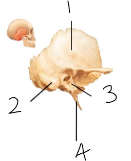
1) squamous part
2) mastoid process
3) tympanic part
4) styloid process
2) mastoid process
3) tympanic part
4) styloid process
26
New cards
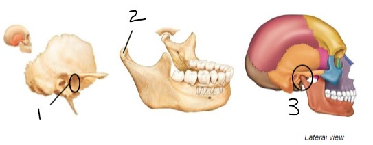
Temporomandibular joint (jaw joint)
condyle of the mandible articulates w/ temporal bone at the mandibular fossa
27
New cards
1) mandibular fossa
2) condyle
3) temporomandibular joint
2) condyle
3) temporomandibular joint
28
New cards
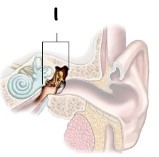
Middle ear cavity (inside temporal bone)
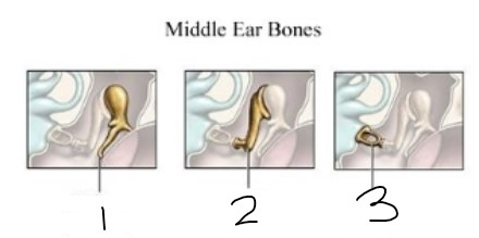
- 3 ear ossicles: malleus, incus, and stapes (small bones)
- sound waves cause vibrations of these bones that are transmitted to the inner ear (cochlea)
- sound waves cause vibrations of these bones that are transmitted to the inner ear (cochlea)
29
New cards
1) middle ear
30
New cards
1) malleus (hammer)
2) incus (anvil)
3) stapes (stirrup)
2) incus (anvil)
3) stapes (stirrup)
31
New cards
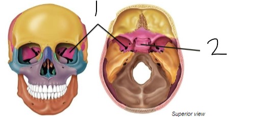
Sphenoid
- large and wing-shaped
- sella turcica = bony depression that holds the pituitary gland
- only cranial bone that articulates with every other cranial bone
- sella turcica = bony depression that holds the pituitary gland
- only cranial bone that articulates with every other cranial bone
32
New cards
1) sphenoid
2) sella turcica
2) sella turcica
33
New cards
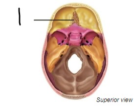
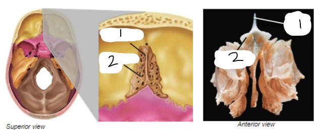
Ethmoid bone
- just anterior to sphenoid
- takes up most of the area between the nasal cavity and the orbits
- forms some boundaries of the nasal cavity and separates the nasal cavity from the brain
- contains crista galli and cribriform plate
- takes up most of the area between the nasal cavity and the orbits
- forms some boundaries of the nasal cavity and separates the nasal cavity from the brain
- contains crista galli and cribriform plate
34
New cards
1) ethmoid bone
35
New cards
Crista galli
- attaches to cribriform plate
- separates nasal cavity from brain
- site of attachment for dura mater (membrane covering brain)
- separates nasal cavity from brain
- site of attachment for dura mater (membrane covering brain)
36
New cards
Cribriform plate
- help form the roof of nasal cavities
- foramina allow passage of olfactory nerves into brain
- foramina allow passage of olfactory nerves into brain
37
New cards
1) crista galli
2) cribriform plate
2) cribriform plate
38
New cards
How many bones are in the facial skeleton?
14 bones
39
New cards
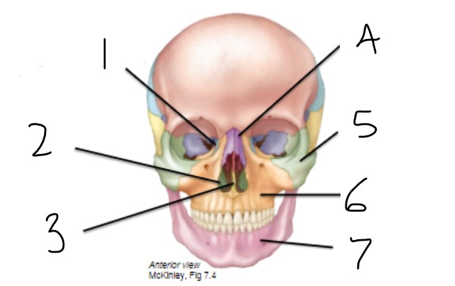
Bones of the facial skeleton
1) mandible (unpaired)
2) vomer (unpaired)
3) 2 nasals (form bridge of nose)
4) 2 lacrimals (allows tears to drain into nasal cavity)
5) 2 maxillae
6) 2 zygomatics (form cheekbones)
7) 2 palatine bones
8) 2 inferior nasal conchae
2) vomer (unpaired)
3) 2 nasals (form bridge of nose)
4) 2 lacrimals (allows tears to drain into nasal cavity)
5) 2 maxillae
6) 2 zygomatics (form cheekbones)
7) 2 palatine bones
8) 2 inferior nasal conchae
40
New cards
1) lacrimal bones
2) inferior nasal concha
3) vomer
4) nasal bones
5) zygomatic bones
6) maxillae
7) mandible
2) inferior nasal concha
3) vomer
4) nasal bones
5) zygomatic bones
6) maxillae
7) mandible
41
New cards
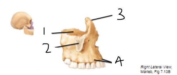
Maxilla(e)
- form upper jaw
- articulate w/ all other facial bones except mandible
- contain alveolar processes (contains teeth), frontal processes (extend upward to reach frontal bone), and zygomatic processes (articulate w/ zygomatic bone)
- articulate w/ all other facial bones except mandible
- contain alveolar processes (contains teeth), frontal processes (extend upward to reach frontal bone), and zygomatic processes (articulate w/ zygomatic bone)
42
New cards
1) orbital surface
2) zygomatic process
3) frontal process
4) alveolar processes
2) zygomatic process
3) frontal process
4) alveolar processes
43
New cards
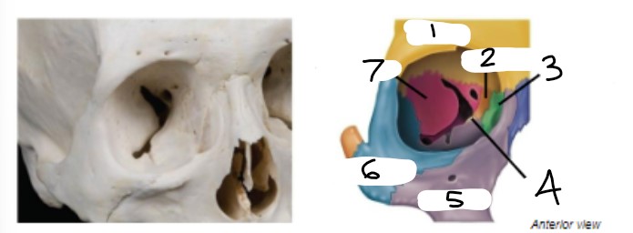
The orbit
- supports the eyes and muscles that move the eyes, contain fat and lacrimal glands (tear-producing glands)
- walls formed by frontal, sphenoid, zygomatic, maxilla, palatine, lacrimal, and ethmoid bones
- walls formed by frontal, sphenoid, zygomatic, maxilla, palatine, lacrimal, and ethmoid bones
44
New cards
1) frontal
2) ethmoid
3) lacrimal
4) palatine
5) maxilla
6) zygomatic
7) sphenoid
2) ethmoid
3) lacrimal
4) palatine
5) maxilla
6) zygomatic
7) sphenoid
45
New cards
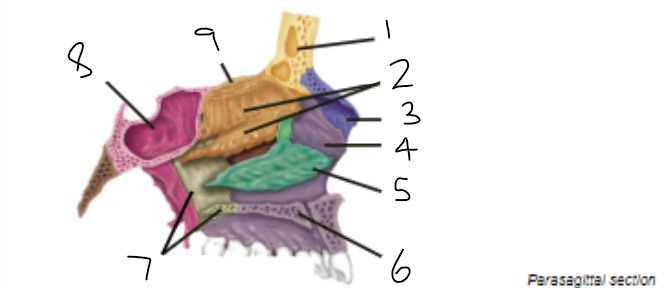
Nasal cavity in sagittal section
- lateral walls: nasal bones, nasal conchae (superior, middle, inferior), maxillae, palatines
- floor = hard palate, formed by palatine process of maxillae, horizontal plate of palatine
- floor = hard palate, formed by palatine process of maxillae, horizontal plate of palatine
46
New cards
1) frontal sinus
2) ethmoid (superior & middle nasal conchae)
3) nasal bone
4) maxilla
5) inferior nasal concha
6) maxilla
7) palatine bone
8) sphenoidal sinus
9) cribriform plate
2) ethmoid (superior & middle nasal conchae)
3) nasal bone
4) maxilla
5) inferior nasal concha
6) maxilla
7) palatine bone
8) sphenoidal sinus
9) cribriform plate
47
New cards
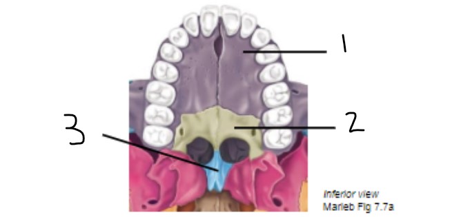
Palate and inferior nasal septum
- hard palate composed of maxillary bones and palatine bone
- vomer forms inferior portion of nasal septum
- vomer forms inferior portion of nasal septum
48
New cards
1) maxilla
2) palatine
3) vomer
2) palatine
3) vomer
49
New cards
Cleft palate
- failure of the 2 sides of the palate to join
- opening between mouth and nasal cavity makes effective nursing difficult
- can be repaired surgically w/ good outcomes
- opening between mouth and nasal cavity makes effective nursing difficult
- can be repaired surgically w/ good outcomes
50
New cards
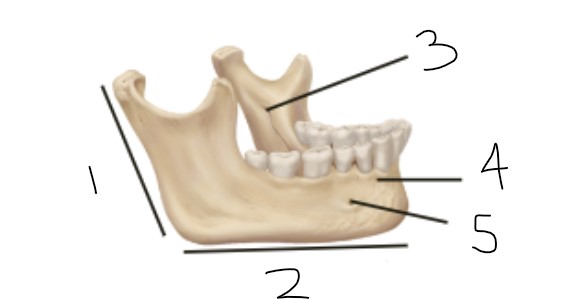
Mandible
- largest, strongest bone in the face
- body contains lower teeth
- alveolar processes = tooth sockets on superior border
- mandibular symphysis = 2 halves of the body join to form chin (mental protuberance)
- vessels and nerves enter via mandibular foramen and exit via mental foramen inferior to teeth
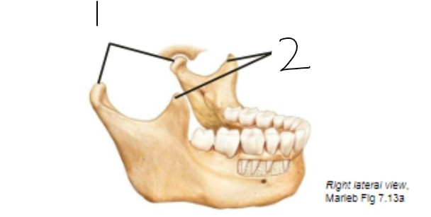
- condyle articulates w/ the temporal bone to form the temporomandibular joint (TMJ)
- coronoid process serves as an attachment site for the temporalis muscle (major chewing muscle)
- body contains lower teeth
- alveolar processes = tooth sockets on superior border
- mandibular symphysis = 2 halves of the body join to form chin (mental protuberance)
- vessels and nerves enter via mandibular foramen and exit via mental foramen inferior to teeth
- condyle articulates w/ the temporal bone to form the temporomandibular joint (TMJ)
- coronoid process serves as an attachment site for the temporalis muscle (major chewing muscle)
51
New cards
1) ramus of mandible
2) body of mandible
3) mandibular foramen
4) alveolar process
5) mental foramen
2) body of mandible
3) mandibular foramen
4) alveolar process
5) mental foramen
52
New cards
1) condyle
2) coronoid process
2) coronoid process
53
New cards
Fetal/infant face
- cranium huge relative to face
- skull is 3/4 adult size by age 2
- age 6-13: face grows outward and develops more "adult" proportions, body size begins to catch up w/ head
- skull is 3/4 adult size by age 2
- age 6-13: face grows outward and develops more "adult" proportions, body size begins to catch up w/ head
54
New cards
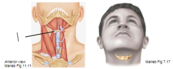
Hyoid
- "free floating" bone in neck inferior to mandible
- only bone in the skeleton that does not articulate w/ any other bone
- acts as base for tongue, site of muscle attachments for muscles that move the larynx
- only bone in the skeleton that does not articulate w/ any other bone
- acts as base for tongue, site of muscle attachments for muscles that move the larynx
55
New cards
1) hyoid
56
New cards
Thoracic cage
- includes thoracic vertebrae, ribs, sternum, and costal cartilages
- protects heart, lungs, and other organs
- supports pectoral girdle and provides attachment points
- intercostal spaces hold muscles that aid in breathing
- protects heart, lungs, and other organs
- supports pectoral girdle and provides attachment points
- intercostal spaces hold muscles that aid in breathing
57
New cards
Sternum
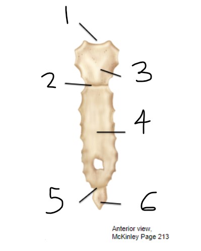
- 3 bones: manubrium, body, xiphoid process
- articulates w/ clavicles & ribs and costal cartilage
- sternal angle is an important landmark for thoracic anatomy
- articulates w/ clavicles & ribs and costal cartilage
- sternal angle is an important landmark for thoracic anatomy
58
New cards
1) jugular notch
2) sternal angle
3) manubrium
4) body
5) xiphisternal joint
6) xiphoid process
2) sternal angle
3) manubrium
4) body
5) xiphisternal joint
6) xiphoid process
59
New cards
Rib cage
- protects internal organs & aids in respiration
- all ribs attach posteriorly to thoracic vertebrae
- all ribs attach posteriorly to thoracic vertebrae
60
New cards
How many pairs of ribs are there?
12 pairs
61
New cards
First 7 ribs (true ribs)
attach to sternum by costal artilages
62
New cards
Ribs 8-10 (false ribs)
do not have a direct attachment to the sternum; attach via a shared costal cartilage
63
New cards
Rib 11-12 (floating ribs)
do not attach anteriorly to the sternum
64
New cards
Rib structure
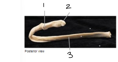
- main part of rib = shaft (body)
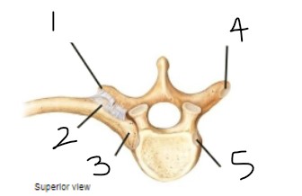
- rib articulates w/ the vertebra at the head and tubercle
- "neck" = thinner region between head and tubercle
- head has 2 "facets"= one articulates w/ the body of its vertebra and one articulates on the body of the vertebra superior to it
- tubercle articulates w/ a facet on the transvers process of the vertebrae
- rib articulates w/ the vertebra at the head and tubercle
- "neck" = thinner region between head and tubercle
- head has 2 "facets"= one articulates w/ the body of its vertebra and one articulates on the body of the vertebra superior to it
- tubercle articulates w/ a facet on the transvers process of the vertebrae
65
New cards
1) tubercle
2) head
3) shaft
2) head
3) shaft
66
New cards
1) transverse process
2) tubercle
3) head
4) transverse costal facet (for tubercle)
5) costal facet
2) tubercle
3) head
4) transverse costal facet (for tubercle)
5) costal facet
67
New cards
How many vertebrae in the vertebral column?
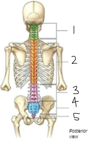
26 vertebrae
- 7 cervical
- 12 thoracic
- 5 lumbar
- 5 sacral (will fuse into 1)
- 4 coccyx (will fuse into 1)
- 7 cervical
- 12 thoracic
- 5 lumbar
- 5 sacral (will fuse into 1)
- 4 coccyx (will fuse into 1)
68
New cards
Vertebral column functions
- protect spinal cord
- supports body axis
- attachment points for ribs and muscles of neck and back
- anchor pectoral and pelvic girdles
- supports body axis
- attachment points for ribs and muscles of neck and back
- anchor pectoral and pelvic girdles
69
New cards
1) cervical
2) thoracic
3) lumbar
4) sacral
5) coccygeal
2) thoracic
3) lumbar
4) sacral
5) coccygeal
70
New cards
Curves of the spine
- vertebrae become larger as they move inferiorly to support weight
- sacrum articulates w/ hip bones of pelvis, passes weight to appendicular skeleton
- curves increase flexibility; also position center of gravity of axis of body
- sacrum articulates w/ hip bones of pelvis, passes weight to appendicular skeleton
- curves increase flexibility; also position center of gravity of axis of body
71
New cards
Scoliosis
lateral curvature of the spine, usually treated w/ body braces or surgery when young
72
New cards
Kyphosis (Dowager's Hump)
excessive curvature of the thoracic spine, typically the result of vertebral body factures caused by osteoporosis
73
New cards
Osteoporosis
- results from imbalance in normal bone building and degradation cycle
- usually after menopause, women don't absorb as much calcium so osteoclasts break bone down to release the calcium into the bloodstream
- usually after menopause, women don't absorb as much calcium so osteoclasts break bone down to release the calcium into the bloodstream
74
New cards
Lordosis
- excessive curvature of the lumbar spine
- usually temporary and resulting from shift for a larger front load (belly in men, pregnant women)
- usually temporary and resulting from shift for a larger front load (belly in men, pregnant women)
75
New cards
Basic vertebral anatomy
- body located anteriorly
- vertebral arch made of lamina and pedicles: creates vertebral foramen (surrounds spinal cord) and stacked vertebrae result in vertebral canal
- spinous processes & transverse process are ligament & muscle attachment sites
- vertebrae articulate at superior and inferior articular processes and facets: each articular process has an articular facet, individual vertebrae articulate w/ the vertebrae just superior and inferior to it, articulation also makes intervertebral foramina between 2 vertebrae
- vertebral arch made of lamina and pedicles: creates vertebral foramen (surrounds spinal cord) and stacked vertebrae result in vertebral canal
- spinous processes & transverse process are ligament & muscle attachment sites
- vertebrae articulate at superior and inferior articular processes and facets: each articular process has an articular facet, individual vertebrae articulate w/ the vertebrae just superior and inferior to it, articulation also makes intervertebral foramina between 2 vertebrae
76
New cards
Vertebral column and spinal cord
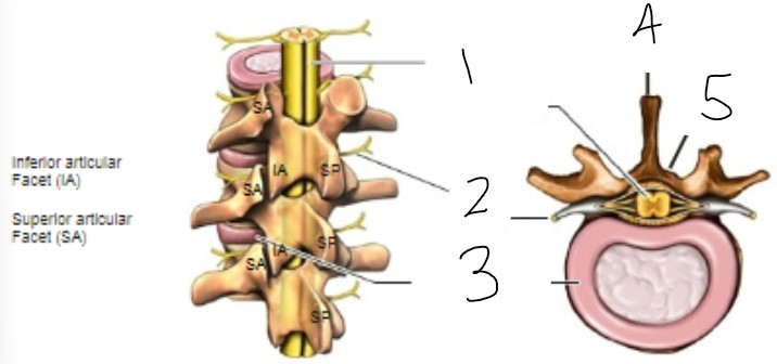
- spinal cord passes through, and is protected within, the vertebral (spinal) canal
- spinal nerves exit between the body of the vertebra and the vertebral arch via intervertebral foramina
- spinal nerves exit between the body of the vertebra and the vertebral arch via intervertebral foramina
77
New cards
1) spinal cord within spinal canal
2) nerve root
3) intervertebral disc
4) spinous process
5) lamina
2) nerve root
3) intervertebral disc
4) spinous process
5) lamina
78
New cards
Seven cervical vertebrae (C1-C7)
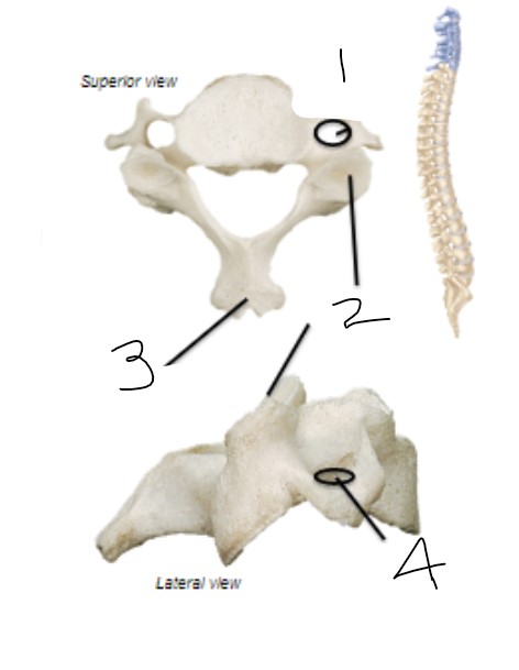
- have transverse foramina
- articular facets face superior/inferior
- bifid (split) spinous processes
- articular facets face superior/inferior
- bifid (split) spinous processes
79
New cards
1) transverse foramen
2) articular facets
3) bifid spinous process
4) transverse foramen
2) articular facets
3) bifid spinous process
4) transverse foramen
80
New cards
Atlas (C1)
- no body or spinous process
- articulates w/ occipital condyles, allows flexion/extension of head (nodding "yes")
- articulates w/ occipital condyles, allows flexion/extension of head (nodding "yes")
81
New cards
Axis (C2)
- dens articulates w/ atlas, allows rotational movement (shaking head "no")
- dens used to be the body of the atlas, but not part of axis
- dens used to be the body of the atlas, but not part of axis
82
New cards
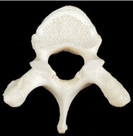
Twelve thoracic vertebrae (T1-T12)
- coastal facets for ribs, located on each body of the thoracic vertebrae
- articular facets face anterior/posterior
- spinous processes are long & project inferiorly
- articular facets face anterior/posterior
- spinous processes are long & project inferiorly
83
New cards
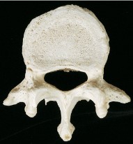
Five lumbar vertebrae (L1-L5)
- large bodies
- articular facets face medial/lateral
- short, flat spinous processes
- articular facets face medial/lateral
- short, flat spinous processes
84
New cards
Sacrum
- 5 fused vertebrae
- forms posterior wall of pelvis
- ala on lateral sacrum articulate w/ hip bones to form sacroiliac joints
- forms posterior wall of pelvis
- ala on lateral sacrum articulate w/ hip bones to form sacroiliac joints
85
New cards
Coccyx
- 3-5 fused vertebrae
- "tail bones"
- "tail bones"
86
New cards
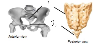
1) sacrum
2) coccyx
2) coccyx
87
New cards
Thoracic vertebra
88
New cards
Lumbar vertebra
89
New cards
Cervical vertebra