Ch. 22 : Attention & Consciousness
 Call Kai
 Learn
 Practice Test
 Spaced Repetition
 Match
 Flashcards
 Knowt Play
1/31

There's no tags or description
Looks like no tags are added yet.
32 Terms
Consciousness vs. Attention
how we select info on which to act & how we select diff behaviours
as sensory info & possible behaviours become more complex/varied → determination on which to select becomes more challenging
Attention = top-down & takes time to focus on a part of the sensory world
narrowing of awareness to a particular part of the sensory environ
parietal lobe is involved in ability to shift attention
an automatic process
conscious operations require attention
Consciousness = provides a summary of all info relevant to the situation
awareness / awareness of awareness
includes things we’re paying attention to & not paying attention to
ex. ducking when a ball almost hits you, when you weren’t paying attention to it
Automatic Processes vs. Conscious operations : reliance on attention
Automatic processes = performed w little focused attention & occur unconsciously
doesn’t require attention, but we can use it
innate or results from training (ex. had to learn how to walk)
bottom-up, THUS driven by sensory input
environ stimuli are sensed by body → body automatically reacts to it
Conscious operations = require focused attention
requires attention
top-down, THUS require cognitive input & control (ex. how you approach writing is based on experience)
→ due to the diff types of processing & input (cognitive vs. sensory), its likely that automatic & conscious processes rely on DIFF brain circuitry - possibly including the circuits related to consciousness
THUS, 2 diff areas should light up when imaging the diff. processes
Visual Processing Tasks (Visual Search)
= have to identify what’s diff & measure the time taken
right-side tasks are more difficult
shows that some visual search tasks are/aren’t automatic
→ visual searches requiring you to look for multiple features (ex. a T and bolded) take longer = feature search
→ visual searches that are simple take less time (ex. looking for a line)
→ visual searches where we add more meaning to an image (ex. seeing these as trees and dining the non tree) are harder & use top-down (THUS require cognitive input & control)
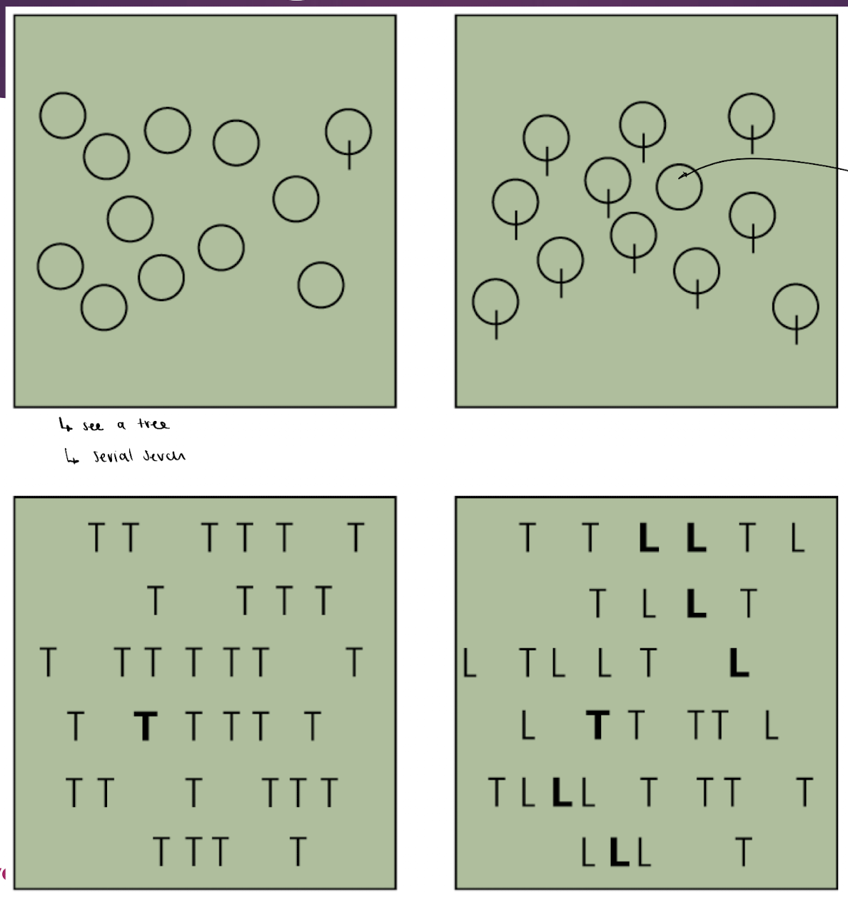
upper left image: serial search, automatic bc looking for simple vertical line
upper right image: harder bc we add meaning to the shapes (trees), thus uses top-down bc we’re relating them to experiences…
bottom left: takes longer bc its combining multiple features ( T and bold)
bottom right image: harder bc were trying to give it meaning, as wer’re looking for words amongst the letters
Research Into Attention: Types of Visual Searches
→ serial & feature search require DIFF amounts of attention
feature search requires MORE attention
certain types of visual search are automatic: addition of a vertical line, or use of bold type
Serial search = slower searches bc they require the combination of multiple features
top-down
Feature Search = strategy for scanning the environ for an object w a specific combination of features
bottom-up process:
object visually perched in V1 → broken down into features to be processed serially by higher visual areas
features of the object in the mental spotlight are recombined (possibly thru reentry processes)
note: areas outside of V1 or V2 should respond differently to a stimulus, depending on whether attention is focused on the object
Treisman’s Model of Feature Search
main idea:
only what’s in your attention spotlight enters object file in memory
if spotlight DOESNT have features we’re looking for → shift visual field
once features are matched → safe object file in memory
proposed process:
V1 encodes basic properties (feature maps)
from retina to v1 (early vision)
attention spotlight = when looking at one spot of visual field
can still see everything else
as we shift attention in visual field → see new feature maps
whatever in attention spotlight is sent to object file (compared to memory to see if features match)
object file is sent to memory to be identified & stored in memory

Study to Determine what a Feature is
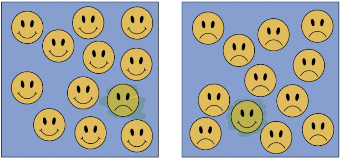
study: found that participants were able to identify sad faces MORE rapidly than happy faces
this occurred regardless of the whether sad face was right-side up, upside-down or an abstract representation
why: its NOT bc of the sad faces lips having extra lines, its bc f the emotion associated with sad
→ THUS, feature DOESNT need to be physical representations, it can be an abstract idea like emotion represented
Neuropsychological Evidence of Attention: Study
note: experiments testing the effects of the focus of attention must use the same stimulus in all conditions
ex. can only use happy or sad face, NOT both
study: varied the location of a stimulus & identified diff responses in higher visual areas (V4, TE) that depended on where the stimulus was presented & what the monkey was trained to attend to
findings:
multiple visual attention systems: one selecting stimuli & the other selecting and directing movements
priority maps encode stimulus location in terms of behavioural significance
maps are found in frontal eye field, lateral intraparietal area & superior colliculus
Demonstrating Selective Attention: Study
study: monkeys were trained to release a bar when a stimulus is presented in a certain location - earned to ignore stimuli in all other locations
results:
before training selective attention: neurons in V4 (visual area for colour) responded to stimuli in all locations
after training: V4 neurons ONLY responded when the visual stimuli was in the rewarded location → showing selective attention
→ THUS, a neuron can learn to respond selectively to info in its receptive field
something before V4 determines whether the info will be perceived
THUS, attention intercepts perception

Divided Attention & Selective Attention
note: most research says we’re NOT good at multitasking (divided attention)
→ mental capacity is limited, thus dividing attention b/w multiple tasks can divide that limited resource & cause all tasks to be performed worse
especially when the tasks require the SAME resources - thus, divided attention is easier is the tasks use DIFF resources
routine tasks can be performed w ease while doing another task - bc they become more automatic w use
→ we can shift our attention to be SELECTIVE
shown by V4 (colour/form) cells being selective abt what they respond to
cells are normally responsive to a combination of orientation & colour, BUT will respond to lines of somewhat diff orientation
ex. when animals discriminate b/w lines of similar orientation: see a decrease in performance accuracy, indicating the task is harder + activity in V4 cells increases to makes the cells more selective abt what they respond to
ex. cells in the pulvinar project, throughout the cortex, may help direct the attention spotlight
Study of Effort & Attention
found that:
cells in V4 only respond to certain lines, but when they go thru discrimination training they become MORE SELECTIVE abt what lines they’ll respond to (trying to make the task easier)
when tasks/discriminaiton becomes more difficult, the cells become more selective

Parallel Processing of Sensory Input
parallel processing of sensory input
with one object in the attention spotlight: visual system can bind all the properties together
with multiple objects in the attention spotlight: cells in areas TE process diff objects in parallel (at same time) to differentiate
→ thus area TE processes what these objects are by identifying differences bw them
attention must be divided across sensory modalitiesfor multi-sensory stimuli → need divided attention to connect senses
→ when attention is directed at one sense: activity increases in the related cortical areas
ex. focusing on the image of a movie → visual areas more active
ex. focusing on the words/sounds → auditory areas more active
→ when attention is divided b/w 2 senses: no change in the individual sensory areas, instead theres an increase in the Left Dorsolateral prefrontal cortex
Left Dorsolateral prefrontal cortex (DLPFC) =F allows us to multitasks when 2 senses/modalities are different (BIOMODAL): by filling in the gap to make multitasking more efficient b/w the 2 diff tasks
ONLY occurs when the 2 tasks use diff resources (ex. auditory & visual tasks at same time)

thus, divided attention recruits DLPFC when bimodal
Functional Imaging & Attention
study: 2 tasks each using the same stimuli, but different attentional demands
attending to the moving light stimulus resulted in increased activity in the posterior parietal cortex
if light was in L-visual field: only the R-parietal cortex was active
if light was in the R-visual field: bilateral parietal cortex was active
→ shows: R-parietal lobe is the main one for paying attention & can fill in for damaged L-partial cortex
each visual field has a diff focus of activation in the R-parietal lobe
note: this is related to contralateral neglect w damage to R-hemisphere
study: has a selective-attention condition & a divided-attention condition
diff types of attention (selective/divided) recruited diff brain areas
attentional tasks activate the anterior cingulate (frontal lobe for decision making), prefrontal cortex, & sensory areas related to the task
ex. if it was an auditory task → activates auditory areas
Shifting Attention
main idea: shifting attention uses the parietal lobe, bc movements of attention is involved
shifting attention is tested covertly, meaning w/o eye movement
→ shifting attention uses diff. brain areas than fixed attention
fixed attention: NO diff b/w hemis parietal lobe activity, bc you’re ignoring activity of the sides
shifting attention: diff b/w hemis parietal lobe activity
L-hemis parietal is ONLY active for shifting on R-visual field
R-hemis parietal is active for shifting on both visual fields

Networks of Attention
attentional system is distinct from the sensory networks that process info (ex. visual movement processing vs. processing movement)
attention ISNT a single process, its a connection of areas throughout the brain
brain regions form 3 diff attention networks:
Altering network : norepinephrine modulates
structures: locus coeruleus, frontal & parietal cortex
Orienting network : acetylcholine modulates
structures: superior parietal, temporal parietal junction, frontal eye fields, superior colliculus, pulvinar
Executive network : dopamine modulates
structures: anterior cingulate, anterior ínsula, basal ganglia
→ each network uses a diff neurotransmitter

Networks of Attention: Alerting Network
Alerting network = ascending reticular activating system from the midbrain, that maintains alertness
changes w age → becomes harder to suppress, leading to sleep disorders
assoc w ADHD
altering network activity SUPRESSES processing in other cerebral networks - thus, when activated it tells other attention networks to stop
→ modulator: norepinephrine
norepinephrine agonist: increased altering network
norepinephrine antagonist = decreased altering network, thus respond less to alerting stimuli
How it works:
noradrenergic neurons project from the locus coeruleus to the forebrain (frontal & prefrontal)
alerts the prefrontal & posterior parietal cortex to rapidly process & respond to stimuli
activity suppresses processing in other cerebral networks
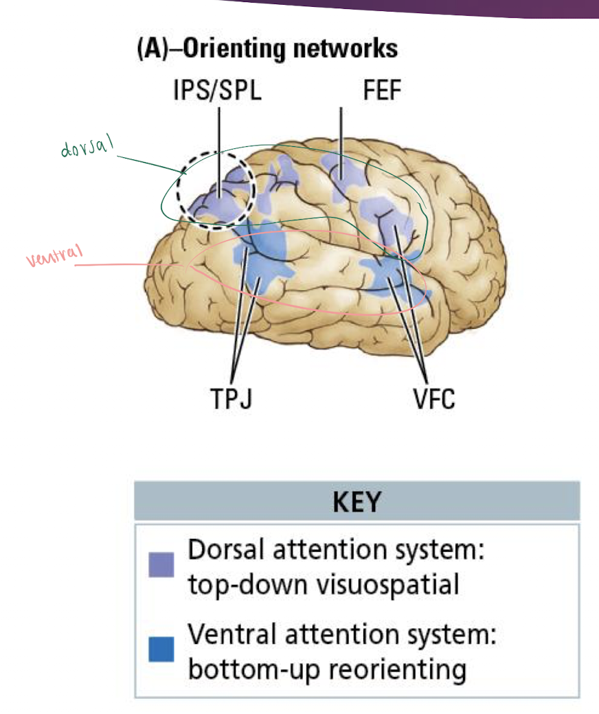
Networks of Attention: Orienting Network
prioritizes ONE sensory modality or a particular location in space
ex. only vision (sense) or only what’s occurring in a location
→ modulator: cholinergic neurons that produce acetylcholine
→ has 2 networks: ideally ACTIVATED @ SAME time
Dorsal orienting network = directs attention twd a specific object in a top-down process
ex. look for Waldo, then break down his features
involves the frontal eye fields & intraparietal sulcus
strongly R-lateralized - more activity in R-hemis
Ventral Orienting network = synchronizes activity w bottom-up incoming sensory info
ex. looks for features until they have all of them
involves the temporoparietal junction (similar to ventral visual stream), ventral frontal cortex & pulvinar (attention & decision making)
→ associated disorders:
autism - hyperfrouc on everything in one location/sense
PTSD - difficulty disengaging from fearful stimuli
neglect - over-orienting on one side (contralateral neglect)
Networks of Attention: Executive Networks
develop as we age, maturing around 3-4yrs old - thus at that age children can control their emotions
can override the alerting or orienting attention networks
talks to orienting, altering networks & others
w increased executive network = MORE control of attention
dopamine modulates
→ 2 executive networks work via top-down influences:
activity in areas WITHIN a network is highly correlated - areas in the 2 networks are all active if one is active
NO correlation b/w activity in the 2 networks - the 2 networks can operate at diff times, thus can be independent
Dorsolateral-prefrontal cortex-parietal network = active at the BEGINNING of a task
ex. active at start of driving
~ex. moment-moment task
Anterior Cingulate/medial frontal-anterior ínsula network = remains active THROUGHOUT the task (so we can continue a task)
ex. takes over after 1-2 min driving & continues for the rest
~ex. task maintenance

→ Associated disorders:
anxiety
depression
OCD - stuck in cingulate network
personality disorders
schizophrenia - lack of inhibition
substance abuse
Attention Networks & Self-control
→ controlling cognition & emotions is a form of self-control
have better self-control when theres MORE control of attention networks
self-control of cognition & emotions is associated w activity in the lateral prefrontal & cingulate regions:
stress changes the structure & gene expression patterns of the prefrontal cortex → deficits in cognitive control of emotion
bc: part of the prefrontal cortex’s job is to inhibit areas that increase emotional rxn, thus when damaged it cant inhibit → increase emotional rxn
ex. psych stress impairs the performance of medical students on attentional tasks - by decreasing activity in the dorsolateral prefrontal cortex, anterior cingulate, premotor & posterior parietal cortex
stress DECREASES activity of the executive control network → less control of attention
Attention Networks: Associated Disorders
→ w/o the control of the executive networks → alerting network goes off all the time

Mechanisms of Attention: Hypothesis of Directing Attention
hypothesis: that the attention system directs attention by inducing synchrony b/w neurons (spatial summation) that assess the sensory signal
thus, synchronizing what diff parts of the brain are doing
adjusts timing of action potentials so the input from 2 neurons arrive at the same time & location → summed → increasing chance of an action potential in the postsynaptic cell (spatial summation)
during a task requiring attention: increased activity in the dorsal executive attention network & decrease in the default network (baseline brain activity)
→ attention increases synchrony in the brain: neurons within a network, even in diff areas, fire at the SAME time
says we’re more likely to reach threshold w summation when attending to something (giving attention), via synchrony of neurons

Inattention
Inattention blindness = person fails to notice something while they’re performs another task
when distracted by a counting task, 70% of participants failed to notice a gorilla suit
Change blindness = ppl routinely fail to notice changes in the environ if they aren’t expecting them
Attentional blink = attending to 1 stimulus prevents the subject from being aware of a 2nd stimulus
using orienting network (prioritizes one sense/location)
→ imaging studies show these unattended items are detected by the sensory systems, BUT are filtered out BEFORE conscious awareness
Sensory Neglect
ex pts w damage to the R-temporoparietal junction DONT attend to the L-side of space around them
Paying attention to objects:
→ objects in the R-visual field can be attended to by the left or R-parietal lobe
both, bc the R-parietal lobe is a backup for the L-parietal
→ objects in the L-visual field are ONLY attended to by the R-parietal lobe
→ damage to the frontal lobes (BA 6) results in contralateral neglect ONLY for objects close enough to be grasped
which hemisphere depends on if damage is to the L or R frontal lobe
Treatment of sensory neglect:
moving head or using prism to shift objects from the L-visual field to the R-visual field for a few min a day → signif greater ability to attend to objects in the L-visual field
reminds brain to scan area
Consciousness
Consciousness = level of responsiveness of the mind to impressions made by the senses - thus, its how much we respond to incoming senses
changes over time:
rxns to stimuli vary overtime
young kids & those w dementia experience a diff level of consciousness
levels of consciousness vary over the day w the transition from sleep → wakefulness (sleep =/ unconscious, its an altered state of consciousness)
can change levels w drugs & hypnosis
reflexive responses to stimuli ARENT considered indications of consciousness
conscious awareness of a movement might only occur AFTER the movement is complete
thus, we can do sensory reflexes and then become aware w didi it
ventral stream (ability to identify) is considered a part of conciousness
dorsal stream (action) ISNT considered a part of consciousness
Consciousness: Dissociating Behaviour & Awareness
study: subjects had to move their hands & grasp the lit rod as fast as possible
some trials had the list rod changing b/w targets → subject had to correct their aim to grab new lit stick
findings:
most participants were already holding the new lit rod BEFORE they could consciously say that it switched (weren’t aware, but still did it)
→ said this shows its possible to dissociate behaviour & conscious awareness
→ say this supports the ventral stream being part of consciousness, whereas the action (dorsal stream) was unconscious

Conciousness: Integrated Information Theory
Integrated Information Theory = attempts to explain consciousness
says consciousness:
exists
is structured & composed of multiple distinctions b/w the phenomena we observe
composed of specific info & and that each experience is DISTINCT from e/o
even if u experience the same info in 2 diff days, still distinct
unified & CANT be broken into multiple subparts: its ALL one thing
made of individual experiences that are separated from other conscious experiences by cause-and-effect relationships
we learn & build on past experiences (related to classical & operant conditioning)
→ can apply this theory to explain diff forms of consciousness in clinical pts
Conciousness: Neural basis
neural basis: consciousness requires the interaction of sensory areas, memory & other systems like emotion & executive fxn - these are connected w networks
consciousness includes 4 processes: arousal, perception, attention, & working memory that use binding to unify
binding synchronizes the activity in relevant networks for a short period (not just the cells in one network syncing up)
rapid synchronization across widely separated brain areas is possible & regularly observed
support: studies of perceiving & responding to faces show patterns of synchronous activity → asynchronous → synchronous activity
showing there seems to be a purpose for both synchronous (cells in diff areas firing at same rate) & asynchronous
shows we’re more consciously aware & pay attention to things perceived as faces
thus certain types of stimuli draw more attention
synchrony & asynchrony are both important for perceiving faces
Conciousness: Neural basis - Shadow of a Perception study
study: tracked neural synchrony in brains perceiving a face vs. those not perceiving a face
findings:
brain w face perceived: synchronous activity over L-hemis → asynchronous in both hemis → synchrony with motor response to pressing button
brain w/o face perceived: asynchronous activity → brief synchronous activity for motor response to press button to say they didn’t perceive a face
shows:
we’re more consciously aware & pay attention to things perceived as faces
thus certain types of stimuli draw more attention
synchrony & asynchrony are both important for perceiving faces

Conciousness: Cerebral Substrates
we dont know the essential neural regions for consciousness
imaging studies study consciousness by contrasting activity when subject in unconscious (coma, vegetative state) with conscious activity
→ dorsolateral prefrontal cortex, medial frontal cortex, posterior parietal cortex, & posterior cingulate cortex are all INACTIVE when unconscious
these can be active when you’re asleep
some parts of the brain increase in activity when asleep
→ a network of frontoparietal areas is correlated during a quiet, resting, conscious state

Clinical studies of Consciousness
pts in a coma or vegetive state (unconscious) produce brain activity in response to verbal commands
fMRI can show diff brain activity from asking questions, thus they can hear
studies suggest theres a bilateral region of the parietal lobe necessary for consciousness
theres NO necessary area in the frontal lobe for consciousness
proposed that the Claustrum is the brain region CRITICAL in binding together info, since is receives connections from nearly the entire cortex

Emotion & Conciousness
→ emotions are related to consciousness, but you can have one w/o the other
overlapping brain regions associated with consciousness & self-awareness:
medial frontal cortex
posterior cingulate cortex
consciousness may NOT be necessary to experience emotions:
non-conscious emotions can be demonstrated by experiments of fear conditioning
Nonconscious Processing
→ blindsight (awareness w/o perception), dorsal-stream processing & other experimental evidence suggests cognitive activity CAN occur on the nonconscious level
Nonconscious Processing = processing sensory info w/o consciousness
when asked to explain their behaviours, ppl often dont recognize & report the actual factors that influenced their behaviours
ex. “idk” → then come up w a reason
sensory systems take in a lot of info to influence decisions, but we are only consciously aware of a small amount of that info:
Automatic process: use system 1 to generate rapid, NONconscious responses to stimuli
ex. pupils constricting to light, raising voice in loud environ
Conscious processes: use system 2 to produce slower, controlled responses based on relevant knowledge & experience
used when learning something new, then we can use automatic
Neuroeconomics
Neuroeconomics = studies how the brain makes decisions
what influences our decisions?
→ influences from neuroscience, cogntive psych, social psych & behavioural economics
→ current research is focus on its application to health psychology
to better understand how ppl make decisions abt health & wellness behaviours -. then we can create better treatment plans for their thinking process