Perio II, Lecture 9 - Mucogingival Deformities (Dr. Ho)
 Call Kai
 Learn
 Practice Test
 Spaced Repetition
 Match
 Flashcards
 Knowt Play
1/71

There's no tags or description
Looks like no tags are added yet.
72 Terms
What has the following characteristics?
- Surface of oral gingival epithelium
- Everything coronal to MGJ
Keratinized tissue width
What has the following characteristics?
- Keratinized Tissue overlying connective tissue that is firmly bound to cementum or buccal bone
- Protective
- Stippled, orange peel look (may be noted in health)
Attached gingiva

ID #1:
Free ginigival margin
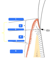
ID #2:
Free gingiva

ID #3:
Free gingival groove

ID #4:
Attached gingiva

ID #5:
Mucogingival junction

ID #6:
Alveolar mucosa

Where is the keratinized tissue width measured from?
Free gingival margin (#1) to mucogingival junction (#5)
How do you calculate the width of attached gingiva?
Keratinized Tissue Width - Free Gingiva
(*sulcular depth often substitutes marginal free gingiva)
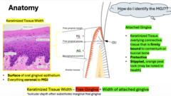
To identify MGJ perform the _______:
Place probe against mucosa and roll upwards—folds along mucogingival junction(MGJ)
(everything coronal to that line is the Keratinized Tissue Width - KTW)
Roll test


If capillaries are visible, what area of the periodontium are you looking at?
Alveolar mucosa (less accurate technique)
T/F: If there is a lack of attached gingiva noted, mark MGD on the periodontal chart
True
Keratinized tissues is important for all of the following reasons EXCEPT:
A) Aids in withstanding frictional forces (e.g. mastication)
B) Promotes retraction of frenum attachments
C) Provides comfort for optimal oral hygiene (i.e. uncomfortable to brush mucosa)
D) Lack of KT and movable mucosa may cause food retention
B- RESISTS retraction of frenum attachments

All surfaces with less than ___ mm of keratinized gingiva exhibited clinical inflammation
2 mm
T/F: There is no significant differences in gingival inflammation or plaque scores in areas with narrow or wide attached gingiva
True
T/F: Keratinized tissue's importance is contentious in literature
True

In the presence of ________ oral hygiene, the width of attached gingiva is not crucial for maintaining gingival health BUT...
1. Increased RISK of recession
2. Discomfort precludes adequate OH
3. Must monitor
Good
Define the following:
The displacement of the soft tissue margin apical to the CEJ with oral exposure of the root surface
Gingival recession
What is the driving force of gingival recession?
Inflammation
Define the following:
Connective tissue destruction with proliferation of outer oral epithelium and sulcular epithelium
Gingival recession
(Why gingival thickness and width - i.e. KTW - matters)
What is the sequence of events in the progression of inflammation-induced gingival recession?
- Normal tissue >
- Inflamed tissue with epithelial proliferation >
- Epithelial tissue begin anastomosis, due to lack of connective tissue core >
- Epithelial tissue merge and form separation of recession

Periodontal phenotype is made up of what three things?
- Ginigval phenotype: KTW and gingival thickness
- Bone morphotype

ID the periodontal phenotype:
Thin scalloped

ID the periodontal phenotype:
Thick scalloped

ID the periodontal phenotype:
Thick flat
ID the periodontal phenotype:
- Slender triangular crown
- Subtle cervical convexity
- Interproximal contact close to incisal edge
- Narrow zone of KT
- Thin, delicate gingiva
- Thin alveolar bone
Thin scalloped
ID the periodontal phenotype:
- Slender Teeth
- Thick fibrotic gingiva
- Narrow zone of KT
- Pronounced gingival scalloping
Thick scalloped
ID the periodontal phenotype:
- Square shaped tooth crowns
- Pronounced cervical convexity
- Large interproximal contacts, more apical
- Broad zone of KT
- Thick fibrotic gingiva
- Thick alveolar bone
Thick flat
Which anatomical landmark is most important for distinguishing altered passive eruption from a thick-flat gingival phenotype?
CEJ
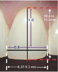
What is the approximate normal width-to-length ratio of a maxillary central incisor?
0.8

ID the Cairo Classification for Gingival Recession:
RT 1

ID the Cairo Classification for Gingival Recession:
RT 2
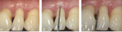
ID the Cairo Classification for Gingival Recession:
RT 3
ID the Cairo Classification for Gingival Recession:
- No loss of interproximal attachment
- Interproximal CEJ not detectable
- 100% root coverage predictable
RT 1
ID the Cairo Classification for Gingival Recession:
- Loss of interproximal attachment
- Interproximal attachment loss less than or equal to buccal recession
- Partial root coverage
RT 2
ID the Cairo Classification for Gingival Recession:
- Loss of interproximal attachment
- Interproximal attachment loss greater than buccal recession
- Complete root coverage not achievable
RT 3
Limitation of root coverage is typically ___mm from tip of papilla
3mm
All of the following are predisposing factors that when in conjunction with inflammation can lead to gingival recession EXCEPT:
A) Aberrant Frenum Pull
B) Abfraction (not proven but anecdotal)
C) Orthodontic movement (in buccal direction)
D) Toothbrush abrasion (i.e. Scrub brush with hard bristles)
E) Tooth Malposition; Root prominence
F) Thick Gingival Phenotype
G) Subgingival restorations
H) Iatrogenic trauma
F - THIN GINGIVA PHENOTYPE (thin CT core)

What most likely caused the gingival recession?
Aberrant frenum pull
What are examples of iatrogenic trauma that are predisposing factors that when in conjunction with inflammation can lead to gingival recession?
- Habits (i.e. nail biting)
- Factitious oral hygiene: toothbrush abrasion, flossing aggressively
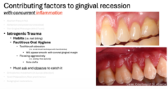

What most likely caused the gingival recession?
Iatrogenic trauma: toothbrush abrasion
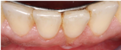
What most likely caused the gingival recession?
Orthodontic movement

What most likely caused the gingival recession?
Tooth malposition

What most likely caused the gingival recession?
Subgingival restorations
What is the gold standard procedure for gingival augmentation/ increasing keratinized tissue?
Free gingival graft
Gingival augmentation takes about how long to heal?
3-4 months
What is the gold standard and most predictable treatment for root coverage?
Connective tissue graft with coronally advanced flaps
What are the two procedures with the highest predictability of success for root coverage of initial recession?
Coronally advance flaps + connective tissue graft
What is the main pathogenic pathway that causes recession and mucogingival deformities?
Inflammation
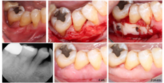
What is the goal of a free gingival graft?
- Improve KT
- Eliminate frenum pull

What is the goal of a lateral pedicle?
- Root coverage
- Improve KT width

What is the goal of a connective tissue graft with tunneling and occlusal guard?
- Improve protective tissue
- Eliminate frenum pull
- Cover exposed roots

What is the goal of a connective tissue graft with Coronally Advanced Flap (CAF)?
- Root Coverage
- Improve Gingival Thickness

What is a challenge of a connective tissue graft with Coronally Advanced Flap (CAF)?
Buccal postion

What is the ideal treatment for peri-implantitis with hard and soft tissue deficiency and an overcountoured restoration?
Since the placement was a problem...
- Implant removal
- Re-bone graft
- Re-do implants
T/F: Friable mucosa causes difficulties for adequate debridement. Movable, allows plaque to freely adhere within sulcular epithelium—little to no connective tissue--risk of recession is high and bone loss is already present in implants
true
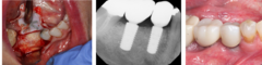
What is the ideal treatment for peri-implant health but a soft tissue deficiency?
- Ensure good OH and monitor

What is the ideal treatment for peri-implantitis and a soft tissue deficiency?
- Modify OH
- Guided Tissue Regeneration with Implantoplasty
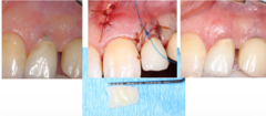
What is the goal of Implant Soft Tissue Augmentation Connective Tissue Graft (CTG) and Vestibular Incision Subperiosteal Tunnel Access (VISTA)?
- Thicken Keratinized mucosa
- Address soft tissue fenestration
When should you refer to a periodontist?
Refer based on chief complaint
Is a referral for mucogingival surgery indicated if a patient experiences discomfort that prevents adequate oral hygiene?
Yes
If the patient is experiencing sensitivity what is your course of treatment?
Treat chemically first, with fluoride or desensitizer
- If that doesn't work, then we can consider root coverage/ improvement in sensitivity

What are three examples of chemical options to treat sensitivity?
- OTC Fluoride toothpaste & make sure they don't rinse after use
- Rx Fluoride gel or paste
- Clinical application of desensitizer (eg Gluma, SDF, Laser treatment, etc)
T/F: Surgical intervention alone may not completely treat sensitivity—if 5mm exposed and 3mm covered, remaining 2mm may still be sensitive. Need to address CC directly/ comprehensively
True
(Once interventions are managed and patient is still experiencing dentinal sensitivity, may refer for further root coverage/improvement in sensitivity)
Is a referral for mucogingival surgery indicated for a patient with gingival recession who has esthetic concerns or is planning future subgingival restorations, buccal orthodontic movement, or an RPD clasp?
Yes
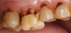
Is a referral for mucogingival surgery necessary solely because a patient fears losing a tooth due to gingival recession?
No
(Bone is still present on other 3 surfaces, so tooth can survive based on attachment. However, increased risk of caries or increased risk of dentinal abrasion)
What is a common area of recession due to tooth malposition and root prominence?
MB root of molars
Thin biotypes pose an increased risk of _____ due to decreased distance from oral epithelium to sulcular epithelium
Recession
Thick biotypes pose an increased risk of _______ due to thicker tissue
Greater pocket depths
The contributing factors to gingival recession include all of the following EXCEPT one. Which is the EXCEPTION?
A) Traumatic occlusion
B) Tooth malposition
C) Thin phenotype
D) Toothbrush abrasion
A) Traumatic occlusion
The mucogingival junction lies between _________ and _________
A) Alveolar mucosa and the alveolar bone
B) Free gingiva and the junctional epithelium
C) Alveolar mucosa and the attached gingiva
D) Attached gingiva and the free gingiva
C) Alveolar mucosa and the attached gingiva