unit 9 - inflammatory diseases of the oral mucosa
 Call Kai
 Learn
 Practice Test
 Spaced Repetition
 Match
 Flashcards
 Knowt Play
1/239

There's no tags or description
Looks like no tags are added yet.
240 Terms
What tissue change defines an erosion of the oral mucosa?
Superficial loss of the epithelium.
What tissue change defines an ulcer of the oral mucosa?
Complete loss of the epithelium.
What is the defining structural feature of a blister in the oral mucosa?
A fluid-filled space within the epithelium.
what helps explain the lesion
The level of tissue damage.
How can a ruptured blister appear?
As an erosion.
Recurrent aphthous ulcers
What are recurrent aphthous ulcers?
Painful, recurrent ulcerative lesions of the oral mucosa.
Recurrent aphthous ulcers
What shape do recurrent aphthous painful ulcers typically have?
Round or oval.
Recurrent aphthous ulcers
What does the base of a recurrent aphthous ulcer typically look like?
Yellow-white and fibrinous base and red borders
Recurrent aphthous ulcers
Which type of oral mucosa is usually affected by recurrent aphthous ulcers?
Non-keratinised mucosa.
Recurrent aphthous ulcers
Which two oral sites are commonly affected by recurrent aphthous ulcers?
The inner lips and cheeks.
Recurrent aphthous ulcers
What happens between episodes of recurrent aphthous ulcers?
The ulcers heal.
Recurrent aphthous ulcers
What is the cause of recurrent aphthous ulcers?
Multifactorial and often unknown.
Recurrent aphthous ulcers
What type of inflammation causes recurrent aphthous ulcers?
Non-infectious inflammation involving an abnormal immune response.
Recurrent aphthous ulcers
What does idiopathic mean in relation to recurrent aphthous ulcers?
No underlying cause is identified.
Recurrent aphthous ulcers
Which two inflammatory diseases are associated with aphthous ulcers?
Behçet disease and Crohn disease.
Recurrent aphthous ulcers
Which infection is listed as an association with recurrent aphthous ulcers?
HIV infection
Recurrent aphthous ulcers
Which blood-related deficiencies/disorders are associated with recurrent aphthous ulcers?
Hematinic deficiencies
Cyclic neutropenia.
Recurrent aphthous ulcers
Which three nutritional deficiencies are associated with recurrent aphthous ulcers?
Iron, vitamin B12 and folate deficiencies.
Recurrent aphthous ulcers
What type of reaction is also associated with recurrent aphthous ulcers?
Hypersensitivity reactions.
Recurrent aphthous ulcers
What does the term complex aphthosis describe?
Aphthous lesions associated with syndromes affecting the skin.
Recurrent aphthous ulcers
In which age group is PFAPA syndrome mainly seen? - IMPORTANT
Children.
Recurrent aphthous ulcers
What does PFAPA stand for?
Periodic fever, aphthous stomatitis, pharyngitis and cervical adenitis.
Recurrent aphthous stomatitis (RAS)
What percentage of adults are affected by recurrent aphthous stomatitis (RAS)?
Approximately 15–20%.
Recurrent aphthous stomatitis (RAS)
Which sex has a slight predominance in recurrent aphthous stomatitis (RAS)?
Females.
Recurrent aphthous stomatitis (RAS)
In which age groups is recurrent aphthous stomatitis (RAS) common?
Adolescents and young adults.
Recurrent aphthous stomatitis (RAS)
How is recurrent aphthous stomatitis (RAS) clinically characterised?
A chronic condition with recurrent, painful oral ulcers.
Recurrent aphthous stomatitis (RAS)
is it recurrent yes or no
yes
Recurrent aphthous stomatitis (RAS)
What type of damage occurs to the oral epithelium in RAS?
Immune-mediated damage.
Recurrent aphthous stomatitis (RAS)
What role do T cells play in the pathogenesis of RAS?
They contribute to epithelial injury.
Recurrent aphthous stomatitis (RAS)
What role does TNF-α play in RAS?
It amplifies local inflammation.
Recurrent aphthous stomatitis (RAS)
What is molecular mimicry in the proposed pathogenesis of RAS?
Similarity between microbial and human proteins.
Recurrent aphthous stomatitis (RAS)
Which two reduced expressions may weaken local protection and immune regulation in RAS?
HSP27 and IL-10.
Recurrent aphthous stomatitis (RAS)
Is the exact cause of recurrent aphthous stomatitis (RAS) known? - IMPORTANT
No, the exact cause remains unclear.

Recurrent aphthous stomatitis (RAS)
difference appearances of aphthous ulcers
read - IMPORTANT ANOUT SIZE AND NUMBER
DONE
Recurrent aphthous stomatitis (RAS)
difference appearances of aphthous ulcers
what are the 3 types
minor aphthae
major aphthae
herpetiform aphthae
Recurrent aphthous stomatitis (RAS)
difference appearances of aphthous ulcers
Which type of aphthous ulcer is the most common?
Minor aphthae.
Recurrent aphthous stomatitis (RAS)
difference appearances of aphthous ulcers
What size are individual minor aphthous ulcers? - imp
3-7mm (less than 1cm)
Recurrent aphthous stomatitis (RAS)
difference appearances of aphthous ulcers
How many minor aphthous ulcers may be present during an episode?
IMP
one (or several ulcers)
Recurrent aphthous stomatitis (RAS)
difference appearances of aphthous ulcers
Which type of oral mucosa is affected by minor aphthous ulcers?
Non-keratinised mucosa.
Recurrent aphthous stomatitis (RAS)
difference appearances of aphthous ulcers
What is the typical appearance of minor aphthous ulcers?
Shallow, rounded ulcers with an erythematous margin and yellowish floor.
Recurrent aphthous stomatitis (RAS)
difference appearances of aphthous ulcers
major
How common are major aphthous ulcers compared with minor aphthous ulcers?
uncommon
Recurrent aphthous stomatitis (RAS)
difference appearances of aphthous ulcers
major
What size are individual major aphthous ulcers?
Several centimetres across.
Recurrent aphthous stomatitis (RAS)
difference appearances of aphthous ulcers
major
How long do major aphthous ulcers persist?
several months
Recurrent aphthous stomatitis (RAS)
difference appearances of aphthous ulcers
what important clinical concern can major aphthous ulcers sometimes resemble?
malignant ulcer.
Recurrent aphthous stomatitis (RAS)
difference appearances of aphthous ulcers
major
What may happen when major aphthous ulcers heal?
Scarring may occur.
Recurrent aphthous stomatitis (RAS)
difference appearances of aphthous ulcers
Herpetiform aphthae
How common are herpetiform aphthous ulcers?
uncommon
Recurrent aphthous stomatitis (RAS)
difference appearances of aphthous ulcers
Herpetiform aphthae
What size are individual herpetiform aphthous ulcers?
1–2 mm across.
Recurrent aphthous stomatitis (RAS)
difference appearances of aphthous ulcers
Herpetiform aphthae
How many - aphthous ulcers may be present in one episode? - IMP
Dozens or hundreds of ulcers.
Recurrent aphthous stomatitis (RAS)
difference appearances of aphthous ulcers
Herpetiform aphthae
Which type of oral mucosa is affected by herpetiform aphthous ulcers?
Non-keratinised mucosa.
Recurrent aphthous stomatitis (RAS)
difference appearances of aphthous ulcers
Herpetiform aphthae
What may happen when herpetiform aphthous ulcers coalesce?
They may form irregular ulcers.

aphthous ulcers : useful distinctions
reaad
done
aphthous ulcers : useful distinctions
What is the typical size of minor aphthous ulcers?
Small, usually less than 1 cm.
aphthous ulcers : useful distinctions
How long do minor aphthous ulcers usually take to heal, and do they scar?
They heal in 1–2 weeks without scarring.
aphthous ulcers : useful distinctions
What is the typical appearance of major aphthous ulcers compared with minor aphthous ulcers?
They are larger and deeper.
aphthous ulcers : useful distinctions
How long can major aphthous ulcers persist, and what may happen when they heal?
They may persist for several weeks and may scar.
aphthous ulcers : useful distinctions
What is the typical appearance of herpetiform aphthous ulcers?
Many tiny ulcers.
aphthous ulcers : useful distinctions
What can happen when herpetiform aphthous ulcers merge?
They may merge into larger ulcers.
aphthous ulcers : useful distinctions
Are herpetiform aphthous ulcers caused by herpes simplex virus?
no
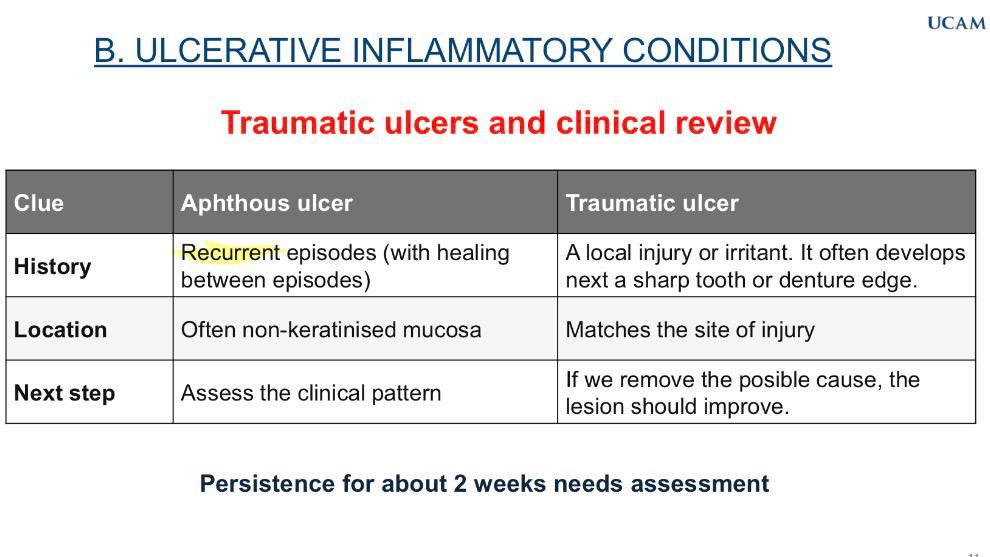
Traumatic ulcers and clinical review
READ
done

Traumatic ulcers and clinical review
What history suggests a recurrent aphthous ulcer rather than a traumatic ulcer?
Recurrent episodes with healing between episodes.
Traumatic ulcers and clinical review
What history suggests a traumatic ulcer rather than a recurrent aphthous ulcer?
A local injury or irritant, often following contact with a sharp tooth or denture edge.
Traumatic ulcers and clinical review
Where do aphthous ulcers commonly occur?
On non-keratinised mucosa.
Traumatic ulcers and clinical review
Where does a traumatic ulcer usually occur?
at the site of the injury.
Traumatic ulcers and clinical review
What is the next step when assessing a suspected aphthous ulcer?
Assess the clinical pattern.
Traumatic ulcers and clinical review
What should happen to a traumatic ulcer if its possible cause is removed?
The lesion should improve.
Traumatic ulcers and clinical review
What should happen if an oral ulcer persists for about two weeks?
It needs assessment.
Ulceration under the microscope
→ Histopathology
What happens to the epithelial surface in an oral ulcer seen histologically?
Loss of the epithelial surface.
Ulceration under the microscope
→ Histopathology
Which materials and cells are found in the ulcerated area histologically?
Fibrin and inflammatory cells.
Ulceration under the microscope
→ Histopathology
What type of tissue forms during the repair of an oral ulcer?
Granulation tissue.
Ulceration under the microscope
→ Histopathology
What type of haemorrhage may be seen in the epithelium of an oral ulcer?
Intraepithelial haemorrhage.
Ulceration under the microscope
→ Histopathology
What may happen to superficial skeletal muscle fibres beneath an oral ulcer?
They may degenerate.
Ulceration under the microscope
→ Histopathology
Are the histological features of ulceration specific to aphthous ulcers?
No. Trauma, immune conditions and other diseases can produce a similar histological appearance.
Ulceration under the microscope
→ Histopathology
Why is the clinical history important when investigating an oral ulcer?
Because different conditions can produce similar histological appearances.
Actinic cheilitis
what does this mean
inflammation of the lip
Actinic cheilitis
What is actinic cheilitis also known as?
other names for the same thing
Actinic keratosis of the lip, solar cheilosis or solar cheilitis.
Actinic cheilitis
Which lip is usually affected by actinic cheilitis?
lower lip.
Actinic cheilitis
What causes actinic cheilitis?
Long-term exposure to ultraviolet (UV) radiation.
Actinic cheilitis
What type of condition is actinic cheilitis?
A precancerous condition caused by damage to the lip epithelium.
Actinic cheilitis
How is actinic cheilitis classified in relation to malignant disease?
An oral potentially malignant disorder.
Actinic cheilitis
What is the significance of actinic cheilitis for skin cancer risk?
It increases the risk of skin cancer.
Actinic cheilitis
What changes can actinic cheilitis cause to the lips?
Dryness, scaling, crusting and loss of definition of the vermilion border.
Actinic cheilitis
What is important when monitoring actinic cheilitis?
Histology and clinical follow-up.
Actinic cheilitis : tissue damage
What histological changes can occur in the vermilion border in actinic cheilitis?
Atrophy to hyperplasia.
Actinic cheilitis : tissue damage
How does keratinisation change in the vermilion border in actinic cheilitis?
The degree of keratinisation varies.
Actinic cheilitis : tissue damage
What changes occur in mitotic activity and inflammatory infiltrate in actinic cheilitis?
Increased mitotic activity and inflammatory infiltrate.
Actinic cheilitis : tissue damage
What cytological change occurs in the epithelium in actinic cheilitis?
Cytological atypia.
Actinic cheilitis : tissue damage
What happens to the basement membrane in actinic cheilitis?
It remains intact.
Actinic cheilitis : tissue damage
What is solar elastosis in actinic cheilitis?
Basophilic degeneration of connective tissue caused by chronic UV exposure.
Actinic cheilitis : tissue damage
Which cells are damaged by chronic UV exposure in solar elastosis?
Dermal fibroblasts.
Actinic cheilitis : tissue damage
What do damaged fibroblasts produce in solar elastosis?
Abnormal elastin and collagen fibres.
Actinic cheilitis : tissue damage
What happens to organised collagen in solar elastosis?
It is replaced by amorphous, basophilic, degraded material.
Periodontal disease
What tissues are affected by periodontal disease?
The gingiva and supporting tissues of the teeth.
Periodontal disease
What is periodontal disease mainly associated with?
Dental bacterial biofilm (plaque) and the host inflammatory response.
Periodontal disease
what are the two main conditions
gingivitis
periodontists
Periodontal disease
What is gingivitis?
Inflammation limited to the gingiva.
Periodontal disease
What is periodontitis?
Inflammation associated with destruction of the periodontal supporting tissues.
Periodontal disease
What is the difference between gingivitis and periodontitis?
Gingivitis affects only the gingiva, whereas periodontitis involves destruction of supporting tissues.
Gingivitis
What is gingivitis, and what is its main cause?
Inflammation of the gingiva, mainly caused by bacterial plaque.
Gingivitis
What are the three main clinical features of gingivitis?
Redness, swelling and bleeding.
Gingivitis
What histopathological changes occur in gingivitis?
Increased blood vessels and inflammatory cells.