8. structure and function of urinary system

0.0(0)
Studied by 0 people
call kaiCall Kai
learnLearn
examPractice Test
spaced repetitionSpaced Repetition
heart puzzleMatch
flashcardsFlashcards
GameKnowt Play
Card Sorting

1/38

encourage image

There's no tags or description

Looks like no tags are added yet.

39 Terms

1
New cards

fluid balance

  • achieved by ensuring water consumed in food and drink (+gen by metabolism) = water excreted

  • regulated by fluid intake + output and hormonal controls


2
New cards

fluid intake

  • reg by behavioural mechanisms incl. thirst and salt cravings

  • RDI

    • 3.7 L/day for adult m

    • 2.7 for f

  • too much water = dilute blood (electrolytes/ sodium)


3
New cards

fluid output

  • homeostatic control mechanisms

  • 1l/d lost thru skin, lungs and feces

  • urinary system = major site of regulated excretion of water (1-2l/d)


4
New cards

hormonal controls - ADH

  • ADH → water reabsorption by kidneys

    • <vol of urine output

    • lowering osmolarity (adding water alone without electrolyte)


5
New cards

hormonal controls.- Renin-Angiotensin II-Aldosterone

  • renal tubes reabsorb > Na and water from urine

  • conserve vol while maintaining osmolarity


6
New cards

kidneys and peritoneum

kidneys lie retro-peritoneally (behind peritoneum) on posterior abdominal wall

outside peritoneum (membrane)


7
New cards

R and L kidney

  • R inferior to L (1cm)

    • bc downward displacement by bulk of liver

  • change in size → kidney failure

  • measurements?


8
New cards

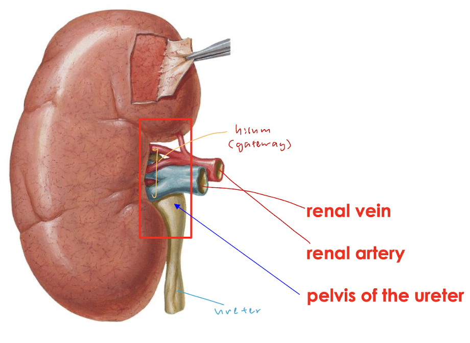
at concave medial margin of each kidney

lies hilum, contains

  • sympathetic nerves and lymphatics

  • renal vein and artery

  • pelvis of ureter


<p>lies hilum, contains</p><ul><li><p>sympathetic nerves and lymphatics </p></li><li><p>renal vein and artery</p></li><li><p>pelvis of ureter </p></li></ul><p></p>
9
New cards

ureter vs urethra

  • ureter: urine from kidney → bladder

  • urethra: bladder → outside


10
New cards

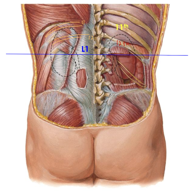
where is hilum and kidneys wrt ribs

  • near transpyloric plane at vertebral level L1

  • posteriorly

    • superior pole/ end of L kidney lies deep within 11th rib

    • R lies deep to 11th intercostal space


<ul><li><p>near transpyloric plane at <strong>vertebral level L1</strong></p></li><li><p>posteriorly</p><ul><li><p>superior pole/ end of L kidney lies deep <strong>within 11th rib </strong></p></li><li><p>R lies deep to <strong>11th intercostal space </strong></p></li></ul></li></ul><p></p>
11
New cards

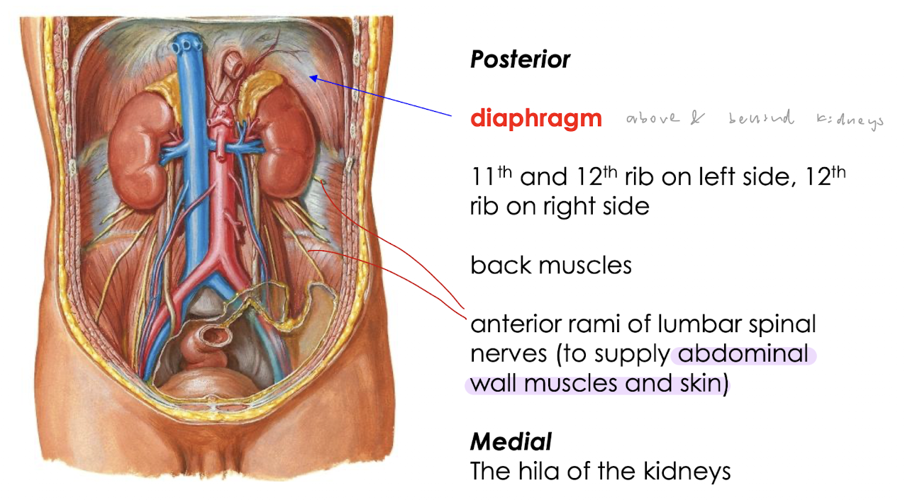
posterior relations of kidney

knowt flashcard image
12
New cards

anterior relations of kidney R

knowt flashcard image
13
New cards

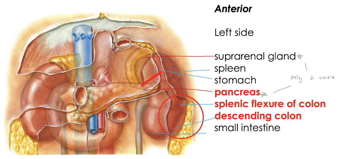
anterior relations of kidney L

knowt flashcard image
14
New cards

kidneys

  • pelvis of ureter divides into 2/ 3 major calyces

    • each divided into minor calyces

      • each indented by renal papilla

        • receives urine from apex of pyramid

        • corresponding to collecting tubules microscopically (what pyramid made of)


<ul><li><p>pelvis of ureter divides into 2/ 3 <strong>major calyces</strong></p><ul><li><p>each divided into <strong>minor calyces</strong></p><ul><li><p>each indented by <strong>renal papilla</strong></p><ul><li><p>receives urine from apex of pyramid</p></li><li><p>corresponding to collecting tubules microscopically (what pyramid made of)</p></li></ul></li></ul></li></ul></li></ul><p></p>
15
New cards

renal corpuscle

glomerular (bowman’s) capsule + glomerulus
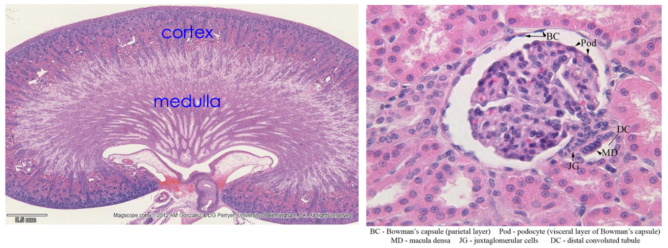
<p>glomerular (bowman’s) capsule + glomerulus </p>
16
New cards

layers surrounding the kidney

  1. renal capsule (fibrous) of kidney

  2. perinephric fat

  3. renal fascia

  4. external to renal fascia = pararenal fat


  • fat + fascia hold kidneys in relatively fixed position but can move about (3cm) during respiration


<ol><li><p>renal capsule (fibrous) of kidney</p></li><li><p>perinephric fat</p></li><li><p>renal fascia</p></li><li><p>external to renal fascia = pararenal fat </p></li></ol><p></p><ul><li><p>fat + fascia hold kidneys in relatively fixed position but can move about (3cm) during respiration </p></li></ul><p></p>
17
New cards

renal capsule

knowt flashcard image
18
New cards

blood vessels in kidneys

  • renal arteries derive from descending aorta

  • renal veins drain into IVC

  • L renal vein longer

    • L artery shorter

    • passes in front of aorta immediately below origin of superior mesenteric artery

  • R renal artery passes behind IVC


<ul><li><p>renal <strong>arteries</strong> derive from descending <strong>aorta</strong></p></li><li><p>renal <strong>veins</strong> drain into <strong>IVC</strong></p></li><li><p>L renal <strong>vein</strong> <u>longer</u></p><ul><li><p>L <strong>artery</strong> <u>shorter</u></p></li><li><p>passes in front of <strong>aorta</strong> immediately below origin of <strong>superior mesenteric artery </strong></p></li></ul></li><li><p>R renal <strong>artery</strong> passes behind <strong>IVC</strong> </p></li></ul><p></p>
19
New cards

lymphatic system

lymphatics drain to paraaortic lymph nodes (1 way/ drain upward)

which drain into cistern chill and then thoracic duct

<p>lymphatics drain to paraaortic lymph nodes (1 way/ drain upward)</p><p>which drain into cistern chill and then thoracic duct </p>
20
New cards

nerve supply

by renal plexus (both sympathetic and para fibres)

<p>by renal plexus (both sympathetic and para fibres)</p>
21
New cards

ureter

made up of:

pelvis of ureter

abdominal portion

pelvic portion

intravesical portion

<p>made up of:</p><p>pelvis of ureter</p><p>abdominal portion</p><p>pelvic portion</p><p>intravesical portion</p>
22
New cards

abdominal ureter

  • lies on transverse process of lumbar vertebrae separated by muscle psoas major

  • crossed anteriorly by testicular arteries in m

    • suspensory ligament in f (water under bridge)

  • cross into pelvis lateral to bifurcation of common iliac artery in front of sacroiliac joint


<ul><li><p>lies on transverse process of <strong>lumbar vertebrae</strong> separated by muscle <strong>psoas major </strong></p></li><li><p>crossed anteriorly by <strong>testicular arteries</strong> in m</p><ul><li><p><strong>suspensory ligament </strong>in f (water under bridge)</p></li></ul></li><li><p>cross into pelvis lateral to bifurcation of common iliac artery in front of sacroiliac joint </p></li></ul><p></p>
23
New cards

pelvic ureter

  • runs on lateral wall of pelvis in front of internal iliac artery

    • then turns forward and medially to enter bladder

  • crossed by uterine artery anteriorly/ superiorly in f

    • (water under bridge)


24
New cards

ureter in m

knowt flashcard image
25
New cards

ureter in f

knowt flashcard image
26
New cards

intravesical ureter

  • passes obliquely (diagonally thru wall of bladder for ~2cm

  • sphincteric arrangement (ring of muscles that squeeze tubes)

    • termination of ureter surrounded by vesicle muscle

    • to avoid reflux of urine into ureter during micturition (urination)


<ul><li><p>passes <strong>obliquely</strong> (diagonally thru wall of bladder for ~2cm</p></li><li><p><strong>sphincteric</strong> arrangement (ring of muscles that squeeze tubes)</p><ul><li><p>termination of ureter surrounded by vesicle muscle</p></li><li><p>to avoid reflux of urine into ureter during micturition (urination)</p></li></ul></li></ul><p></p>
27
New cards

ureter receives segmental blood supplies from

knowt flashcard image
28
New cards

ureter has nerve supplies form

knowt flashcard image
29
New cards

urinary bladder

  • pelvic retroperitoneal structure in adults

    • in pelvis, outside peritoneal sac

  • when urinary retention, adult bladder can distend into abdomen, stripping peritoneum upwards

    • (ave opacity = 300ml)

  • 3yo bc pelvis small, urinary bladder = abdominal organ


<ul><li><p><strong>pelvic retroperitoneal </strong>structure in adults </p><ul><li><p>in pelvis, outside peritoneal sac</p></li></ul></li><li><p>when urinary retention, adult bladder can distend into abdomen, stripping peritoneum upwards</p><ul><li><p>(ave opacity = 300ml)</p></li></ul></li><li><p>3yo bc pelvis small, urinary bladder = abdominal organ </p></li></ul><p></p>
30
New cards

parts of urinary bladder

knowt flashcard image
31
New cards

bladder - trigone, bladder wall, urethral sphincter

trigone

  • triangle formed by 2 ureteric orifices and internal urethral orifice

bladder wall

  • made up of detrusor muscle


circular component of it condenses as involuntary internal urethral sphincter around internal urethral orifice

  • external = skeletal muscle (voluntary)

  • internal = smooth (involuntary)


<p><strong>trigone</strong></p><ul><li><p>triangle formed by 2 ureteric orifices and internal urethral orifice </p></li></ul><p><strong>bladder wall</strong></p><ul><li><p>made up of detrusor muscle</p></li></ul><p></p><p>circular component of it condenses as <strong>involuntary internal urethral sphincter </strong>around internal urethral orifice </p><ul><li><p>external = skeletal muscle (voluntary)</p></li><li><p>internal = smooth (involuntary)</p></li></ul><p></p>
32
New cards

bladder anterior

knowt flashcard image
33
New cards

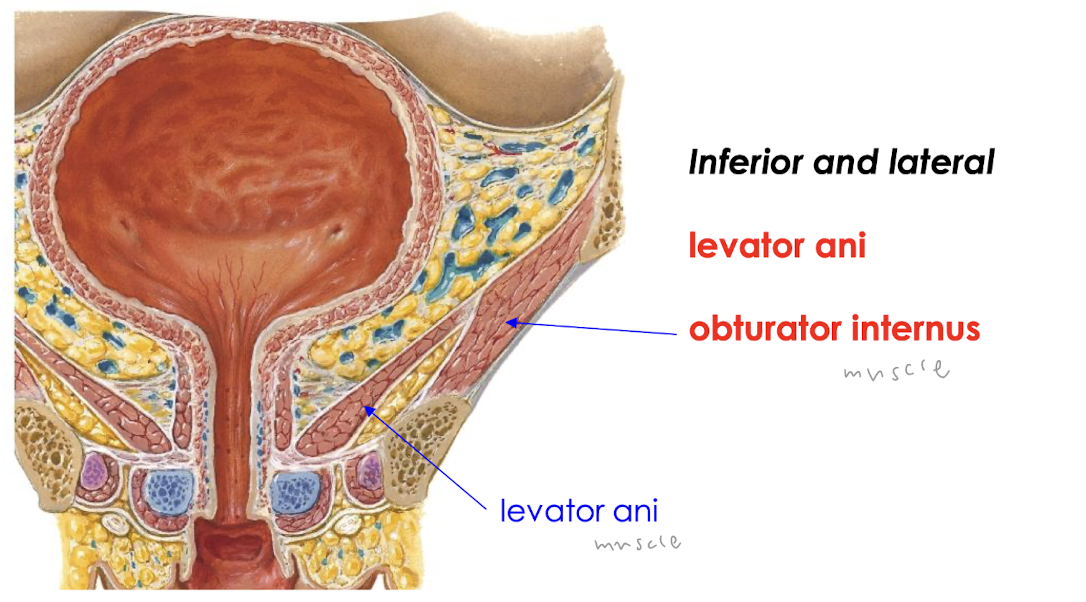
bladder inferior and lateral

knowt flashcard image
34
New cards

bladder superior

covered by peritoneum

<p>covered by peritoneum </p>
35
New cards

bladder posterior m

knowt flashcard image
36
New cards

bladder posterior f

knowt flashcard image
37
New cards

bladder blood supply

knowt flashcard image
38
New cards

lymphatic drainage in bladder

knowt flashcard image
39
New cards

urinary bladder has nerve supplies from

knowt flashcard image