neuroimaging flashcards week 1-4
 Call Kai
 Learn
 Practice Test
 Spaced Repetition
 Match
 Flashcards
 Knowt Play
1/161

There's no tags or description
Looks like no tags are added yet.
162 Terms
what are 4 ways to characterise and differentiate techniques?
Structure vs. function
Temporal (relies on electromagnetic) vs. spatial resolution (relies on blood flow)
Degree of invasiveness
Correlation vs. causation
what are 6 features of neuroimaging?
Focus on humans
In vivo
Non-invasive or minimally invasive
Correlational
Systems-level explanations
Multiple approaches to look at the brain
The brain has face-specific activity which can be identified from what?
- Prosopagnosia p's
- Single cell recordings (from animals)
- Hemodynamic activity
- Electrical activity
what were the 4 key landmarks of the history of neuroimaging dating back 10,000 years ago?
Skulls found with circumscribed holes.
Evidence of trepanning (purposely making incisions).
Indicates that there was a special interest in the head.
Treating the body, mind or soul?
what did the ancient greeks introduce to the development of neuroimaging?
they started to think about the concept of the mind (psyche)
up until the 1800's, what did the key questions about neuro/neuroimaging focus on?
Questions about where does the mind reside? Brain? Body? Non-physical?
in the 1800s, what was a key question about neuro/neuroimaging?
ow does the brain enable the mind? Tissue? Ventricles? Localisation? Mass action?
between the 1800s-1900s, what developments were made in the field?
Better tools -> more experiments -> better understanding of the brain
Late 1970s onwards: breakthroughs in measuring the living brain
where are we now in the field?
Neuroimaging remains important for mind-brain questions
Focus changing (localisation ➞ connectivity, computational approaches, networks, more structural imaging)
fMRI criticized; other techniques becoming more popular (EEG, eye tracking, optical imaging)
Continued development of (complex and sophisticated) methods
what is dualism?
that the mind and the brain are separate entities (not viable in cognitive neuro)
what is monism?
that the mind and the brain are the same
what is the dominant view in cog neuro of mind-brain relationships?
Physicalism
what is physicalism?
the view that all mental states, thoughts, and consciousness are rooted entirely in physical brain activity, neurochemistry, and biological processes
what is the reductive and non-reductive aspect of physicalism?
reductive: mental states are reducible to physical brain states.
non-reductive: mental and physical states are linked but not identical.
what are philosophical issues in the convo about mind-brain relationships?
Defining core concepts (cognition, function, brain patterns).
Choosing appropriate level of explanation (regions or systems; segregation or integration).
what is forward inference?
infer brain activity from a known cognitive function; you only look at function and work out what brain bit this corresponds to
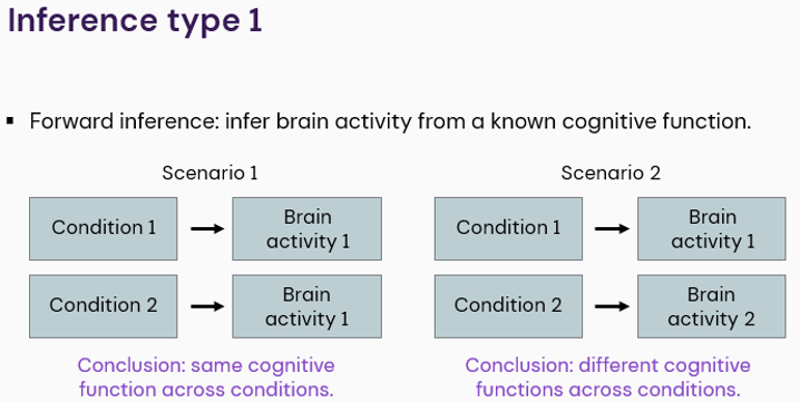
what is reverse inference?
infer cognitive function from observed brain activity

what is one of the key problems of reverse inference?
the validity of the conclusion depends on the validity of the link/previous work. you can only draw a valid conclusion if you are 100% sure about the link (which is like never the case as theres always other things)
is one-to-one mapping possible under reductive physicalism?
no
do brain regions often have one-to-one relationships with mental processes?
no
what is apparent diffusion coefficient (ADC)?
a DTI measure of water molecule diffusion within brain tissue
what is axial diffusivity (AD)?
a DTI measure of water molecule diffusion parallel to the nerve tract
what is beta decay?
radioactive decay characterised by emission of an electron
what is the BOLD signal?
the change in fMRI signal between oxygenated and deoxygenated haemoglobin
what is cerebral blood flow?
the amount of blood supplied to the brain at any given moment
what is cerebrospinal fluid?
clear, colourless fluid that surrounds the brain and spinal cord
what do structural MRI’s show?
neurological structures with clarity and precision; detailed, static anatomical brain pictures
what is task-evoked fMRI?
fMRI technique that measures brain activity in response to a stimulus
what is resting state fMRI?
fMRI technique that measures brain activity while at rest
what is T1 weighting?
measures how fast protons return to their resting state using short repetition times (TR) and short echo times (TE)
white matter appears bright (and fat tissue)
white matter appears light grey
CSF (and water) appears black
used for looking at normal body anatomy, brain structures, and contrast-enhanced tumours
what is T2 weighting?
It measures how long protons stay in sync before losing their signal using long repetition times (TR) and long echo times (TE)
grey matter appears light grey
white matter appears dark grey
CSF appears bright
brain matter appears dark
best for finding pathology, injuries, fluid build-up, and diseased or inflamed tissue
what is T2* weighting?
It is a faster variation of T2 imaging that includes natural, tiny imperfections (inhomogeneities) in the main magnetic field.
Blood products (like old or new haemorrhages/bruising), iron deposits, and calcium appear dark
Best used for detecting bleeds in the brain, stroke evaluations, or spotting iron and calcifications in tissues

what weighting is this mri?
T1
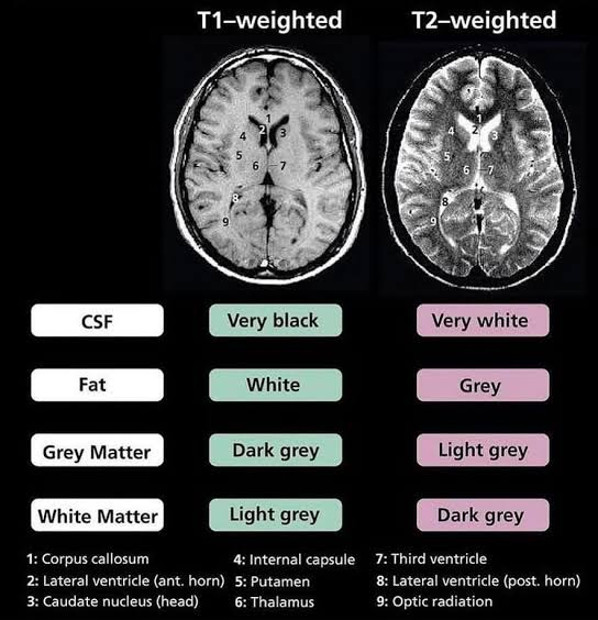

what weighting is this mri?
T2
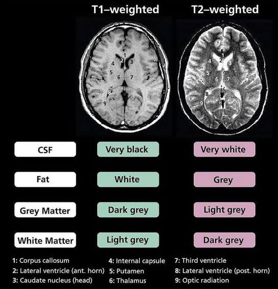
what does T1 weighting describe?
the spin relaxation characteristics or the rate of decay of applied magnetisation of the excited protons of water molecules in the brain
how do mris localise tissue?
by manipulating and measuring behaviour of protons
what is the step-by-step to how mris work?
Tissue -> water molecules -> hydrogen atoms -> protons. Protons spin -> magnetic moment -> behaves like tiny magnet
In a magnetic field, protons’ magnetic moments precess (wobble) at a characteristic frequency. A brief, specific radiofrequency pulse (B1) transfers energy to the protons. Net magnetisation tips away from equilibrium (flip), producing excitation
After the radiofrequency pulse, spins gradually realign with the main magnetic field, releasing energy (T1 relaxation). Different tissues have different relaxation times. Time measures (ms) used to generate images: T1 / T2 / T2*
fill in the blanks: ___, ___ and ___ are measures of the time it takes to get back to the magnetic state
T1, T2 and T2*
is spin-lattice relaxation T1, T2 or T2*?
T1
what is spin-lattice relaxation?
recovery of longitudinal magnetisation to the static magnetic field
what is spin-spin relaxation?
decay of transverse magnetisation as spins lose synchrony
is spin-spin relaxation T1, T2 or T2*?
T2
what is effective transverse relaxation?
decay of transverse magnetisation due to spin-spin interactions and magnetic field variations (faster de-phasing, T2* < T2)
is effective transverse relaxation T1, T2 or T2*?
T2*
is T2* used for functional or structural images?
functional
in what weighting is fat bright and fluids dark?
T1
in what weighting is fat dark and fluids bright?
T2
what is mri homogenisation (otherwise known as image harmonisation or B1/B0 field homogenisation)?
the process of correcting, standardising or translating mri images to remove technical and hardware-induced variations
what is magnetic field homogeneity (B0/B1 field)?
Refers to the uniformity of the main magnetic field (B₀) or the radiofrequency transmit/receive field (B₁) inside the scanner. Inhomogeneities—especially at high-field (7T) or ultra-high-field systems—cause shading artifacts, signal dropouts, or geometric distortions
what are some hardware and physical fixes of magnetic field inhomogeneities (such as shading artifacts, signal dropouts or geometric distortions)?
multi-layer coupled coils
high-permittivity materials (HPM) and mode-shaping
parallel transmit and pre-emphasis pulse adjustments
what is dataset and protocol harmonisation (image-to-image translation)?§ ): In clinical data warehouses and multi-center studies, differences in scanners, field strengths, and contrast agents (such as gadolinium) create heavy data variability (Dataset and Protocol Harmonization (Image-to-Image Translation)). What kind of modern approaches are used to help this, and how does it help?
Modern approaches use deep learning (such as variational autoencoders and CycleGANs or 3D U-Nets) to translate or normalize images into a standard/common feature space.
This ensures that downstream automated tools (like tumor segmentation or brain classification algorithms) do not pick up scanner-specific bias instead of actual pathology.
fill in the blank: - Gradient coils produce controlled variations in the magnetic field; These variations make the MRI signal dependent on its ______; The scanner uses this information to determine where signals come from
location
what is diffusion tensor imaging (DTI)?
MRI technique that measures the strength and direction of water molecule diffusion along nerve fibres, allowing direct visualisation of white matter bundles (i.e. axons) in the brain
when is DTI used/what is it useful for?
Useful for disease processes with impaired WM integrity such as Alzheimer’s, MS or mental health disorders
what is the basic principle/mechanism behind DTI?
water molecules will diffuse at different rates along nerve fibre tracts, depending on the integrity of the nerve fibre and the presence of barriers. Water molecule diffusion becomes altered in areas of impaired WM integrity, allowing DTI to detect alterations to WM not observable in many other forms of MRI
Altered diffusion of water molecules in white matter tracts can be measured via fractional anisotropy and quantified on a scale of 0 to 1; to what end of the scale do values indicate nerve fibre integrity?
1
what are two things fractional anisotropy values are sensitive to?
the degree of myelination
axonal density of white matter
what does the apparent diffusion coefficient (ADC) (a common measure in DTI) measure?
the amount of diffusion of water molecules within brain tissue; The ADC can be used to identify areas of damage to white matter caused by insults such as ischemia (i.e., low ADC values) or edema (i.e., high ADC values). The diffusion-weighted images provided by DTI measure the magnitude of water molecule diffusion in multiple directions (e.g., parallel to nerve fibre tract, perpendicular to nerve fibre tract) at a single point in the brain.
what does axial diffusivity measure?
the rate of diffusion parallel to the nerve fibre tract
what does radial diffusivity measure?
the diffusion perpendicular to the nerve fibre tract
fill in the blank: DTI provides a ___, ___-___ method to study ___ integrity without the use of contract or chemical tracers
sensitive; non-invasive; WM
what are 3 key difficulties of DTI
the inherent complexity of the analysis
the inherent complexity of the interpretation of results
methods high sensitivity to movement (which may limit the ability to use it in some vulnerable populations)
what does a fMRI sequence measure?
changes in deoxygenated haemoglobin (Hb) concentration to indirectly reflect brain metabolic demand over time; neural activity or associated physiological changes
how does fMRI work?
Deoxygenated Hb exhibits paramagnetic (a material that is weakly attracted to an external magnetic field and does not retain its magnetic properties once the field is removed, ai definition) characteristics when an external magnetic field is applied. Because of this characteristic, molecules of deoxygenation Hb align with the applied magnetic field of the MRI machine
This shift in alignment allows for the concentration of deoxygenated Hb to be measured, resulting in an MRI signal known as the blood oxygen-level dependent (BOLD) signal
This modality offers researchers a real-time depiction of the brain’s activity, whether at rest or attending to a task or stimulus
what is the BOLD signal?
This shift in alignment allows for the concentration of deoxygenated Hb to be measured, resulting in the BOLD MRI signal
Deoxyhaemoglobin distorts the magnetic field, increasing field inhomogeneity and causing faster spin dephasing (shorter T2*). More neural activity → less deoxyhaemoglobin → longer T2* → stronger signal
what is a key problem of the use of gradient magnets?
it makes it more dangerous in the presence of prostheses or devices that are only conditionally safe at lower MRI magnetic field strengths
how does neural activity change the blood supply?
the brain has high metabolic demands, when brain regions become active, they need more oxygen and glucose, local blood flow and volume increase, this exceeds metabolic demand, these changes can be measured to infer regional neural activity
do oxygenated and deoxygenated haemoglobin affect MRI signals differently or the same?
differently
what is the spatial resolution of fMRI?
~1mm
fill in the blank: Whole brain can be measured, but some regions ____ to image
difficult
does movement lead to substantial artefacts across images?
yes
how does task-evoked fMRI work?
Functional imaging typically evokes changes in a participant’s cerebral blood flow through a series of tasks or stimuli. As the research participant engages these stimuli, activated brain regions experience a greater degree of change in deoxygenated Hb concentration, as oxygen is delivered to satisfy increased metabolic demands. This results in greater BOLD signal relative to the surrounding tissue.
fill in the blank: The choice of stimulus is an ___ consideration when performing task-evoked fMRI, as the ___ signal is the result of a hemodynamic process that reaches its peak a few ___ after stimulus onset and exhibits as low decline during which the stimulated brain region’s ___ signal returns to baseline
important; BOLD; seconds; BOLD
what is resting-state fMRI concerned with?
the changes in brain activation in the absence of a stimulus or task
as the sheer volume of data from rs-fMRIs may be the most difficult barrier, and the study of whole-brain connectivity through voxel-to-voxel analysis is a conceptually and computationally challenging starting point, what is a simpler approach?
a priori selection of regions of interest (ROIs) in networks described by the literature and ROI-to-ROI connectivity analysis, minimizing the computational workload and allowing clearer interpretation of results
what is a distinct advantage of rs-fMRI over task-evoked fMRI?
A distinct advantage of rs-fMRI over task-evoked fMRI is evident when imaging research participants in vulnerable populations or participants who may otherwise be unable to tolerate or perform a stimulus or task such as frail, elderly subjects or cognitively impaired individuals.
when is positron emission tomography (PET) used?
May be used for in vivo images of biological, biochemical and physiological processes
what is administered to the participant in PET studies?
a radionuclide tracer
Radionuclides used in PET emit positrons when they decay through a process known as betadecay
fill in the blank: Positron emissions are detected by cameras called ___
scintillators
fill in the blank: A modern PET machine consists of any number of scintillators, each connected to ___ ___, which amplify the signal of the emitted positrons
photomultiplier tubes
do radionuclides require stimulation to emit a positron?
no, so there is a constant source of signal that may be collected
is PET a poor or strong modality for structural imaging?
poor; This is often remedied by registering PET images with anatomical CT images or simultaneous collection of both modalities using hybrid PET/CT systems
what are two key disadvantages of PET scans?
poor modality for structural imaging
radionuclide acquisition. Tracer molecules do not exist in nature and must have a short half-life or time in which half of the substance decays to limit harmful radiation exposure to both the research participant and the researchers. (can cause damage to tissue and DNA)
how does PET work?
Radioactively labelled tracer is injected into the blood stream -> Tracer decay produces two gamma rays travelling in opposite directions, detected by the PET scanner -> More neural activity → more blood flow → more tracer
what is the spatial resolution of PET?
~10mm
can the whole brain be measured using PET?
yes
briefly, how does EEG and MEG work?
they map brain activity by detecting signals of ionic currents from biochemical processes occurring at the cellular level during neural activation. Sensor arrays are used to record these ionic currents, which are electrical fields in EEG and magnetic fields in MEG
in reference to EEG and MEG, what are oscillations?
The frequency, bandwidth, and amplitude of the electrical or magnetic signals are recorded and called oscillations. Oscillations are not just correlates of brain activation but are considered a representation of underlying neural mechanisms of cognitive processes
what are three things/barriers that can distort the signal detection of EEG sensors?
CSF, bones of skull and tissues of scalp
why are the magnetic fields detected by MEG less distorted?
because of the constant magnetism permeating brain tissue
what is the key similarities about the information yielded from EEG and MEG?
both EEG and MEG ultimately yield nearly the same neural information and their localization to a specific brain region is similar enough to be considered almost interchangeable
Both EEG and MEG can be used to examine and investigate cognitive processes such as perception and memory, brain attentional systems, transfer of information, and the organization and consolidation of memories
Both yield information on the dynamic interaction between areas of the brain with the specificity of milliseconds
EEG and MEG use neuronal populations which generate electromagnetic fields. what is the electrical component?
EEG
EEG and MEG use neuronal populations which generate electromagnetic fields. what is the magnetic component?
MEG
how does volume conduction work in reference to EEG and MEG?
Volume conduction: neuronal activity generates currents that spread through brain tissue, cerebrospinal fluid, skull and scalp – this results in measurable electrical potentials and magnetic fields on the scalp
what are the 4 key requirements of EEG and MEG?
the electromagnetic field must be large (to be detectable on the scalp)
requires the summated activity of many neurons (>10,000)
the neurons must be active synchronously: action potentials (~1ms) unlikely; graded post-synaptic potentials most likely
the neurons must have an open field spatial configuration
fill in the blank ___ detects deep and shallow, radial and tangential (touching lightly)
EEG
fill in the blank: ___ detects mainly superficial, tangential
MEG
does EEG detect a broader range of brain activity than MEG?
yes
is MEG or EEG easier to localise and why?
MEG is easier to localise because signals are less distorted by volume conduction
it is debated if it can actually get deep structures like the amygdala or if its just the surrounding areas its picking up on
is EEG or MEG more expensive?
MEG