lecture 18 - spinal cord and spinal nerves
 Call Kai
 Learn
 Practice Test
 Spaced Repetition
 Match
 Flashcards
 Knowt Play
1/105

There's no tags or description
Looks like no tags are added yet.
106 Terms
regions of vertebrae (superior to inferior)
cervical region
thoracic region
lumbar region
sacral + coccygeal regions

vertebrae are stacked onto one another to create the _________ ______
vertebral column

vertebral foramen
an opening near the spiny processes of each vertebrae
successive holes in each vertebrae create the vertebral canal

vertebral canal
where the spinal cord is located

how do we keep the spinal cord in the middle of the vertebral column?
meninges (of spinal cord)
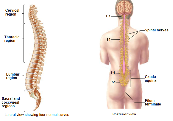
intervertebral foramina
spaces b/w the vertebrae
where the spinal nerves will exit the vertebral canal

the numbering of the spinal nerves is based on..
which level of the vertebral canal they exit through the intervertebral foramina

true or false: the spinal cord is longer than the vertebral canal
false: the spinal cord is shorter than the vertebral canal

why is the spinal cord shorter than the vertebral canal?
difference in speed of growth during development

the spinal cord stops growing in length around ages _-_. the vertebral column continues to elongate until early __________
4-5, adulthood
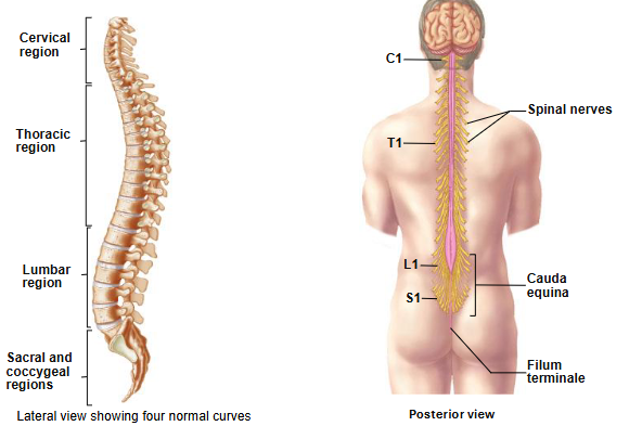
the spinal cord extends from the ________ ______ to L_
foramen magnum, 2
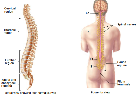
foramen magnum
very large circular hole in base of skull (occipital bone)

where the ______ _______ ends at the level of the foramen magnum, the spinal cord will begin
medulla oblongata

where can you landmark L2 at?
most inferior rib
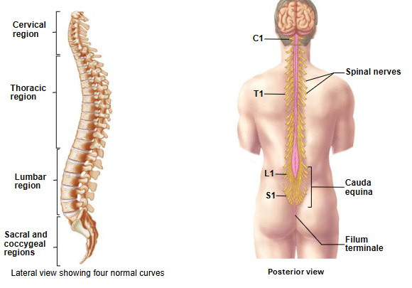
spinal cord
slightly flat on one side
not uniform in diameter
has 2 enlargements

2 enlargements of spinal cord
cervical enlargement
lumbosacral enlargement
associated with extra nervous tissue needed to supply the structures of upper/lower limbs

cervical enlargement
enlargement on spinal cord
occurs b/w vertebrae C4 and T1

lumbosacral enlargement
enlargement on spinal cord
occurs b/w vertebrae T9 and T12

conus medullaris
inferior end of spinal cord

sacral foramina
holes in the sacral bone that nerves lower down can exit through

cauda equina
hair-like nature of nerves coming out of bottom of spinal cord
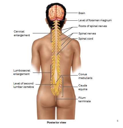
filum terminale
extension of the pia mater
anchors the spinal cord to the coccyx
prevents movement of the spinal cord in the superior direction

what is the most superficial layer of the meninges?
dura mater

which meningeal layer is the thickest and strongest?
dura mater

what is different about the dura mater in the spinal cord compared to in the brain?
there is a space b/w the dura mater and the periosteum (connective tissue layer on bone) called the epidural space

epidural space
space b/w the dura matter and periosteum
filled w/ fat, blood vessels + areolar CT
protects spinal cord + holds it in place

epidural
anesthesia injected into the epidural space to block pain receptors

meninges superficial to deep
dura mater
arachnoid mater
pia mater

arachnoid mater
deep to dura mater
thin, avascular
simple squamous epithelial cells

subdural space
space b/w the dura mater and arachnoid mater
contains a little serous fluid

pia mater
deepest layer
tightly adheres to spinal cord
has small extensions (denticulate ligaments) that connect itself to the dura mater

denticulate ligaments
small extensions in the pia mater that are connected to the dura mater
occur b/w each root that exits the spinal cord
help anchor spinal cord laterally (prevent side-to-side movement)

subarachnoid space
b/w arachnoid and pia mater
contains CSF

2 major portions of spinal cord
outer white matter
inner gray matter

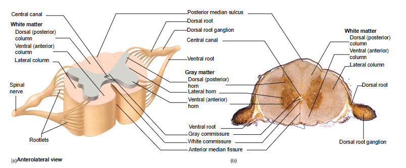
posterior median sulcus
narrow groove on posterior side of spinal cord

anterior median fissure
groove on anterior side of spinal cord

the posterior median _____ and anterior median ______ partially divide the spinal cord into 2 halves
sulcus, fissure

white matter of spinal cord
composed of myelinated axons (run the length of the spinal cord)
can either be ascending or descending

columns
nerve tracks (white matter) organized together

3 white matter columns
posterior column
anterior column
lateral column

in each white matter column, there will be groups of either _________ or __________ tracts that are bundled together because they have similar ________ or serve a similar area of the body
ascending, descending, functions

white commissures
axons that connect L and R sides of spinal cord in white matter region
allows signals to move from one side of the body to another

3 horns of gray matter of spinal cord
posterior horn
lateral horn
anterior horn

the lateral horns (gray matter region of spinal cord) are found only in the ______ and ______ regions
thoracic, lumbar

lateral horn
gray matter
very small
associated w/ neurons of ANS

posterior horn
gray matter
where axons of sensory neurons enter spinal cord

true or false: sensory nerve somas are only found in the dorsal root ganglion (outside CNS)
true

the cell bodies in the posterior horn are all the cell bodies of the __________ that are going to transmit info around in the gray matter area
interneurons

anterior horn
gray matter
contains cell bodies for somatic motor neurons
largest horn

the anterior horn is also referred to as the
motor horn

gray commissure
axons that connect L and R side of spinal cord in gray matter region
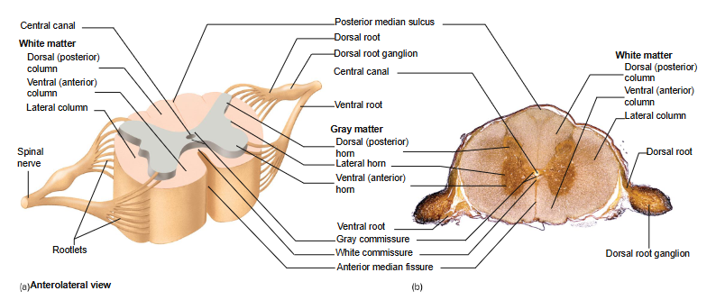
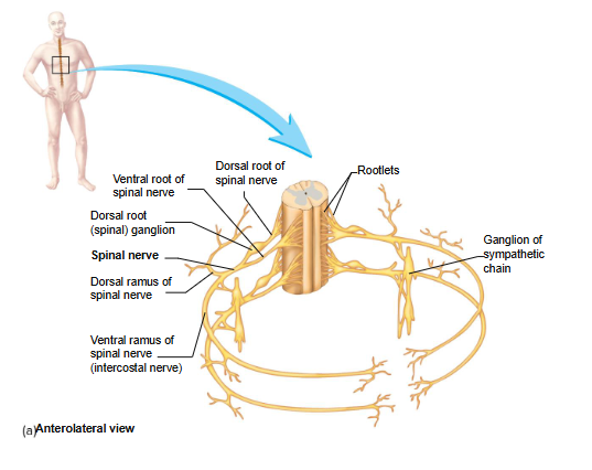
rootlet
leave or enter the spinal cord
part of PNS

ventral root
motor neurons exit spinal cord

dorsal root
sensory neurons enter spinal cord

dorsal root ganglion
contains cell bodies of sensory neurons
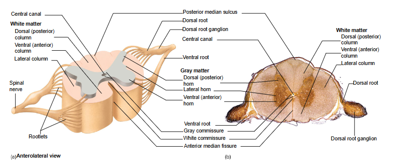
spinal nerve
dorsal and ventral roots merged together
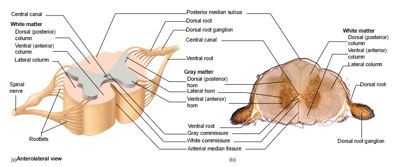
all spinal nerves have _____ function
mixed (both sensory and motor)
nerve tracts are also known as
fascicles
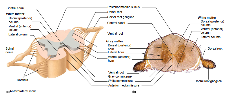
polio
Degeneration of gray matter within anterior horn
Virus attacks motor neurons
Motor loss + paralysis

ALS (amyotrophic lateral sclerosis) or Lou Gehrig’s disease
Degeneration of anterior horn + descending motor axons
Loss of speech, swallowing and breathing

how many pairs of spinal nerves are there
31

where does the first pair of spinal nerves exit the vertebral column?
b/w skull and first cervical vertebrae

the spinal nerves are typically located _____ their respective vertebrae
above (e.g. spinal nerve C1 would be above vertebrae C1)

how many cervical vertebrae are there
7

how many pairs of cervical spinal nerves are there
8

the 8th cervical nerve exits ______ vertebrae C7
below
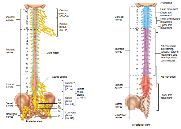
from the 8th cervical nerve on, spinal nerves are represented by the vertebrae _________ to them
superior (e.g. above vertebrae C7 is spinal nerve C7, below it is spinal nerve C8)
(next is the vertebrae T1, with spinal nerve T1 below it)
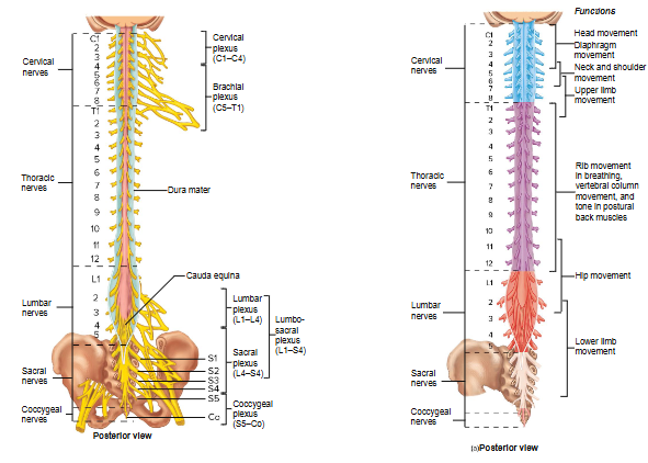
there are _ pairs of spinal nerves that exit via the sacral foramina. the rest of the spinal nerves exit through the _____________ __________
4, intervertebral foramina

how many pairs of thoracic spinal nerves are there
12

how many pairs of lumbar spinal nerves are there
5

how many pairs of sacral spinal nerves are there
5

how many pairs of coccygeal spinal nerves are there
1

a complete cut in the cervical region of the spinal cord results in
quadriplegia (all 4 limbs)

a complete cut in the upper thoracic region results in
paraplegic (only legs)

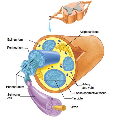
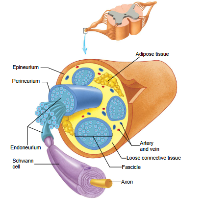
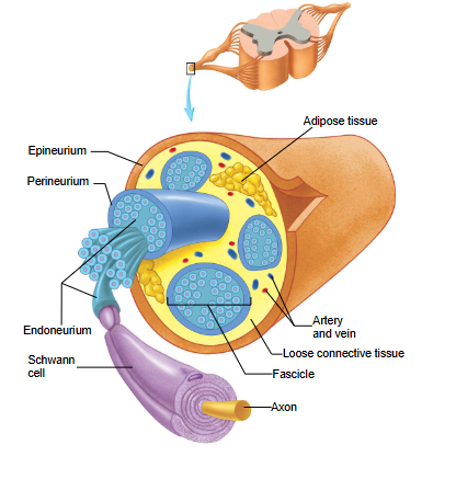
endoneurium
CT
surrounds the myelinated axons
separates axons from each other

perineurium
CT
surrounds bundles of myelinated axons

what surrounds the fascicles (bundles of neurons) other than the perineurium?
arteries and veins that provide blood supply
adipose tissue

the groups of fasicles are bound together to form a _____ _____
spinal nerve

epineurium
CT
surrounds spinal nerve
continuous with the dura mater of the brain

CT layers of spinal nerve from superficial to deep
epineurium
perineurium
endoneurium
about how many rootlets merge to form dorsal/ventral roots?
6-8

the dorsal and ventral roots pass through the ___________ space, pierce the arachnoid mater and join to form a ______ _____
subarachnoid, spinal nerve
each dorsal root contains a _______
ganglion
rami
split in spinal nerve shortly after the dorsal and ventral roots merge
there is posterior and anterior rami

2 branches of rami
posterior ramus
anterior ramus

posterior ramus
innervates deep muscles of back responsible for movements of the vertebral column (also skin + CT on back)

anterior ramus
where these nerves go depend on where you are in the body
from T2-T12 → form the intercostal nerves
remaining anterior rami → roots of plexuses
from C1-C4 → cervical plexus
from C5-T1 → brachial plexus
from L1-L4 → lumbar plexus
from L4-S4 → sacral plexus
S5 and coccygeal nerve → coccygeal plexus

roots of plexuses
intermingling of nerves that exit the spinal cord
formed from remaining anterior rami (5 plexuses)

intercostal nerves
formed by anterior ramus
innervate the intercostal muscles (involved in breathing)
cervical plexus
most superior plexus
roots: C1-C4
innervate superficial neck structures, skin of neck, posterior portion of head
contains phrenic nerve

phrenic nerve
part of cervical plexus
roots: C3-C5
innervates diaphragm

brachial plexus
roots: C5-T1
3 trunks, 6 divisions, 3 cords, 5 branches
contains axillary, radial, musculocutaneous, ulnar and median nerve
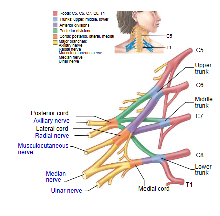
axillary nerve
part of brachial plexus
innervates deltoid + teres minor muscles
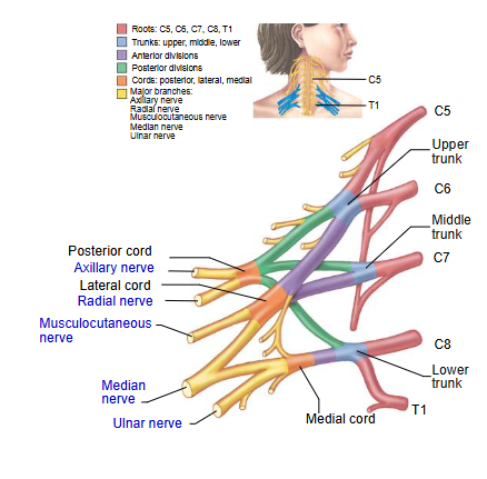
radial nerve
part of brachial plexus
innervates muscles on back of arm + forearm
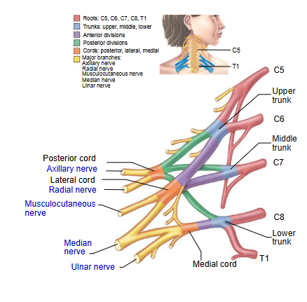
musculocutaneous nerve
part of brachial plexus
innervates biceps brachii

median nerve
part of brachial plexus
innervates most of anterior forearm + some hand muscles associated w/ thumb

ulnar nerve
part of brachial plexus
innervates forearm + most hand muscles

lumbar plexus
L1-L4
contain the femoral and obturator nerves

femoral + obturator nerves
part of lumbar plexus
innervate lateral abdominal walls, external genitals + parts of lower limbs

sacral plexus
L4-S4
contain common fibular and tibial nerve (which make up sciatic nerve)
