Signalling in the Nervous System (II)
 Call Kai
 Learn
 Practice Test
 Spaced Repetition
 Match
 Flashcards
 Knowt Play
1/50

Earn XP
Description and Tags
Week 12
51 Terms
action potential
a rapid, transient and self-propagating change in the membrane potential of an excitable cell (neuron or muscle fibre)
*Vm = resting membrane potential
temporary rise - once initiated, can affect an entire excitable membrane!
unlike a graded potential, action potentials do not diminish as they move away from the source; impulse travels along the axon to axon terminal
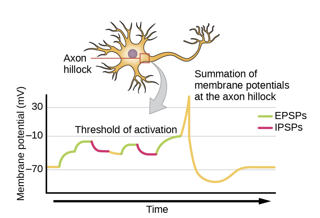
excitatory post-synaptic potential (EPSP)
a temporary, local depolarisation of a postsynaptic neuron’s membrane potential that makes the cell more likely to fire an action potential
summation of EPSPs and IPSPs at Axon Hillock
threshold
the membrane potential at which an action potential begins
usually between -60 mV and -55 mV (all-or-none principle)
graded potential v action potential


generating an action potential
1) depolarisation to threshold
2) activation of voltage-gated Na+ ion channels and rapid depolarisation
3) inactivation of voltage-gated Na+ ion channels and activation of voltage-gated K+ ion channels, beginning depolarisation
4) closing of voltage-gated K+ ion channels producing a brief hyperpolarisation (due to a lag in total closing of channels), and the a return to the resting membrane potential

refractory period
the time interval after an action potential is initiated during which it is impossible or more difficult for a neuron to fire a second action potential

function of Na+/K+ ATPase
pumps 2 extracellular K+ ions into the the neuron cell, while pumping 3 intracellular Na+ ions out of the cell
requires ATP
energy acquired from splitting a phosphate group from a molecule of ATP, forming ADP
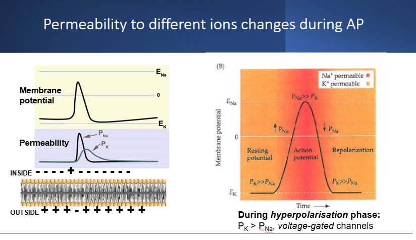
permeability of different ions during an action potential
at resting membrane potential:
low permeability for K+ (leak channels)
almost zero permeability for Na+
during depolarisation:
high permeability for Na+ (voltage-gated channels)
less for K+ , but slightly increased from previous stage
during repolarisation:
decreasing permeability for Na+
increasing permeability of K+
during hyperpolarisation:
high permeability of K+ (voltage-gated channels)
almost zero permeability of Na+
an action potential is generated at a small portion of the total plasma membrane surface
how is the action potential spread along the entire excitable membrane?
(propagation of action potential)
when Na+ ions move into an axon and depolarize adjacent sites, this triggers the opening of additional voltage-gated Na+ ion channels
result:
a chain reaction that spreads across the surface of the membrane
action potential is propagated along the length of the axon, until it reaches the axon terminal
continuous propagation
occurs in unmyelinated axon
action potential begins at axon’s initial segment
for a brief moment the membrane potential becomes positive rather than negative
Steps:
1) as Na+ ions begin moving from extracellular fluid into cytosol, a local current develops
2) the local current spreads in all direction, depolarizing adjacent portions of the membrane
3) action potential proceeds away from the site of generation
4) eventually reaching axon terminal
these steps repeat along the axon membrane
*action potential is not affected by distance
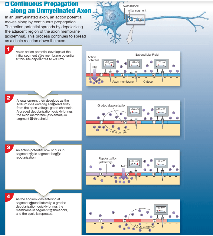
when a local current develops, the action potential moves forward, but not backward - why?
the previous segment of the axon is still in absolute refractory period
saltatory propagation
occurs in myelinated axons
more rapid than continuous propagation
uses less energy than continuous propagation
ions can cross the axolemma at nodes of ranvier (via voltage-gated ion channels) in response to a stimulus
myelinated sections cannot respond to stimulus
steps:
1) action potential generated at the initial segment of a myelinated axon
2) local current skips the internodes (myelinated sections) and depolarises the closest node to threshold
3) action potential continues “jumping” from node to node rather than moving along the axon in a series of tiny steps

why can’t myelinated section of the axon respond to stimulus and fire an action potential?
myelin increases the resistance to the flow of ions across the membrane
why does saltatory propagation use less energy than continuous propagation
less surface area is involved and fewer sodium ions must be pumped out of the cytoplasm (via Na+/K+ ATPase)
propagation speed in relation to axon diameter
larger diameter = less resistance to ion movement* = faster propagation
*ions must move through the cytosol to depolarize adjacent portions of the plasma membrane


different structures and propagation speeds of each type of fibre lend themselves to different functions - give examples
Type A Fibres:
carry sensory information about position & balance to CNS
sensory information about touch and pressure sensations from skin surface to CNS
efferent neurons that send motor commands from CNS to skeletal muscles
relays urgent sensory info about things that threaten survival
Type B & C Fibres:
carry information to and from CNS
deliver temperature, pain and general touch, pressure sensations
carry motor instructions to smooth muscle, cardiac muscle, glands and other peripheral effectors
less urgent sensory info and motor commands
*only one-third of axons carrying sensory information are myelinated
* most sensory information arrives over slender Type C fibres
electrical synapse
direct physical contact between presynaptic and postsynaptic membranes of two communicating cells
membrane proteins, called connexons, bind the two cells at gap junctions
connexons form pores, allowing ions to pass between cells
as if the two cells share a common membrane
changes in membrane potential in one cell, produces local currents that affect the other cell
propagate action potentials quickly and efficiently
extremely rare in adults

chemical synapse
one neuron (presynaptic cell) sends chemical signals (neurotransmitters) to another postsynaptic cell
both cells are separated by synaptic cleft
The presynaptic cell is not always a neuron
e.g. specialised receptor cells (presynaptic cell) can form synaptic connections with the dendrites of neurons (postsynaptic cell)

type of postsynaptic cells
neuron
muscle cells
glandular cells
axoaxonic synapse
a synapse that occurs between the axons of two neurons

axosomatic synapse
a synapse that occurs between the axon terminal of one neuron with the cell body of another neuron

axodendritic synapse
synapse between the axon terminal of one neuron and the dendrite of another neuron

neuromuscular junction
a synapse between a neuron and skeletal muscle cell
neuroglandular junction
a chemical synapse or connection point between a neuron and a gland cell
neurotransmitters are contained in…
synaptic vesicle
a synaptic cleft separates…
the presynaptic membrane and the postsynaptic membrane

postsynaptic membranes bears …….. for neurotransmitters
receptors
contents on axon terminal
mitochondria
vesicles filled with neurotransmitters
Receives continuous supply of neurotransmitters, enzymes and lysosomes that are synthesised in the cell body (anterograde flow)
why axon terminals have mitochondria
provide a local, continuous supply of ATP for high-energy processes
high-energy processes:
priming and recycling of synaptic vesicles
releasing neurotransmitters into synaptic cleft
powering ion pumps (e.g. Na+/K+ ATPase
axon terminals are far away from the soma and energy molecules cannot diffuse fast enough from that distance - local mitochondria provide a readily-available supply of ATP
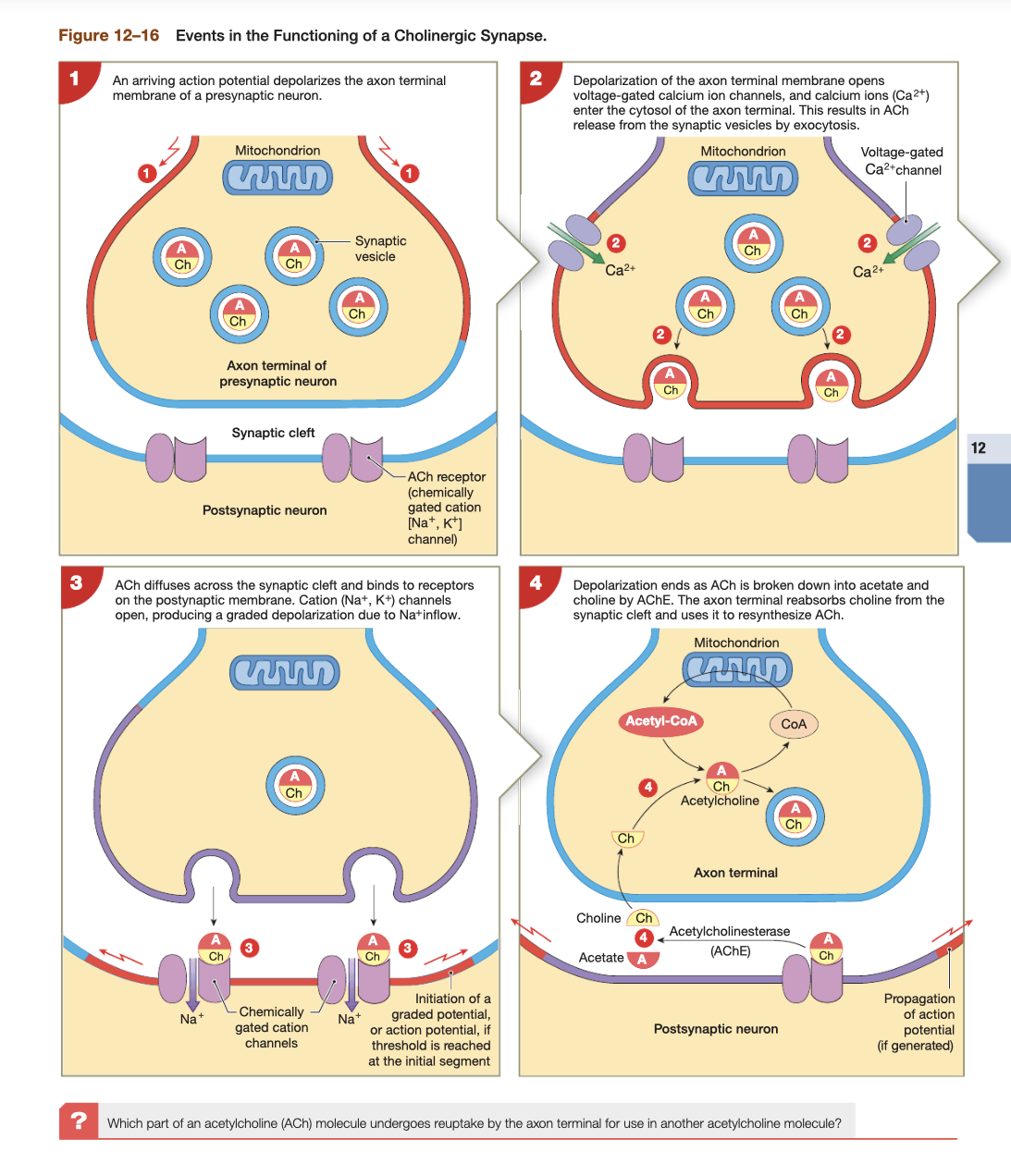
Ca2+ buffering - when an action potential reaches the axon terminal, voltage-gated Ca2+ ion channels open. Mitochondria rapidly absorbs and buffers the temporary influx of Ca2+ . Prevents toxic over-accumulation
journey of neurotransmitters
presynaptic cell releases neurotransmitters into synaptic cleft
bind to receptors on postsynaptic membrane
change its permeability to ions, inducing a graded potential
different in propagation across electrical synapse and chemical synapse
an action potential that reaches an electrical synapse is always propagated to the next cell
an action potential that reaches a chemical synapse may or may not release enough neurotransmitter to bring the postsynaptic neuron to threshold h
cholinergic synapse
synapses that release acetylcholine
present at:
all neuromuscular junctions
some CNS synapses
all neuron-to-neuron synapses in PNS
all neuroglandular junctions in parasympathetic division of ANS
main steps in the release of neurotransmitters (cellular level)
e.g. acetylcholine (ACh)
an action potential arrives at the presynaptic axon terminal and depolarises the membrane
voltage-gated calcium ion channels open at presynaptic terminus - extracellular calcium ions enter the axon terminal, initiating vesicular release of neurotransmitter (exocytosis)
ACh diffuses across the synaptic cleft & binds to receptors (chemically-gated ion channels) on the postsynaptic membrane and depolarises the membrane (increases membrane’s permeability to Na+ ions)
the depolarisation is a graded potential: more ACh released by presynaptic membrane, more cation channels in postsynaptic membrane (receptors) open, larger depolarisation, action potential occurs if threshold is reached
ACh is removed from the synaptic cleft by acetylcholinesterase (present in synaptic cleft and in postsynaptic membrane
how does the presynaptic neuron cell deal with Ca2+ ion influx to prevent toxic over-accumulation at the axon terminal
active transport mechanisms pump Ca2+ out of the cell
mitochondria absorbs Ca2+ (calcium stimulates dehydrogenases involved in ATP production)
recycling of neurotransmitter (acetylcholine) from synaptic cleft
acetylcholinesterase hydrolyses acetylcholine in synapse & postsynaptic membrane
products: acetate + choline
choline is actively absorbed by the axon terminal and used to synthesise more acetylcholine
acetate diffuses away from the synapse, can be absorbed and metabolised by postsynaptic cell
synaptic delay
a delay that occurs between the arrival of an action potential at the axon terminal and the effect on the postsynaptic membrane due to the time involved in calcium ion influx and neurotransmitter release
not due to neurotransmitter diffusion across synapse
synaptic delays in relation to reflexes
reflexes involve few synapses
fewer synapses = shorter total synaptic delay
result: rapid, automatic response
fastest reflexes have just one synapse: between a sensory neuron directly controlling a motor neuron
excitatory neurotransmitters
cause depolarisation and promote the generation of action potentials

inhibitory neurotransmitters
cause hyperpolarization and suppress the generation of action potentials

the effect of a neurotransmitter on the postsynaptic membrane depends on …
the properties of the receptor, not on the nature of the neurotransmitter
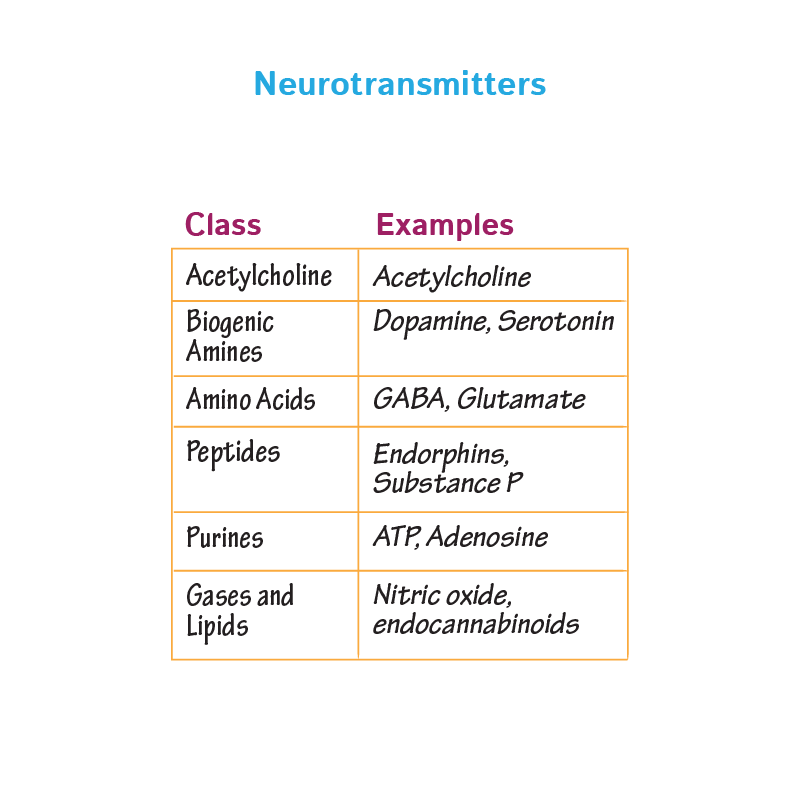
major classes of neurotransmitters
biogenic amines
amino acids
neuropeptides
dissolves gases
more varieties
biogenic amines (neurotransmitters)
norepinephrine (also known as noradrenaline)
brain + autonomic nervous system neurotransmitter
released at adrenergic synapses
typically has an excitatory, depolarising effect
dopamine
CNS neurotransmitter
inhibitory & excitatory effects
inhibitory: prevent overstimulation of neurons that control skeletal muscle tone
excitatory: stimulant drug cocaine inhibits removal of dopamine from synapses in the brain, responsible for the “high” experienced
serotonin
CNS neurotransmitter
inhibitory & excitatory effects
related to attention and emotional states
antidepressant drugs inhibit reabsorption of serotonin by axon terminals, increased serotonin concentrations at synapses, may relieve symptoms of depression

amino acid (neurotransmitters)
gamma aminobutyric acid (GABA)
CNS neurotransmitter
generally inhibitory
its function is not fully understood
GABA release appears to reduce anxiety

neuromodulators
compounds that alter the rate of neurotransmitter release by the presynaptic neuron or change the postsynaptic cell’s response to neurotransmitters

neuropeptides
(a subclass of neuromodulators)
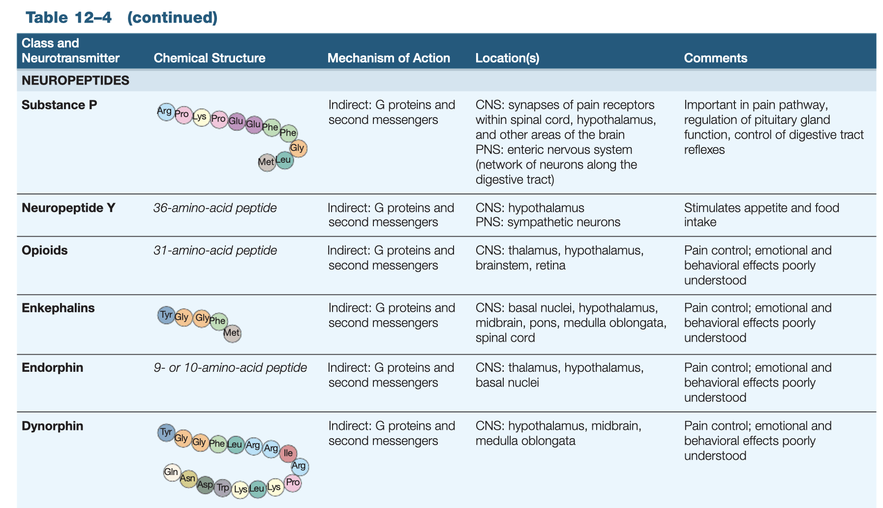
small peptide chains synthesised and released by the axon terminal
how do neuromodulators act to alter neuronal activity
either:
bind to receptors in presynaptic
or postsynaptic membrane
and:
activate cytoplasmic enzymes
in general:
have a long-term effect that’s relatively slow to appear
triggers responses that involve several steps and intermediary compounds
may affect presynaptic membrane, postsynaptic membrane or both!
can be released alone or along with neurotransmitter
opioids
a type of neuromodulator
bind to the same group of postsynaptic receptors as drugs like opium and morphine
three main classes:
1) enkephalins
2) endorphins
3) dynorphins
primary function:
probably to relieve pain
mechanism:
inhibit the release of the neurotransmitter substance P at synapses that relay pain sensations
dissolved gases (neurotransmitters)
nitric oxide (NO)
peripheral nervous system (PNS) neurotransmitter
generated by axon terminals that innervate smooth muscle in the walls of blood vessels in PNS
carbon monoxide (CO)
brain neurotransmitter
generated by specialised axon terminals

functionally, neurotransmitters and neuromodulators fall into one of three groups:
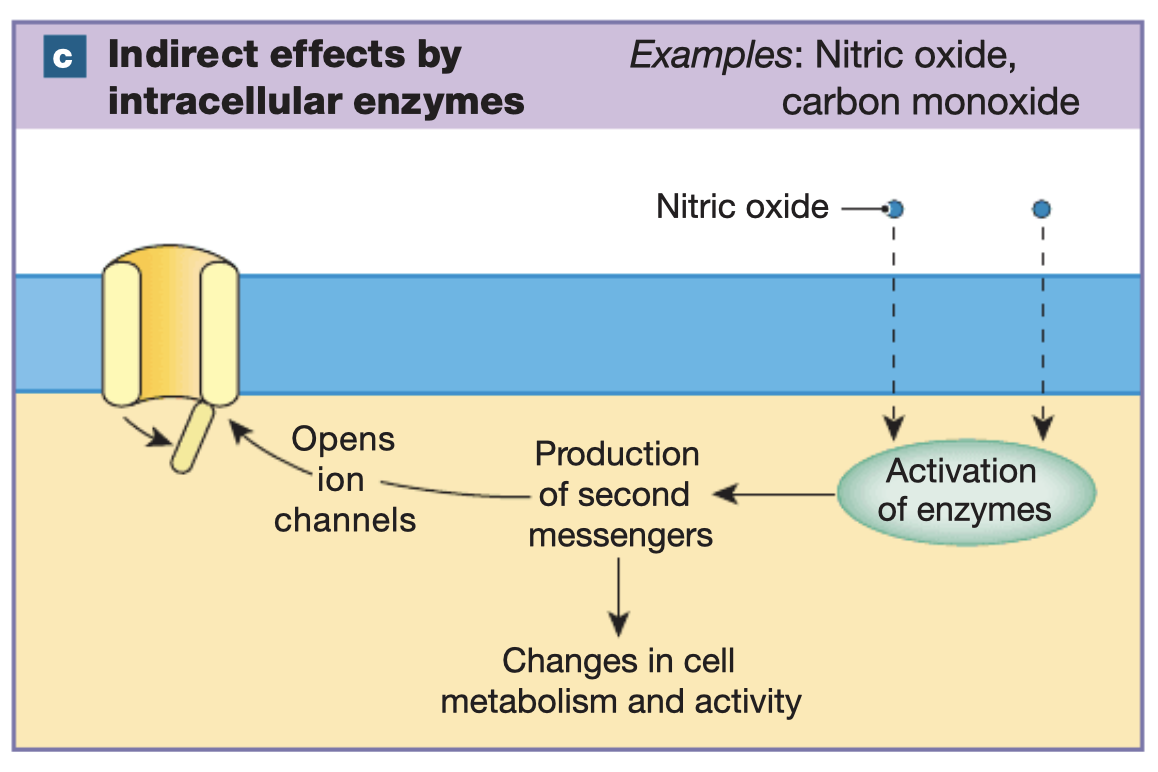
1) compounds that have a direct effect on membrane potential
2) compounds that have an indirect effect on membrane potential through G proteins
3) lipid-soluble gases that have an indirect effect on membrane potential through intracellular enzymes
explain function 1
compounds that open or close chemically gated ion channels receptors on postsynaptic membrane
e.g ACh, glutamate and aspartate
these neurotransmitters alter ion-movement across the postsynaptic membrane (ionotropic effect)
a few neurotransmitters (e.g. GABA, norepinephrine & serotonin) have both direct and indirect effects because these compound target two different classes of receptors

functionally, neurotransmitters and neuromodulators fall into one of three groups:
1) compounds that have a direct effect on membrane potential
2) compounds that have an indirect effect on membrane potential through G proteins
3) lipid-soluble gases that have an indirect effect on membrane potential through intracellular enzymes
explain function 2
the neurotransmitter represents a first messenger
the first messenger delivers a message to receptors on the plasma membrane (or within the cell)
second messengers are ions or molecules produced/released inside the cell when a first messenger binds to the receptor
many neurotransmitters bind to receptors in the plasma membrane called G protein-coupled receptors (GPCRs)
the G protein is an enzyme complex coupled to a membrane receptor
several types of G proteins exist (each type includes an enzyme that is “turned on” when an extracellular compound binds to it associated receptor)
FIGURE:
1) neurotransmitter (first messenger) binds to receptor
2) enzyme adenylate cyclase is activated
3) adenylate cyclase converts ATP to cyclic AMP (second messenger)
4) cyclic AMP may open membrane ion channels / activate intracellular enzymes / both

unctionally, neurotransmitters and neuromodulators fall into one of three groups:
1) compounds that have a direct effect on membrane potential
2) compounds that have an indirect effect on membrane potential through G proteins
3) lipid-soluble gases that have an indirect effect on membrane potential through intracellular enzymes
explain function 3
two lipid-soluble gases: nitric oxide (NO) + carbon monoxide (CO)
function as neurotransmitters in specific region of the brain
these gases diffuse through lipid membranes
bind to intracellular enzymes on the inner surface of plasma membrane / elsewhere in cytoplasm
these enzymes promote the appearance of second messengers that can affect cellular activity (e.g. open ion channel)