3.1.6 Uses both a non-contact and contact tonometer to measure intra-ocular pressure and analyses and interprets the results.
 Call Kai
 Learn
 Practice Test
 Spaced Repetition
 Match
 Flashcards
 Knowt Play
1/16

There's no tags or description
Looks like no tags are added yet.
Name | Mastery | Learn | Test | Matching | Spaced | Call with Kai | Chat |
|---|
No analytics yet
Send a link to your students to track their progress
17 Terms
Currently, NICE guidelines [NG81], last updated on 26th January 2022, states…
that an IOP measurement using Goldmann-type applanation tonometry must be recorded before referring patients for chronic open angle glaucoma.
Principles of GAT
The Imbert-Fick principle
“A small force applied to a sphere will exert a force back related to the internal pressure of the sphere.”
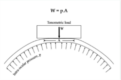
The Imbert-Fick principle relies on several assumptions regarding the properties of the eye:
1. The cornea is infinitely thin and flexible
2. The tear film has no pulling effect on the probe
3. Changes in the glove volume and IOP from contact with the probe are negligible
4. All anterior eye structures have the sae uniform elastic properties
In 1957 Goldmann concluded that …
A probe tip of 3.06mm in diameter allowed for 1g of pressure to equal 10mmHg.
This sized probe was chosen so the surface tension attraction from the tear film is equal and opposite to the corneal rigidity force.
Limitations of GAT
No account for CCT
Thin corneas underestimate IOP
Thick corneas overestimate IOP
Requires local anaesthetic 1mmHg error for every 4D of corneal astigmatism.
Incorrect NaFL conc. affects results
Interpreting IOP values
The normal range is between 10-21mmHg and varies diurnally.
NICE guidance requires IOP >24mmHg to be referred for specialist review.
IOP >21mmHg on two occasions using contact tonometry can be classified as ocular hypertension.
IOP needs testing at different times and is often higher in the morning than later in the day.
Suspicious IOPs are values about 25mmHg in the absence of any other ocular signs, and/or a difference of >4mmHg between the eyes.
GAT
Patient Records:
Record what type of contact tonometry you used. Drug information!
Example:
1% oxybuprocaine, 1 drop per eye, inserted at 10:55am BN H8978, Expiry 05/2027
• You need to check any staining before and after applanation.
• Check to make sure the patient hasn’t had any adverse reactions to drops previously
• Note that you have explained to the patient that the “Drops last for 30 minutes, and you are advised not to rub your eyes in case they get scratched.”
• Important to show you have given the patient an information leaflet about the drops
“Why do we take 3 readings for NCT and 1 reading for GAT?”
GAT takes into account the ocular pulse which is why you see the mires pulsating at you so the one measurement you get is already an average reading.
With NCT it doesn’t take into account the ocular pulse so you have to take 3-4 readings to get an average.
Performing GAT
Procedure
Preparation
Calibration
Patient explanation & consent
Anaesthetic & Fluorescein Instillation
Slit lamp set up
Applanation
Repeatability
Aftercare
Performing GAT
Preparation
• Wash hands and sanitise station
• Insert disposable probe onto Goldmann tonometer, aligning the biprisms horizontally
• Check tonometer calibration (often done at start of clinic)
Performing GAT
Calibration
• With no calibration bar attached, set dial at 0mmHg
The arm should just oscillate freely either side of zero
• Attach calibration bar at “20” notch
With the dial at 20mmHg, the arm should balance evenly, falling equally either side
• Attach calibration bar at “60” notch
Set dial to 60mmHg and repeat same test
Interpretation
Acceptable error margin is usually:
±0.5 mmHg at 0
±1 mmHg at 20
±2 mmHg at 60
If outside this, the tonometer must be serviced before use.
GAT
Patient explanation & consent
Explain procedure to patient
“We’ll measure the pressure inside your eye using a blue light and a gentle instrument. It may feel slightly cold or uncomfortable, but it won’t hurt. We need to put a small drop of local anaesthetic in both your eyes, this will stingy briefly for a couple seconds and will last for 30 minutes, its important you don’t rub your eyes for 30 minutes so you don’t damage you eyes.”
GAT
Anaesthetic & Fluorescein Instillation
• Instil topical anaesthetic (oxybuprocaine, proxymetacaine, lidocaine)
• Instil fluorescein
GAT
Slit lamp set up
Sit patient at slit lamp and the eye level marker is perfectly aligned.
Adjust illumination
Cobalt blue filter
Moderate beam width
45-60º angle to tonometer arm
Magnification 10x is sufficient to view mires clearly
Set tonometer dial to around 10mmHg
Ask pt to look straight ahead and keep both eyes open
GAT
Applanation
Pull the joystick of the slit-lamp back and slowly move the entire slit-lamp toward the cornea
When the probe biprisms are approximately 3-5mm away from the cornea, look through eyepieces and gently push the joystick to bring the biprisms into contact with the cornea
Observe the fluorescein mires
Adjust the dial until the inner edges of the two mires just touch (overlap by one thickness of the line)
Read the scale (dial x10 = IOP in mmHg)

GAT
Repeatability
Take at least two readings, especially if values differ
GAT
Aftercare
• Tell patient vision may be slightly blurred from anaesthetic and fluorescein
• Check staining from probe contact
• Reaffirm how the anaesthetic lasts 30 minutes and patients must not rub their eye for this duration.