EKG Strips - HAVE TO LOOK AT
 Call Kai
 Learn
 Practice Test
 Spaced Repetition
 Match
 Flashcards
 Knowt Play
1/34

There's no tags or description
Looks like no tags are added yet.
35 Terms
Atrial Flutter -has a "saw tooth or picket fence" atrial pattern (no P waves, F-waves in stead that may be in a ratio) between the QRS complexes.

Atrial Fibrillation-Has "chaotic" atrial electrical activity (no P waves, f-waves instead) with irregular R-R intervals.

PJCs cause the rhythm to be irregular. The P wave is inverted if it can be seen. It may occur before, during or after the QRS. The P wave will not be seen due to its low voltage.

Junctional Rhythm may have an inverted or absent P wave. The P wave may occur before, during or after the QRS complex. The rate for this rhythm is 40-60 bpm.

Accelerated Junctional Rhythm may have an inverted or absent P wave. The P wave may occur before, during or after the QRS complex. The rate for this rhythm is 60-100 bpm.

Junctional Tachycardia Rhythm may have an inverted or absent P wave. The P wave may occur before, during or after the QRS complex. The rate for this rhythm is 100-180 bpm

Supraventricular Tachycardia presents with a "normal-narrow" appearing QRS complex and a rate of greater than 150 bpm. Remember for heart rates approaching 150 or higher, it will be very likely that the P wave will be buried. Be prepared to increase the paper speed for this rhythm.

First Degree AV Block, the PR interval is constant and measures greater than 0.20 second.

Second Degree AV Blocok Mobitz I (Wenkebach) has a cyclical prolonging PR interval until the QRS is dropped. Then the cycle begins again. ***Remember the clue "lengthen, lengthen drop Wenkebach.

Second Degree (AV) Block has a constant PR interval with blocked QRS complexes

Second Degree (AV) Block has a constant PR interval with blocked QRS complex (2)

Third Degree (Complete) Heart Block, the P-P and R-R intervals are regular (constant) but firing at different rates.

T wave inversion, or negatively deflected T wave indicates ischemia and is usually seen with ST segment elevation.

T wave inversion with ST elevation on a 12-lead ECG is considered myocardial injury, acute injury pattern, or acute

Physiologic Q wave is normal and the width measures less than 0.04 second and the depth measures less than one-third of the height of the R wave in that lead.

Pathologic Q wave indicates tissue death (infarction) and is defined as measuring 0.04 second and/or greater than or equal to one third the height of the R wave in that lead tracing.
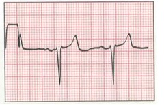
Ventricular Tachycardia has wide and bizarre QRS complexes with a classic "sawtooth" appearance, a rate in excess of 100 bpm, with no P wave.

Ventricular fibrillation is the absence of organized electrical activity. There are no P waves, QRS complexes or T waves. The tracing has a chaotic or disorganized appearance.

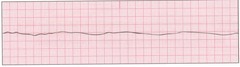
Asystole is absence of ventricular activity and depolarization. Often this is called "the straight flat line" of rhythms. No electrical activity is present. This rhythm is neither regular or irregular. It is simply absent!
Atrial Pacemaker Rhythm is identified by the presence of a pacing spike immediately prior to the atrial depolarization (P wave)

Ventricular Pacemaker Rhythm is identified by the presence of a pacing spike immediately prior to the ventricular depolarization (QS complex)

Atrioventricular Pacemaker Rhythm is identified by the presence of a pacing spike immediately prior to the atrial depolarization (P wave) and the ventricular depolarization (QS complex)

A rhythm containing Bundle Branch Block will retain its own usual features with the only change being the QRS complex now measure 0.12 second or greater

An ST segment below the isoelectric line is known as ST segment depression and may indicate myocardial ischemia

A PVC is an early QRS complex that is wide (0.12 second or greater) and has a bizarre appearance. There is no P wave. PVC's may come in different shapes - describe them appropriately. remember to identify the underlying rhythm when providing the interpretation.

Agonal Rhythm has an absence of P waves, a ventricular rate of less than 20 bpm and wide-bizarre QRS complexes

Idioventricular Rhythm has an absence of P waves, slow ventricular rate of 20 to 40 bpm, and wide-bizarre QRS complexes

Accelerated Idioventricular Rhythm has an absence of P waves, a ventricular rate of 40 to 100 bpm, and wide-bizarre QRS complexes.

Sinus Dysrhythmia, the P-P and R-R intervals will progressively widen then narrow following the patient's breathing pattern.

Sinus Arrest has regularly occurring PQRST's both before and after the arrest period. No electrical activity during the arrest period. Remember to report frequency and duration of Sinus Arrest!

A Premature Atrial Complex (PAC) is a complex that occurs sooner than it should with a positively deflected P wave. Remember to analyze and report the underlying rhythm along with the PAC.

Wandering Atrial Pacemaker (WAP) has at least three different shaped P waves. the rhythm may be regular or irregular. Rate is typically between 60-100 bpm

Sinus Rhythm is the only rhythm for which all five steps are within normal limits.

Sinus Bradycardia, the heart rate is less than 60 and all other measurements are within normal limits.

Sinus Tachycardia, the heart rate is greater than 100 and all other measurements are within normal limits.
