Anatomy Exam 4
 Call Kai
 Learn
 Practice Test
 Spaced Repetition
 Match
 Flashcards
 Knowt Play
1/194

There's no tags or description
Looks like no tags are added yet.
195 Terms
What are the two main circulatory systems?
Pulmonary circulation: Right side of the heart pumps blood to the lungs for oxygenation.
Systemic circulation: Left side of the heart pumps oxygenated blood throughout the body.
What is a portal system, and what are the types?
Portal system: Blood vessels connecting two capillary beds.
Venous: Hepatic portal system and hypophyseal portal system.
Arterial: Efferent arteriole in the kidney.
What is the azygos vein?
drains deoxygenated blood from the thoracic wall and parts of the abdomen into the SVC, while also providing an alternate route for blood to return to the heart
Provides a potential connection between the superior and inferior vena cava creating an anastomotic route.
What is an anastomosis, and how does it relate to the azygos vein?
Allows blood to take an alternate route back to the heart if normal venous flow is blocked (azygos vein is an example of that)
What is cardiac tamponade?
Accumulation of fluid or blood in the pericardial cavity that compresses the heart, preventing proper ventricular filling during diastole and decreasing cardiac output.
What is jugular venous distention (JVD), and what does it indicate?
Visible distention of the jugular veins due to elevated venous pressure.
Can indicate impaired venous return or right-sided cardiac filling, such as in cardiac tamponade.
What is the mediastinum, and what are its main divisions?
Central compartment of the thoracic cavity between the lungs.
Superior: Trachea and great vessels.
Middle: Heart.
Posterior: Esophagus and descending aorta.

Where is the heart located?
In the middle mediastinum, approximately between the sternal angle and 5th intercostal space.
About 2/3 of the heart is left of the midline.

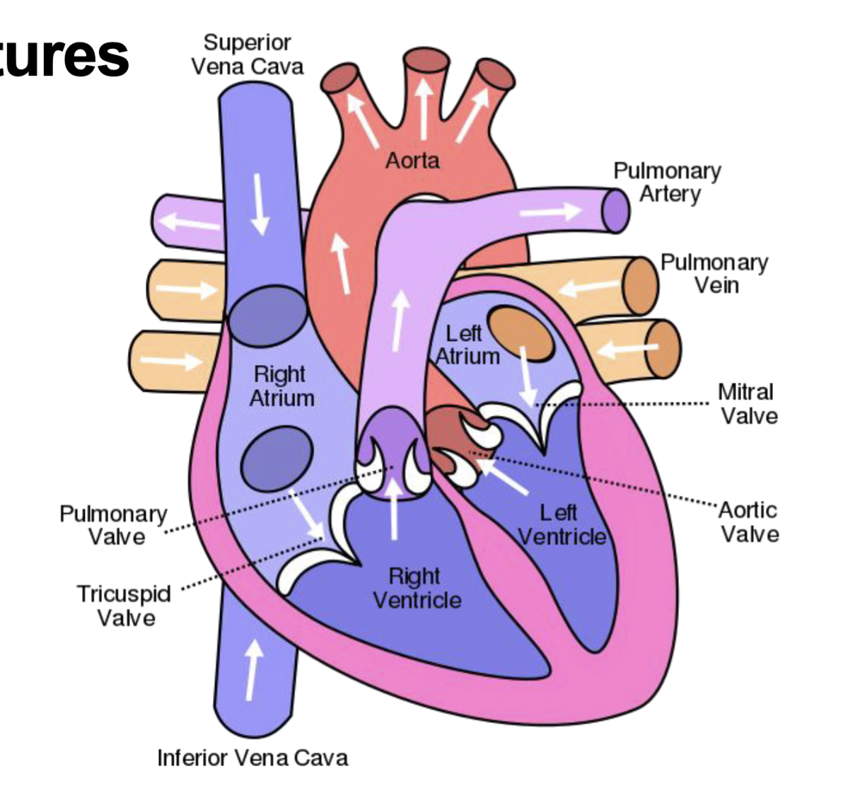
What are the four chambers of the heart and their functions?
Right atrium: Receives deoxygenated blood from the body.
Right ventricle: Pumps blood to the lungs.
Left atrium: Receives oxygenated blood from the lungs.
Left ventricle: Pumps blood to the body.
What is the difference between systole and diastole?
Systole is contraction of the heart, pumping blood out of the ventricles
Diastole is relaxation of the heart, allowing the ventricles to fill with blood
What three vessels empty into the right atrium?
Superior vena cava
Inferior vena cava
Coronary sinus
What are the major structures of the right ventricle?
Tricuspid valve: Prevents backflow into right atrium.
Chordae tendineae: Connect valve cusps to papillary muscles.
Papillary muscles: Prevent valve prolapse.
Pulmonary semilunar valve: Prevents backflow from pulmonary trunk.

What are the major internal features of the right atrium?
Auricle
Crista terminalis: A muscular ridge in the right atrium that separates its smooth posterior wall from its rough anterior wall. sinoatrial nerve located here
Musculi pectinati
Fossa ovalis
Tricuspid valve

What do the three cusps of the tricuspid valve do during circulation?
During diastole: The cusps open, allowing blood to flow from the right atrium → right ventricle.
During systole: The cusps close, preventing blood from flowing backward into the right atrium.
The cusps are attached to chordae tendineae, which connect to papillary muscles. These prevent the cusps from prolapsing into the atrium during ventricular contraction.
Why is the left ventricular wall thicker than the right?
Left ventricle pumps blood through systemic circulation, requiring higher pressure.
Right ventricle pumps blood through lower-pressure pulmonary circulation.
What is the interventricular septum?
Muscular wall separating the right and left ventricles.
What are the four heart valves and their locations?
Valve | Location |
|---|---|
Tricuspid | Right atrium → Right ventricle |
Pulmonary semilunar | Right ventricle → Pulmonary trunk |
Mitral (bicuspid) | Left atrium → Left ventricle |
Aortic semilunar | Left ventricle → Aorta |

What is the difference between AV valves and semilunar valves?
AV valves (filling): Tricuspid and mitral; prevent backflow into atria.
Semilunar valves (ejecting): Pulmonary and aortic; prevent backflow into ventricles.
All valves open and close in response to pressure differences
LUB = AV valves close. DUB = Semilunar valves close
What are the first arteries that the aorta supplies?
coronary arteries
What are the three branches of the aortic arch, and what do they supply?
Brachiocephalic trunk: Right head/neck and right upper limb through its branches
Left common carotid artery: Left head and neck
Left subclavian artery: Left upper limb

What is the difference between coronary arteries and the coronary sinus?
Coronary arteries deliver oxygenated blood from the aorta to the myocardium, while the coronary sinus collects deoxygenated blood from cardiac veins and drains into the right atrium.
What is the intrinsic conduction system of the heart?
Specialized (modified) cardiac muscle cells that generate and conduct electrical impulses.
Allows rhythmic contractions without direct nervous system stimulation.
Where is the pace of the heart initiated at?
Sino-Atrial node (SA node) located at junction of superior vena cava and right atrium
What is the function of the AV node and Bundle of His?
AV node: Receives the impulse from the SA node and delays its transmission to the ventricles.
Bundle of His: Conducts impulses toward the ventricles through right and left bundle branches.
What is the pathway of electrical conduction through the heart?
SA node → AV node → AV bundle (Bundle of His) → Right and left bundle branches → Purkinje fibers.

What are Purkinje fibers, and where are they located?
Specialized cardiac muscle fibers that rapidly conduct electrical impulses through the ventricles.
Located in the subendocardial layer of the heart wall.
What causes the waves seen on an electrocardiogram (ECG)?
Depolarization and repolarization of cardiac tissue associated with electrical conduction
The SA node starts the electrical impulse, depolarization activates cardiac cells, and repolarization resets those cells for the next heartbeat
How does the autonomic nervous system regulate the heart?
Sympathetic: Increases heart rate and contractility.
Parasympathetic (CN X, vagus nerve): Decreases heart rate
What is the difference between baroreceptors and chemoreceptors?
Baroreceptors: Detect changes in blood pressure or tension
Chemoreceptors: Detect changes in blood oxygen and carbon dioxide levels
Both send sensory information to the brain to help regulate cardiovascular and respiratory functions
CN X (Vagus) and CN IX (Glossopharyngeal) carry signals from different parts of the body but hopefully not imp for exam
What are the three layers of the heart wall?
Epicardium: Outer layer of mesothelium and connective tissue.
Myocardium: Middle layer of cardiac muscle.
Endocardium: Inner endothelial and connective tissue lining.
What is the subendocardial layer?
Deeper connective tissue beneath the endocardium.
Contains components of the cardiac conduction system, including Purkinje fibers.
What is the pericardial sac?
Protective sac surrounding the heart.
Consists of an outer fibrous pericardium and inner serous pericardium
different from pleural sac

What is the difference between visceral and parietal pericardium?
Visceral pericardium: Directly covers the heart; also called the epicardium.
Parietal pericardium: Lines the inside of the fibrous pericardium.
Pericardial fluid between the layers reduces friction.
both serous pericardium

What is the difference between arteries and veins?
Arteries: Carry blood away from the heart.
Veins: Carry blood toward the heart.
Arteries and veins often travel together within the same connective tissue sheath
Arteries and veins are classified by the direction blood travels relative to the heart, NOT by oxygen content
What are the functions of arterioles, capillaries, and postcapillary venules?
Arterioles: Regulate blood flow into capillary networks.
Capillaries: Allow exchange of substances between blood and tissues.
Postcapillary venules: Return fluid to circulation and allow WBCs to migrate into tissues.
Normal circulation v portal circulation
Normal: Artery → Arteriole → Capillaries → Venule → Vein → Heart
Portal: Artery → Arteriole → Capillaries → Portal vein → More capillaries → Veins → Heart
What are the three layers (tunics) of blood vessels?
Tunica intima (interna): Innermost layer containing endothelium.
Tunica media: Middle layer containing smooth muscle.
Tunica adventitia (externa): Outer connective tissue layer.

What are the major functions of endothelial cells?
Maintain selective permeability and prevent unwanted clotting.
Regulate blood flow, vascular resistance, and immune responses.
Regulate cell growth, extracellular matrix, and lipoproteins.
What type of epithelium lines blood vessels?
Simple squamous epithelium (endothelium).
All blood vessels have endothelium.
Capillaries contain only an endothelium
What is the composition of each vascular tunic (different layers of tissue that form the wall of blood vessels)?
Intima: Endothelium, loose connective tissue, and elastic lamina.
Media: Smooth muscle, elastic fibers, type III collagen, proteoglycans, and glycoproteins.
Adventitia: Type I collagen and elastic fibers.
What is the vasa vasorum?
Small blood vessels that supply the walls of large arteries and veins.
Necessary because diffusion from the lumen (Hollow space inside a blood vessel where blood flows) cannot nourish the entire vessel wall.
How do arteries and veins differ structurally?
Arteries: Thicker tunica media, more smooth muscle and elastic tissue, generally smaller and rounder lumen (need to withstand high pressure of being pushed out to the body by the heart which is why its thicker)
Veins: Thinner tunica media, relatively thicker adventitia, larger and often irregular lumen.
What are the two major types of arteries?
Elastic arteries: Large arteries such as the aorta and pulmonary trunk; stretch and recoil with pressure changes.
Muscular arteries: Distribute blood to organs and tissues; contain more smooth muscle.
What is the difference between elastic and muscular arteries?
Elastic: Expand during systole and recoil during diastole to maintain blood flow.
Muscular: Use smooth muscle to regulate blood distribution to organs.
Muscular arteries are common sites of atherosclerotic lesions (a localized buildup of fats, cholesterol, calcium, and other substances within the inner lining of an artery)
What are arterioles, and why are they important?
Small arteries containing approximately 1–2 layers of smooth muscle.
Regulate blood flow into capillary beds.
Provide the greatest resistance to blood flow in circulation.
How does blood pressure change throughout the circulatory system?
Highest in large arteries.
Drops significantly across arterioles because of their high resistance.
Continues decreasing through capillaries, venules, and veins.
What are the major characteristics of capillaries?
Approximately 5–10 μm in diameter.
Composed of simple squamous endothelium and a basement membrane.
Slow blood flow allows exchange between blood and tissues.
What are the three types of capillaries and their locations?
Type | Characteristics / Location |
|---|---|
Continuous | Most common; continuous endothelial lining |
Fenestrated | Pores in endothelium; endocrine organs and kidneys |
Sinusoidal | Large gaps; liver, spleen, and bone marrow |

What are pericytes and pinocytotic vesicles?
Pericytes: Cells surrounding capillaries that provide support and participate in vessel repair (can continue to divide)
Pinocytotic vesicles: Transport larger molecules across endothelial cells in continuous capillaries.
How is blood flow regulated through capillary beds?
Arterioles regulate how much blood enters the capillary network.
Precapillary sphincters, where present, regulate entry into individual capillaries.
What are precapillary sphincters?
Rings of smooth muscle that regulate blood flow into true capillaries.
What happens when precapillary sphincters relax and contract?
Relax: Blood flows into the true capillaries, allowing oxygen, nutrient, and waste exchange with surrounding tissues.
Contract: Blood bypasses much of the capillary bed through the metarteriole and thoroughfare channel, reducing exchange with tissues.

What are the functions of venules?
Collect blood from capillaries.
Postcapillary venules allow white blood cells to migrate from the bloodstream into tissues (mostly in postcapillary venules)
What is the difference between superficial and deep veins?
Superficial veins are located in subcutaneous tissue and generally do not accompany arteries. Deep veins are located beneath deep fascia and often accompany arteries.
What is the pathway of oxygenated blood from the placenta to the fetal heart?
Placenta → Umbilical vein → Ductus venosus → Inferior vena cava → Right atrium.
Some blood also enters the liver before returning to the inferior vena cava.

How does fetal blood bypass the lungs?
Foramen ovale: Right atrium → Left atrium.
Ductus arteriosus: Pulmonary trunk → Aorta.
These shunts allow most blood to bypass the fetal lungs.
What are the three major fetal circulatory shunts?
Ductus venosus: Bypasses much of the liver.
Foramen ovale: Connects right atrium to left atrium.
Ductus arteriosus: Connects pulmonary trunk to aorta, bypassing the lungs.
What happens to the umbilical arteries after birth?
Distal portions: Become medial umbilical ligaments.
Proximal portions: Remain patent and contribute to the superior vesical arteries.

Why does fetal circulation change after birth?
Newborn begins breathing, increasing pulmonary blood flow.
Pressure changes promote closure of the foramen ovale.
Increased oxygen promotes closure of the ductus arteriosus.
Placental circulation ends.
What is the function of the lymphatic system?
Monitors the body's surface and interior for potentially harmful substances.
Initiates immune responses against harmful substances.
Consists of immune cells, lymphatic tissues, organs, and vessels.
Where does lymph originate, where does it drain, and why is this clinically important?
Lymph originates as excess interstitial fluid in connective tissue spaces. It enters lymphatic vessels, passes through lymph nodes, and eventually returns to the bloodstream through the right lymphatic duct and thoracic duct. This is clinically important because cancer cells can travel through lymphatic vessels, enter the bloodstream, and spread to other parts of the body (metastasis)
What are the two major lymphatic ducts, and where do they drain?
Right lymphatic duct: Drains lymph from the right upper portion of the body.
Thoracic duct: Drains lymph from most of the rest of the body.
Both return lymph to the venous circulation near the junctions of the internal jugular and subclavian veins, where the brachiocephalic veins form.
How does lymph move through lymphatic vessels if there is no central pump?
Lymph moves slowly under low pressure through skeletal muscle contractions, respiratory pressure changes, and smooth muscle contractions in lymphatic vessels. One-way valves prevent backflow.
What are the major cells found in lymphatic tissue?
Lymphocytes
Plasma cells
Mast cells
Neutrophils
Eosinophils
Phagocytes
All originate from hematopoietic cells in red bone marrow.
What are antigen-presenting cells (APCs)?
Cells that capture antigens and present them to helper T cells.
Include B lymphocytes, macrophages, dendritic cells, and Langerhans cells (specialized type of dendritic cell found in the skin)
What is the difference between an antigen and an antibody?
Antigen: Molecule recognized by immune cells that can trigger a specific immune response (because it shouldnt be there).
Antibody: Protein that binds to an antigen and helps mark invaders for destruction.
IgG: Most common antibody class.

What is the complement system?
A series of proteins activated in a cascading reaction.
Helps the immune system eliminate foreign substances (compliments the work of the antibody)
What are B-cell lymphocytes and their function?
Develop in bone marrow and recognize specific antigens (B stands for bone marrow)
When activated, differentiate into plasma cells that produce antibodies
can also act as APCs by presenting antigens to helper T cells using MHC II
What are natural killer (NK) cells?
Immune cells that attack virus-infected cells, cancer cells, and some transplanted cells
What are the three types of T lymphocytes and their functions?
T stands for thymus. In the thymus it goes through a selection process and become either a helper or cytotoxic t-cell
Helper T cells: Coordinate immune responses and activate other immune cells.
Cytotoxic T cells: Kill infected or abnormal cells.
Regulatory T cells: Help regulate and suppress excessive immune responses.
How do antigen-presenting cells activate an immune response?
Capture antigens and present them to helper T cells.
This interaction helps initiate a coordinated immune response.
What is the difference between innate and adaptive immunity?
Innate: Fast, nonspecific response involving complement proteins, neutrophils, macrophages, mast cells, and NK cells.
Adaptive: Slower initially, antigen-specific, involves B and T cells, and produces memory cells (basis for immunization)
What are the two types of adaptive immune responses?
Humoral immunity: Activated B cells become plasma cells that produce antibodies.
Cell-mediated immunity: T cells coordinate immune responses and attack infected or foreign cells.
What is the function of cytokines in adaptive immunity? (different from cytotoxic T cells)
Signaling molecules that activate and coordinate B cells, T cells, and inflammatory cells (instead of a single police officer you call in an army)
What are naïve B and T lymphocytes?
Lymphocytes that have not yet encountered their specific foreign antigen.
B cells mature in bone marrow; T cells undergo maturation and selection in the thymus.
What is the major histocompatibility complex (MHC)?
Cell-surface proteins involved in presenting antigens to T cells.
MHC genes vary between individuals, making them important in tissue transplantation and graft rejection.
What is the difference between MHC I and MHC II?
MHC I | MHC II |
|---|---|
Found on all nucleated cells | Found on antigen-presenting cells, including B cells and macrophages |
Recognized by CD8 cytotoxic T cells | Recognized by CD4 helper T cells |
Involved in identifying infected or abnormal cells | Involved in activating helper T cells |
What is the difference between CD4 and CD8 T cells?
CD4 (helper T cells): Bind to MHC II and help coordinate immune responses.
CD8 (cytotoxic T cells): Bind to MHC I and can kill infected or abnormal cells.
How does MHC II present antigens to CD4 helper T cells?
Antigen-presenting cells engulf and break down foreign material, attach antigen fragments to MHC II molecules, and display them on their surface. CD4 helper T cells use their T-cell receptors to recognize the antigen-MHC II complex.
What is T-cell selection, and where does it occur?
Occurs in the thymus.
Eliminates T cells that fail appropriate recognition tests or react strongly against self-antigens.
Only approximately 2% survive selection
What is co-stimulation in immune responses?
Additional signaling required for full activation of B or T lymphocytes.
Helps ensure immune cells respond appropriately to antigens.
How do helper T cells (CD4) become activated, and what do they do?
APCs present antigens using MHC II, which CD4 helper T cells recognize through their TCRs.
Activated helper T cells release cytokines (including IL-2) that help stimulate other immune cells.
Produce activated and memory helper T cells
How do cytotoxic T cells (CD8) become activated, and what do they do?
Recognize antigens presented on MHC I using their TCRs.
Signals including IL-2 from helper T cells help stimulate their proliferation.
Produce activated and memory cytotoxic T cells that can kill infected cells
What is lymphoid tissue composed of?
Loose connective tissue containing lymphocytes, other immune cells, and reticular fibers.
May be diffuse or organized into lymphoid nodules.
Nodules contain primarily B lymphocytes
What is the function of the thymus?
Site of T-cell maturation and selection.
B cells mature in red bone marrow.
What are the two regions of the thymus?
Cortex: Outer region containing many immature T lymphocytes.
Medulla: Inner region where thymocytes continue maturation and self-reactive T cells are eliminated.
What is mucosa-associated lymphoid tissue (MALT)?
immune tissue that protects areas of your body exposed to the outside environment, such as your digestive and respiratory tracts
Contains B lymphocytes, dendritic cells, APCs, and IgA-secreting plasma cells.
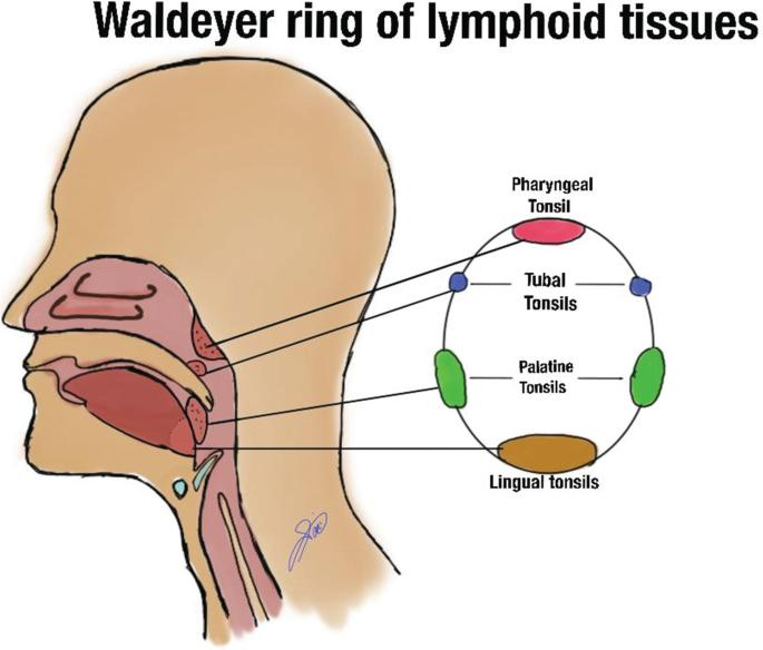
What are the four groups of tonsils?
Palatine tonsils
Lingual tonsils
Pharyngeal tonsil
Tubal tonsils
Together they form the tonsillar ring.
What are the major histological features of tonsils?
Lymphoid nodules (follicles) containing B lymphocytes.
Crypts, especially prominent in palatine tonsils.
Germinal centers where activated B cells proliferate.
What is the primary function of lymph nodes?
Filter lymph as it travels through lymphatic vessels.
Provide sites where immune cells encounter antigens.
What are the three main regions of a lymph node?
Cortex: Primarily B lymphocytes.
Paracortex: Primarily T lymphocytes.
Medulla: Inner region containing lymphatic sinuses and immune cells.
How does lymph travel through a lymph node, and where are B and T cells located?
Lymph enters through afferent lymphatic vessels, flows through sinuses in the cortex and medulla, and exits through efferent lymphatic vessels. B cells are primarily in the cortex, while T cells are primarily in the paracortex.
What are high endothelial venules (HEVs)?
Specialized postcapillary venules in lymph nodes.
Allow lymphocytes to move from the bloodstream into lymph nodes for immune surveillance.
What is blood, and what is it composed of?
Blood is a specialized connective tissue that makes up approximately 8% of body weight.
Contains cells suspended in a fluid extracellular matrix called plasma.
Plasma proteins participate in blood clot formation.
What are the major components of blood?
Plasma: Liquid extracellular matrix (contributes to the formation of lymph)
Erythrocytes (RBCs): Transport oxygen.
Buff Coat:
Leukocytes (WBCs): Immune defense
Platelets (thrombocytes): Blood clotting

What is hemopoiesis, and where does it occur?
Formation of blood cells in red bone marrow.
Pluripotent stem cells give rise to progenitor cells that differentiate into blood cells.
What are the two major blood cell lineages?
Myeloid lineage: Produces RBCs, platelets, and certain WBCs (granulocytes and monocytes) (what most cells become)
Lymphoid lineage: Produces lymphocytes (B cells, T cells, and NK cells), which are WBCs involved in immune defense.
What are the three major types of blood cell production?
Erythropoiesis: Production of RBCs (myeloid lineage).
Thrombopoiesis: Production of platelets (myeloid lineage).
Leukopoiesis: Production of WBCs (myeloid and lymphoid lineage).
How do newly formed blood cells enter circulation from bone marrow?
Red bone marrow contains sinusoidal capillaries.
Mature blood cells pass through the sinusoidal endothelium into the bloodstream
What are the major structural characteristics of erythrocytes?
Biconcave discs: Increase surface area and flexibility. (extra SA means maximum ability to exchange H20 with tissue)
Hemoglobin: Carries oxygen to cells
No nucleus: Terminally differentiated cells (means they can’t divide)
Lifespan: Approximately 120 days.