Superficial & Small Parts Ultrasound II- FINAL Exam Review
 Call Kai
 Learn
 Practice Test
 Spaced Repetition
 Match
 Flashcards
 Knowt Play
1/19

There's no tags or description
Looks like no tags are added yet.
20 Terms
An echo-free structure is described as:
heterogeneous.
sonolucent.
homogeneous.
echogenic.
b
Select the term that describes a diffuse disease process or metastatic disease.
Shadowing
Homogeneous
Infiltrating
Echogenic
c
Identify the shape of a curved array transducer image.
Annular
Octagon
Pie
Square
c
Doppler imaging demonstrates flow toward and away from the transducer. Identify the direction the spectral tracing displays with flow toward the transducer.
Below the baseline
Bidirectionally
Both sides of the baseline
Above the baseline
d
Which of the following transducer movements is the best to image the abdominal aorta to the bifurcation?
Fan
Slide
Angle
Rotate
b

What scanning plane is this taken in?
inferior
sagittal
superior
transverse
b

This following is the correct standard protocol to obtain transverse images of the right lobe of thyroid.
true
false
a
What is the best transducer to scan pediatric hip?
Curved array 2.5-5.0 MHZ
Phased sector 2.5 -5.0 MHZ
Endocavitary array 5.0-7.5MHZ
Linear array 3.5-7.5 MHZ
d
What artifact is present when the sound beam is attenuated by solid or calcified structure?
Posterior enhancement
Reverberation
Posterior shadowing
d
When a pathology is very close to the normal parenchyma echogenicity pattern it is called:
Anechoic
Hypoechoic
Isoechoic
Hyperechoic
c
During ultrasound examination, color Doppler is used for?
Determine presence or absence of blood flow within blood vessel
Determine direction of blood flow
All are correct
Differentiate blood vessel from nonvascular structure
c
When obtaining ultrasound images, an established protocol (such as established by the AIUM/ACR) should be utilized.
true
false
a
Without internal echoes; the structure is fluid filled and transmits sound easily, it would be:
hypoechoic
Isoechoic
Hyperechoic
Anechoic/sonolucent
d
Color doppler evaluation should be done on any pathology seen during a ultrasound exam.
True
False
a

What type of blood flow demonstrated by this Spectral Doppler?
Venous
arterial
Arterial and venous
Cannot be determined
b

What is the best description of the mass that is being measured?
homogenious
isoechoic
heterogeneous
anechoic
c
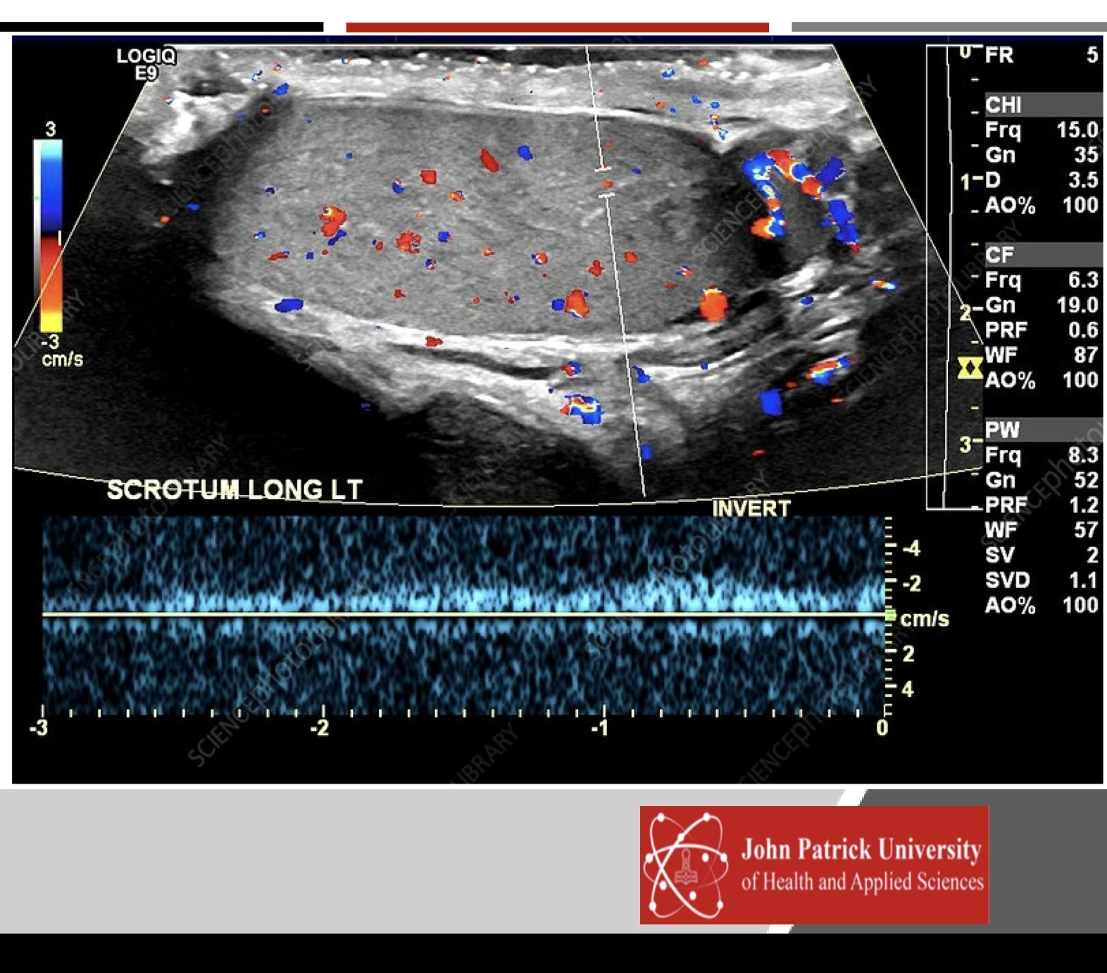
What is type of blood flow is demonstrated by Spectral Doppler?
arterial and venous
arterial
cannot be determined
venous
d
The beam is ideally reflected when the transducer is _______________
to the surface of the structure being imaged.
Diagonal
oblique
parallel
Perpendicular
d

How to describe structure that have been measured?
hyperechoic/echogenic
anechoic
hypoechoic
sonolucent
a

What blue arrow is pointing to?
Reverberation
Shadow
Isoechoic structure
enhancement
d