CVM 712 Lab 10
 Call Kai
 Learn
 Practice Test
 Spaced Repetition
 Match
 Flashcards
 Knowt Play
1/21

There's no tags or description
Looks like no tags are added yet.
22 Terms
Intestinal Mucosa
Simple columnar epithelium
Goblet cells
Increase in number aborally
Lamina propria – loose connective tissue that holds the epithelium to the muscularis mucosa
Muscularis mucosa – thin layer of smooth muscle under mucosa
Intestinal Submucosa
Collagenous connective tissue
Intestinal Muscularis externa
Smooth muscle
One layer circular and one layer longitudinal
Intestinal Serosa
Outer layer of connective tissue
Small Intestine
Villi – only in small intestine
Carnivores – long and slender
Ruminants – short and thick
Crypts – at the base of the villi
Progenitor cells to replace lost cells
Brunner’s glands
Duodenum submucosa
Mucous glands
Peyer’s patches
Ileum and other parts of small intestine
Lamina propria
Large intestine
Mucosa is flat (no villi)
Crypts are longer
Horses and pigs have longitudinal bands of smooth muscle and elastic fibers
Taeniacoli – bands in the colon
Taeniaceci – bands in the cecum
Rectum and Anal Canal
Stratified squamous epithelium
Nonkeratinized orally
Keratinized aborally
Carnivores and pigs have tubuloacinaranal glands
Apocrine glands in dogs
Apocrine and sebaceous glands in cats
In submucosa and muscularis of anal canal, near anal sphincter
Empty into anal sacs in carnivores
Keratinized stratified squamous epithelium
Dogs also have circumanal glands or “hepatoid” glands
Sebaceous and nonsebaceous
Look similar to hepatocytes
Liver
Outer capsule
Lobes are divided into lobules
Hepatocytes in cords
Sinusoids
Portal tracts or portal triad
Portal vein
Hepatic artery
Bile ductile
Lymphatic vessel
Central vein
Bile is secreted by hepatocytes
Travels through bile canaliculi to bile ducts and stored in gallbladder
Except horse, rat, etc.
Bile ducts have simple cuboidal to simple columnar epithelium
Portal triads
Some bile ducts have goblet cells
Kupffer Cells (Macrophages)
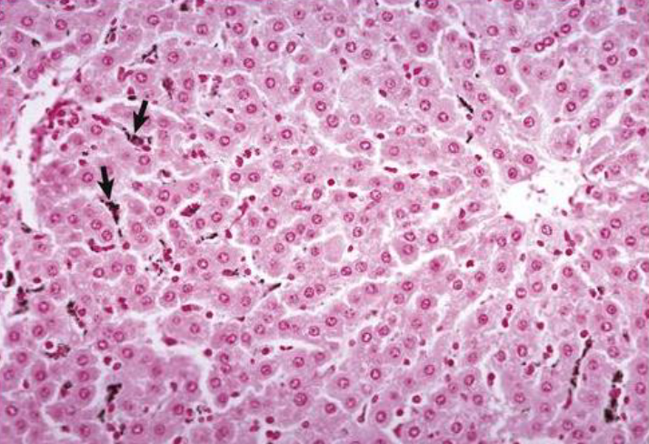
Gallbladder
Stores bile
Simple columnar epithelium
Cattle and goats have goblet cells too
Smooth muscle

Pancreas
Exocrine
Glands that secrete enzymes to help with digestion
Simple cuboidal
Drain to ducts
Simple cuboidal
Endocrine
Islets of Langerhans
Produce insulin
Pacinian corpuscles
Prominent in cats, sometimes in dogs
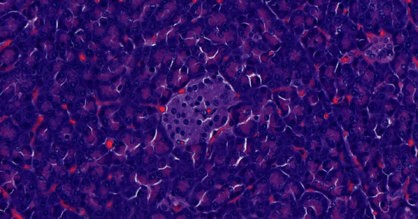
What is in the center of this image from the pancreas?
Islet of Langerhans
Pacinian corpuscle

Villi of duodenum
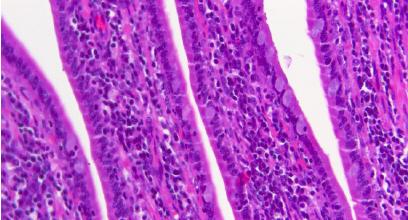
Microvilli of duodenum


What ring-like structure can be found in the submucosa in this picture of the duodenum?
Brunner’s glands (mucus producing)
Small intestine
Villi

Large intestine
Flat

Cecum
No villi, increased goblet cells, longer crypts
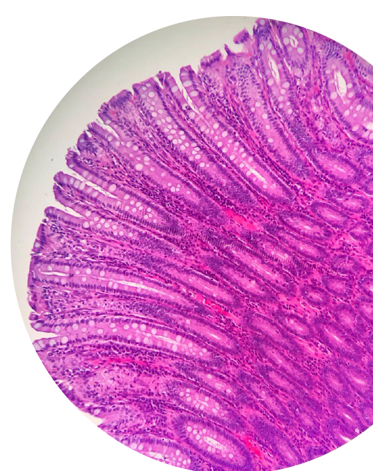
Central vein (Liver)

Portal triad (Liver)

Gallbladder
Much thinner wall compared to the other gastrointestinal tissues
Storage organ for bile.
Columnar epithelium in the mucosa, which is thrown into multiple folds, and a very thin muscular layer.
