immunology test 2
 Call Kai
 Learn
Learn Practice Test
 Spaced Repetition
 Match
Match Flashcards
 Knowt Play
1/215

There's no tags or description
Looks like no tags are added yet.
Name | Mastery | Learn | Test | Matching | Spaced | Call with Kai | Chat |
|---|
No analytics yet
Send a link to your students to track their progress
216 Terms
commensalism
neither organism is significantly helped or harmed
mutualism
both the host and microorganism benefit
parasitism
the microorganism benefits while harming the host
symbiosis
host and microorganism live together long term
normal biota DOES NOT = infection
infectivity
ability to establish an infection
can it infect?
pathogenicity
ability to cause disease
can it cause disease?
virulence
extent of damage/pathology produced
how severe is the damage?
knowledge check: a factor that helps a bacterium evade phagocytosis and cause more severe disease is a…
virulence factor
bacterial virulence factors
capsule - resists phagocytosis/ immune interactions
pili/fimbriae - attachment to host cells
flagella - motility; may aid adherence
endotoxin (LPS) - strong inflammatory cytokine release
exotoxins - secreted proteins that damage/ alter host cells
bacterial virulence factors - immunology connection
endotoxin → cytokine release
capsule → phagocytosis/complement
how does the immune system respond to EXTRACELLULAR BACTERIA?
antibodies
complement
phagocytosis
inflammatory cytokines
how does the immune system respond to INTRACELLULAR BACTERIA?
cell-mediated immunity
CD4+ T-cell cytokine responses
cytotoxic T cell responses
*type of immune response depends partly on where the organism is located
bacteria can evade host defenses
prevent effective chemotaxis
interfere with phagocyte attachment
resist destruction after phagocytosis
interfere with complement
degrade IgA
alter or mask surface antigens
*host response and laboratory findings may VARY during infection
ask FIRST before choosing laboratory detection method:
are we detecting the pathogen…
OR
are we detecting evidence of the host response?
what can the laboratory detect?
living organism → culture
morphology → microscopy/staining
bacterial antigen → immunologic antigen detection
DNA or RNA → molecular testing
patient antibody → serology
bacterial proteins → proteomic methods (gene sequence)
direct detection
“can i detect the PATHOGEN or one of its components?”
culture → organism
antigen assay → bacterial antigen
PCR → bacterial nucleic acid
serology
“has the patient produced ANTIBODIES against the organism?”
detects the HOST immune response
does NOT directly detect the bacterium
*Positive antibody DOES NOT equal direct detection of the organism
why detect antibody instead of the organism?
difficult to culture: some organisms are difficult to recover in routine culture
hazardous to culture: serology may provide safer indirect evidence
ORGANISM MAY BE GONE: complications can appear after the original infection has resolved
evidence of exposure: antibody can document a host immune response
*TRADEOFF: antibody production takes time
timing matters in serologic testing
exposure/infection → early infection (pathogen may be present/antibody undetectable) → immune response (antibody becomes detectable) → convalescence (antibody may persist)
serologic results must be interpreted in relation to TIME
early negative antibody result DOES NOT mean we rule out infection
IgM and IgG think pattern (not just pos/neg)
IgM: often appears earlier, may support RECENT exposure/infection
IgG: usually develops LATER, may persist AFTER INFECTION, may reflect PAST exposure or a LATER immune response
IgM = recent & IgG = past is NOT a universal rule
sometimes one specimen is not enough
acute specimen: collected EARLY in the illness
convalescent specimen: collected later, after the immune response has had time to develop
COMPARE ANTIBODY LEVELS - look for a significant change in atnibody titer (usually 4-fold)
what is a fourfold rise in titer?
Acute - 1:40 → 1:160 in convalescent
40 × 4 = 160 → fourfold rise
1:40 → 1:80 is NOT fourfold, instead is twofold
usually not considered significant
why can serology be misleading?
too early: antibody not yet detectable
cross reactivity: antibody reacts with related antigens
vaccination: may affect some antibody results
past exposure: antibody may persist
immunosuppression: weak or absent antibody response
single specimen: may not show when exposure occurred
*bottom line = interpret the result in context
what are febrile agglutinins?
PATIENT ANTIBODIES that react with standardized bacterial antigens
a reactive antigen—antibody rxn produces visible agglutination
why were/are febrile agglutinins used?
relatively rapid and simple serologic testing
historically useful when culture was difficult/slow/hazardous
still performed in some clinical laboratories
important limitations of febrile agglutinins
antibodies take time to develop
cross reactivity may cause false positives
a single reactive result does not prove active infection
more specific testing is preferred when available
common febrile agglutinin tests
Widal test for Salmonella
O (somatic) and H (flagellar) antigens used
detects antibodies reacting with salmonella antigens
Weil-Felix test for Rickettsial disease
Proteus antigens used: OX19, OX2, OXK
detects cross reacting antibodies associated w some rickettsial infections
Huddleston test for Brucella
Brucella antigen used
detects antibodies reacting with brucella
Francisella tularensis test
F. tularensis antigen used
detects antibodies reacting with F. tularensis
knowledge check: a patient is tested for antibodies to a bacterial pathogen very early in the illness. the antibody test is negative. why can this be?
the patient may not yet have produces detectable antibody
4 infections = 4 diagnostic problems
Group A Strep (GAS): the organism may be gone when immune mediated sequelae appear → detect antibody to a previous infection
Mycoplasma pneumoniae: specific antibody and nonspecific cold antibodies can coexist → know what the antibody is actually binding
Rocky Mountain Spotted Fever (RMSF): the patient may be very ill before antibodies become detectable → timing can make serology negative
Tuberculosis: antibody is not the useful immune marker → detect T cell sensitization/ IFN-gamma response
Group A streptococcus - PRIMARY infection
pharyngitis or impetigo/pyoderma
M protein
major virulence factor
inhibits phagocytosis
limits C3 deposition (innate immune)
lipoteichoic acid + Protein F
allow for attachment in skin or pharyngeal area
SpeA/SpeC
superantigens
SpeB
tissue invasion/injury - destroy host cell
Group A Strep - immune targets
extracellular products can stimulate antibodies
streptolysin O
DNase B
hyaluronidase
streptokinase
NADase
these antibody responses become useful AFTER the primary infection
GAS: what can the immune system recognize?

GAS: primary infection vs immune mediated sequences
GAS infection: pharyngitis or impetigo/pyoderma
immune response: antibodies and cellular responses develop
infection resolves: GAS may no longer be detectable at the original site
later: sequelae
acute rheumatic fever
cross reactive immune response → inflammation of joints and heart → possible chronic VALVULAR DAMAGE/ RHEUMATIC HEART DISEASE
poststreptococcal glomerulonephritis
immune complex deposition + complement activation in glomeruli → hematuria, proteinuria, edema, hypertension, decreased GFR
acute rheumatic fever (molecular mimicry)
Previous GAS (esp. after pharyngeal infection) → immune response (antibodies/cellular responses to GAS antigens) → cross-reactivity (SOME GAS EPITOPES RESEMBLE HOST TISSUE ANTIGENS)
molecular mimicry leads to inflammatory joint pain, carditis, possible rheumatic heart disease/valvular damage
post strep glomerulonephritis: immune complex disease
previous GAS (pharyngitis/skin) → antigen + antibody (immune complexes form) → glomeruli (immune complexes DEPOSIT) → inflammation (complement activation + tissue injury)
TYPE III HYPERSENSITIVITY
pt experiences: hematuria, proteinuria, edema hypertension, decreased GFR
ASO vs Anti-DNase B (site of infection matters)
ASO
target: streptolysin O
commonly elevated after PHARYNGITIS
peak response: 3-6 wks
post BOC association: recent GAS, especially THROAT
Anti-DNase B
target: DNase B
commonly elevated after pharyngitis
especially useful after PYODERMA/IMPETIGO
peak response: 6-8 wks
best BOC association: recent GAS after SKIN INFECTION/POST STREP GLOMERULONEPHRITIS
ASO testing - rapid slide
latex agglutination
streptolysin O antigen is attached to latex particles
patient ASO cross-links particles → VISIBLE AGGLUTINATION
rapid, simple, affordable, still used in clinical labs
ASO testing - automated methods
nephelometry → measures scattered light
immunoturbidimetry → measures decreased transmitted light
provides QUANTITATIVE ASO results
both ASO testing methods…
same antigen-antibody rxn
different ways of detecting immune complexes
IMPORTANT DISTINCTION: ASO latex agglutination detects ASO specifically
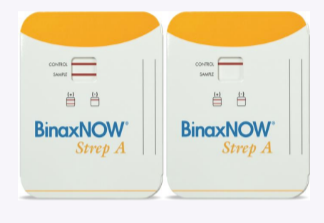
historical note: streptozyme
what it was: qualitative screening agglutination assay designed to detect one or more anti-strep antibodies ( ASO, Anti-DNase B, Anti-hyaluronidase, Anti-streptokinase, Anti-NADase)
how to teach it now:
NO LONGER ROUTINELY AVAILABLE/MANUFACTURED
knowledge check: a child develops periorbital edema, dark urine, and hypertension 3 wks after impetigo. which result best supports recent GAS infection?
elevated anti-DNase B
IMPETIGO = main clue
skin GAS may produce little ASO, Anti-DNase B is especially useful after pyoderma and supports PSGN history
Mycoplasma pneumoniae - what makes it different?
very small bacterium
NO CELL WALL
respiratory infection
gradual onset
persistent cough
fever, headache, malaise
often called “WALKING PNEUMONIA”
Mycoplasma pneumoniae - what happens immunologically ?
infection stimulates:
M. pneumoniae-specific antibodies
cytokine/chemokine response
antibodies, including COLD AGGLUTININS
Mycoplasma pneumoniae - possible extrapulmonary effects
stevens-johnson syndrome
raynaud phenomenon
COLD AGGLUTININ-ASSOCIATED RBC AGGLUTINATION/HEMOLYSIS
the immune response can extend beyond the lungs
M. pneumoniae - specific antibody response
infection → patient makes antibodies against M. pneumoniae antigens → specific IgM/IgG
these antibodies support organism specific serology
M. pneumoniae NONSPECIFIC cold agglutinin
infection → IgM cold agglutinins develop → IgM binds RBC I antigen → RBC agglutination at colder temps
NOT M. pneumoniae specific
what is happening in cold agglutinins?
antibody: usually IgM, directed against RBC I antigen, pentameric IgM efficiently bridges RBCs and leads to strong agglutination
RBC effect: IgM binds RBCs best at COOLER temps, visible RBC agglutination can occur
if clinically significant: complement can be activated and lead to RBC destruction + hemolytic anemia
are cold agglutinins good for diagnosing mycoplasma?
NOT BY THEMSELVES
why not?
cold agglutinins develop in only abt HALF of pts
NOT SPECIFIC for M. pneumoniae
better approach:
M. pneumoniae specific IgM/IgG serology
paired specimens when serology is used
molecular testing for current infection
mycoplasma serology: IgM vs IgG
specific IgM = supports current/recent infection
IgG alone = may reflect previous exposure; adults may show IgG on reinfection
rising IgG in paired sera = supports recent infection
early negative antibody = does NOT exclude infection
rocky mountain spotted fever - what happens?
R. rickettsii
tick borne, obligate intracellular bacterium
replicates INTRACELLULARLY
targets vascular endothelial cells
endothelial injury increases vascular permeability
clinical effects
leaky vessels can lead to:
edema
hypovolemia (losing fluid to extracellular space)
hypotension
overall picture:
fever
severe headache
GI symptoms
rash
potentially severe disease

RMSF: clinical timing matters
tick bite (exposure) → 2-14 days later (symptoms begin - fever, headache, GI symptoms) → 3-5 days after fever (rash may appear in hand/soles/trunk)
the pt may be seriously ill BEFORE detectable antibodies develop
RMSF serology timing problem
first week
pt may have RMSF but antibody may still be undetectable
7-10 days
antibodies become detectable in many patients
later
serology becomes more useful for confirmation
* BOC TRAP: early negative IFA does NOT rule out RMSF
IFA: serologic gold standard for RMSF
R. rickettsii antigen is fixed to a slide
add pt serum; specific patient antibody binds to antigen
wash, then add fluorescent ant-human immunoglobulin
fluorescence under microscope = patient antibody detected
*INDIRECT immunofluorescence → the labeled reagent is the secondary anti-human antibody
paired titers confirm RMSF
acute specimen
early illness
example titer - 1:32
convalescent specimen
2-4 weeks later
example titer - 1:128
1:32 → 1:128 = FOURFOLD RISE
supports recent infection
RMSF older vs current testing
Weil-Felix
detects cross reacting antibody using proteus OX antigens
main issue: poor sensitivity/specificity - largely replaced
IFA
detects R. rickettsii-specific IgM/IgG
main issue: early specimen may be negative
Molecular Testing
detects rickettsial DNA
main issue: direct organism detection - availability/specimen timing matter
knowledge check: a pt has a fever, severe headache, rash, and recent tick exposure. symptoms began 5 days ago. RMSF antibody IFA was negative. what is the best interpretation?
antibodies may not yet be detectable
RMSF antibodies are often undetectable during the first week. timing is the trap
Mycobacterium tuberculosis - what happens after exposure
inhalation: aerosolized MTB reaches alveoli
macrophage: alveolar macrophages phagocytose MTB. the organism can survive intracellularly
cell mediated immunity: T-cell response develops and attempts to contain infection
possible outcomes: clearance, latent TB infection (dormant), active TB, later reactivation
TB belongs in immunology bcs control depends heavily on cell mediated immunity
tuberculosis - why T cells matter
intracellular MTB: macrophage contains MTB antigens
CD4+ T cell: recognizes presented MTB antigen
Th1 response: produces IFN-gamma which calls macrophages
macrophage activation: improved intracellular control of TB
*immune response is measured by PPD (TB skin test) and IGRA (Interferon Gamma Release Assay)
granuloma
containment, not necessarily elimination
purpose
contain the organism and limit spread - contained does NOT equal eliminated - MTB may persist and can cause latent (dormant) TB infection
if immune control weakens
granuloma containment may fail → MTB replicates → reactivation TB

PPD/TST: measuring cell mediated immunity in vivo
PPD (pure protein derivative) injected intradermally → previously sensitized T cells recognize antigen → cytokines recruit inflammatory cells → INDURATION develops and is read 48-72 hrs after
TYPE IV - DELAYED HYPERSENSITIVITY - T CELL MEDIATED
PPD false positive considerations
BCG vaccinations
some nontuberculous mycobacteria
T cells may recognize related antigens
positive TST without MTB infection
PPD false negative considerations
testing too soon after exposure
immunosuppression/inadequate immune response
patient may be infected but unable to produce detectable induration
*TST detects immune sensitization - not the organism itself
interferon gamma release assay
patient blood
bring patient’s blood into the lab
MTB antigens
expose sensitized T cells to MTB-specific peptides
T-cell response
T cells release IFN-gamma (macrophages engulf and present to T cells)
lab readout
measure IFN-gamma response
*IGRA measures a T cell response - NOT MTB DNA and NOT anti-MTB antibody
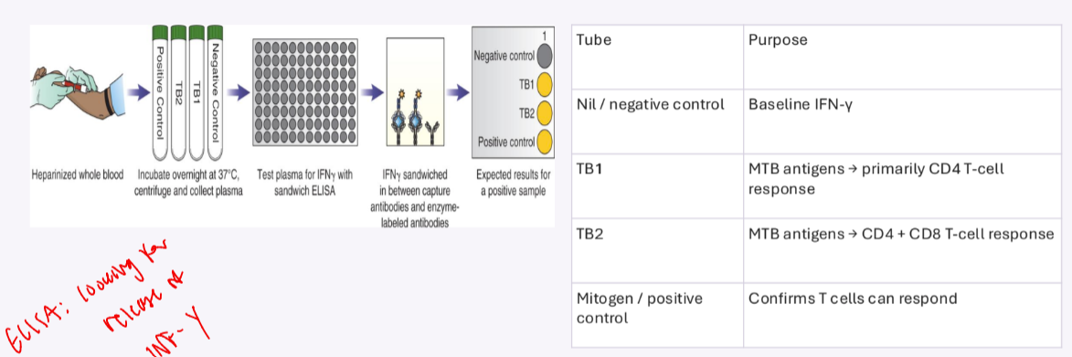
QuantiFERON-TB
Nil/NEG control tube
baseline IFN-gamma
TB1 tube
MTB antigens → primary CD4 T cell responses
TB2 tube
MTB antigens → CD4 + CD8 T cell response
Mitogen/POS control tube
confirms T cell can respond
after incubation: collect plasma → measure IFN-gamma by SANDWICH ELISA
looking for release of IFN-gamma
why do the QuantiFERON controls matter?
positive IGRA
Nil: low
TB antigen: HIGH
mitogen: responds
T cells specifically responded to MTB antigens
negative IGRA
Nil: low
TB antigen: NO response
mitogen: STRONG response
T cells can respond, but did NOT respond to MTB antigens
intermediate
Nil: low
TB antigen: NO response
mitogen: NO response
cannot tell whether there is no sensitization or the T cells failed to respond
PPD vs IGRA
both detect immune sensitization - neither directly demonstrates viable MTB

knowledge check: an asymptomatic healthcare worker has a positive QuantiFERON-TB gold plus result. what did the test demonstrate?
the patient’s T cells recognized MTB antigens and released IFN-gamma
a positive IGRA demonstrates immune sensitization. it does not by itself distinguish latent infection from an active disease
final comparison: 4 infections, 4 immunologic testing problems
GAS vs M. pneumoniae vs RMSF vs TB
WHAT are we detecting → WHEN was the specimen collected → WHAT does the result actually mean

rapid fire wrap up
pyoderma/impetigo → Anti-DNase B
mycoplasma cold agglutinin → IgM anti-I
early negative RMSF IFA → does NOT rule it out
positive IGRA → T cell sensitization; NOT proof of active TB
spirochetes big picture
long, slender, helically coiled bacteria w/ axial filaments (endoflagella)
characteristic corkscrew motility
ROUTINE CULTURE IS DIFFICULT
SEROLOGY therefore plays an important part in diagnosis
Treponema pallidum → syphilis
Borrelia burgdorferi → lyme disease
Leptospira spp. → leptospirosis
other Borrelia spp. → relapsing fever

Treponema pallidum
SYPHILIS
thin, tightly coiled spirochete
outer membrane has few exposed proteins and LACKS LPS (usually immunogenic marker)
cannot be routine cultured in the clinical lab
transmission: primarily SEXUAL CONTACT; can cross the PLACENTA
4 stages of syphilis
PRIMARY - painless chancre @ site of inoculation (10-90 days after infection)
SECONDARY - systemic dissemination; lymphadenopathy, fever, malaise, nonpruritic rash on PALMS/SOLES (hallmark)
LATENT - no clinical symptoms (early: <1 yr) (later: >1 yr)
TERTIARY - gummas (big nodules @ site of infection), cardiovascular disease, neurologic complications

neurosyphilis
do NOT memorize neurosyphilis as a tertiary only complication
CNS involvement can occur during ANY STAGE
early manifestations may include meningitis, cranial nerve abnormalities, or stroke
ocular syphilis and otosyphilis can also occur at ANY STAGE
how can we detect syphilis?
DIRECT DETECTION
detect the organism itself
dark field microscopy - direct fluorescent method
NONTREPONEMAL SEROLOGY
detect reagin antibodies associated w/ lipid antigens/tissue damage
RPR, VDRL
TREPONEMAL SEROLOGY
detect antibodies against T. pallidum
enzyme immunoassays - FTA/ABS, TP-PA, EIA/CILA
direct detection of T. pallidum
DARK FIELD MICROSCOPY can examine serous fluid from an active lesion
collected from chancre
look for characteristic morphology and corkscrew/flexing motility
specimen should be examined RAPIDLY
do NOT use darkfield microscopy on ORAL lesions
nonpathogenic oral treponemes may resemble T. pallidum

nontreponemal tests: what is cardiolipin?
CARDIOLIPIN = phospholipid associated w/ lipid antigens released/exposed during tissue damage and infection
the immune response produces antibodies historically called REAGIN
may include IgM and IgG
RPR and VDRL detect these anti-lipid/REAGIN antibodies - NOT T. pallidum specific antibody
the test antigen contains cardiolipin + cholesterol + phosphatidylcholine (lecithin)
RPR and VDRL: flocculation tests
both are NONTREPONEMAL tests that DETECT REAGIN
antigen-antibody interaction produces flocculation
RPR uses CHARCOAL particles so the reaction can be read MACROscopically
VDRL is read MICROscopically; SERUM requires HEAT INactivation
VDRL has an important role in CSF testing
ONLY METHOD FDA APPROVED FOR NEUROSYPHILIS DIAGNOSIS
NOT a (+) or (=) report
report as: REACTIVE, WEAK REAC., OR NONREACTIVE

why can RPR/VDRL be false positive?
nontreponemal tests are NOT SPECIFIC for T. pallidum (detects only reagin/damage to cell)
transient biologic false positives may occur w PREGNANCY, VACCINATION, or INFECTIONS
chronic false positives may occur w SLE/AUTOIMMUNE disease, IV drug use, ADVANCED AGE, or malignancy
a reactive nontreponemal test screen must be interpreted w a treponemal test
Prozone false negative
SECONDARY syphilis may produce very HIGH ANTIBODY concentrations → prozone rxn; sample must be diluted
antibody excess prevents optimal lattice formation
little/no visible flocculation → falsely nonreactive result
solution: perform serial dilutions of the patient’s serum
why report an RPR/VDRL titer?
reactive samples can be tested in serial twofold dilutions - 1:2 → 1:4 → 1:8 → 1:16 → 1:32…
titer = highest dilution that remains ACTIVE
nontreponemal titers usually DECLINE W/ SUCCESSFUL TREATMENT
a FOURFOLD change is CLINICALLY SIGNIFICANT (successful treatment)
example - 1:32 → 1:8 = fourfold increase

treponemal tests
now we get specific
detect ANTIBODIES directed against T. pallidum ANTIGENS
examples: FTA-ABS, TP-PA, automated EIA/CILA
usually become reactive EARLY and remain reactive LONG TERM
often remain active AFTER successful TREATMENT
therefore: NOT useful for MONITORING RESPONSE to THERAPY
FTA-ABS: indirect fluorescence
ABS = absorption: patient serum is preabsorbed with NONPATHOGENIC REITER TREPONEMAL ANTIGENS to remove cross-reactive antibodies
patient antibody binds fixed T. pallidum on the slide
fluorescent anti-human immunoglobulin binds the patient’s antibody
fluorescent spirochetes indicate a reactive test
LABEL IS ON ANTI-HUMAN Ig → INDIRECT fluorescence
TP-PA: Particle Agglutination
colored gelatin particles are coated with TREPONEMAL ANTIGENS
patient ANTI-T. pallidum ANTIBODIES cross link sensitized particles
POSITIVE → diffuse lattice/mat (pink spreads throughout well)
NEGATIVE → compact button (bright pink circle does not spread out in the well)

automated treponemal immunoassays
high volume labs may use EIA or chemiluminescent immunoassays (CLIA) for TREPONEMAL antibody screening
automation supports high throughput testing
treponemal immunoassays are central to the REVERSE screening algorithm

nontreponemal vs treponemal
NONTREPONEMAL: RPR/ VDRL
reagin against cardiolipin-containing antigen → lower specificity → useful for treatment monitoring
TREPONEMAL: FTA-ABS/ TP-PA/ EIA or CLIA
anti-T. pallidum antibody → higher specificity → usually remains active
traditional syphilis algorithm
SCREEN w/ RPR or VDRL
if reactive → confirm w TREPONEMAL test
reactive nontreponemal + reactive treponemal SUPPORTS syphilis
reactive nontreponemal + nonreactive treponemal suggest a BIOLOGICAL FALSE POSITIVE

reverse syphilis algorithm
screen w/ AUTOMATED TREPONEMAL EIA/ CLIA
if reactive → perform quantitative RPR or another NONTREPONEMAL test
if TREPONEMAL (+) and RPR (=) → perform DIFFERENT TREPONEMAL TEST, commonly TP-PA
*less time consuming, used in large hospital settings

discordant reverse results
EIA/CLIA (+) and RPR (=) can reflect:
previous treated syphilis
late/latent infection
very early infection
false positive initial treponemal screen
resolve discordance w a different treponemal test such as TP-PA
neurosyphilis CSF testing
specimen: cerebrospinal fluid
CSF-VDRL is highly SPECIFC when reactive - only FDA approved test
a positive CSF-VDRL strongly supports neurosyphilis
SENSITIVITY IS LIMITED: a negative CSF-VDRL does NOT exclude neurosyphilis
CSF lymphocytes and protein can provide supportive information
*Positive is Powerful BUT negative does NOT rule it out
syphilis: don’t get tricked
secondary symptoms + negative RPR → think PROZONE and DILUTE
positive RPR → confirm w TREPONEMAL testing
monitor treatment → QUANTITATIVE RPR/ VDRL
treponemal test (+) AFTER treatment → can be EXPECTED
EIA (+) / RPR (=) in reverse algorithm → perform TP-PA
suspected neurosyphilis → CSF-VDRL (negative does NOT exclude)
Borrelia burgdorferi
LYME DISEASE
loosely coiled spirochete transmitted by IXODES TICK
humans = accidental hosts
nymphs cause many human infections
transmission generally requires PROLONGED TICK ATTACHMENT
white footed mouse = important RESERVOIR
deer = important tick HOST

lyme disease clinical progression
EARLY LOCALIZED
erythema migrans (EM) + flu like symptoms
pt may be otherwise symptomatic
EARLY DISSEMINATED
multiple lesions + facial palsy + neurologic involvement + carditis + migratory pain
HEMATOGENOUS SPREAD TO MULTIPLE ORGAN SYSTEMS
LATE
ARTHRITIS + PERIPHERAL NEUROPATHY + ENCEPHALOMYELITIS

erythema migrans - why early serology can be negative
EM may appear before a measurable antibody response develops
early serology can therefore be nonreactive despite infection
testing is measuring the HOST antibody response - NOT directly detecting the ORGANISM
clinical presentation and timing matter
early localized disease may be present w/ EM while the patient is otherwise asymptomatic
Lyme antibody response
antibody may not be detectable until several WEEKS AFTER the infection
IgM appears FIRST; IgG follows and peaks LATER
early antibiotic treatment may delay or inhibit antibody development
IgG may persist LONG TERM
serology therefore does NOT reliably indicate CURRENT activity or TREATMENT SUCCESS
the 30 day rule for Lyme disease
symptoms < 30 days: standard immunoblot interpretation may evaluate IgM + IgG
symptoms > 30 days: rely on IgG
isolated IgM is NOT used to support late disease
two tier testing for Lyme disease
standard two tier testing (STTT)
first tier EIA/ IFA → immunoblot if positive/equivocal
modified two tier testing (MTTT)
first EIA → second, different EIA if positive, equivocal
both approaches use sequential testing to improve interpretation

standard two tier testing (STTT)
STEP 1: EIA or FIA for B. burgdorferi ANTIBODY
negative → usually NO FURTHER TESTING
consider TIMING if very EARLY DISEASE
positive/equivocal step 1 → STEP 2:
<30 days → IgM + IgG immunoblot
>30 days → IgG immunoblot
what does a Lyme immunoblot band mean?
Borrelia proteins are represented by molecular size kDa
patient serum is added to the antigen strip/membrane
if patient antibody recognizes a particular Borrelia antigen, it binds there
detection reagent makes that antibody—antigen location visible as a BAND
the kDa number is the approximate molecular mass of the antigen - NOT the antibody level

lyme immunoblot: reading the pattern
IgM positive pattern: 2 of 3 designated bands
23 kDa (OspC)
39 kDa
41 kDa (flagellin)
IgG positive pattern: 5 of 10 designated bands
18, 23, 28, 30, 39, 41, 45, 58, 66, 93 kDa
DO NOT interpret a single reactive band as diagnostic
why isn’t the 41-kDa band enough?
41 kDa = flagellin, an immunodominant antigen
flagellar proteins can share antigenic similarity among spirochetes
cross-reactive antibodies may occur w/ other Borrelia species or T. pallidum
therefore one 41-kDa band alone is not sufficient evidence of Lyme disease