Chapter 6 Anatomy and Physiology
 Call Kai
 Learn
Learn Practice Test
 Spaced Repetition
 Match
Match Flashcards
 Knowt Play
1/86

There's no tags or description
Looks like no tags are added yet.
Name | Mastery | Learn | Test | Matching | Spaced | Call with Kai | Chat |
|---|
No analytics yet
Send a link to your students to track their progress
87 Terms
Dermatology
Scientific study and medical treatment of the integumentary system.
Inspection of the skin, hair, and nails is significant part of a
physical exam
Skin is the most vulnerable organ
• Exposed to radiation, trauma, infection, and chemicals
• Receives more medical treatment than any other organ
system
Integumentary system
An Organ system that consists of hair, skin, nails, and associated glands.
The skin is the body’s largest and heaviest organ: Covers area2
1.5 to 2.0 m, and Composes 8% of body weight
What are the two main layers of the skin
Epidermis: stratified squamous epithelium
Dermis: deeper connective tissue layer
Below the dermis is the hypodermis, not part of the skin but usually
studied with it.
Thick skin (anatomy, layers, thickness in mm and location)
Covers palms, soles, and corresponding surfaces of fingers and toes
Has sweat glands, but no hair follicles or sebaceous (oil) glands.
Epidermis is about 0.5 mm thick, primarily due to the thicker stratum
corneum, and the additon of the stratum lucid.
Layers in thick skin: Stratum corneum, lucidum, granulosum, spinosum, basale.
Thin skin (Anatomy, thickness in mm, locatiuon, and layers)
Location: Covers rest of the body.
Anatomy: Has hair follicles, sebaceous glands, and sweat glands.
Thickness: Epidermis about 0.1 mm thick due to a thin stratum corneum and NO stratum lucidum.
Layers: stratum corneum, granulosum, spinosum, and basale.
List the 6 functions of the skin and relate them to its
structure.
Protection: Resistance to trauma and infection: Keratin provides physical protection. Dermcidin, defensins, and acid mantle protect against bacteria. Other barrier functions: Barrier to water, ultraviolet (UV) rays, harmful chemicals.
Excretion and absorption: eliminates small amount of waste products through sweat and can absorb topical substances
Vitamin D synthesis: Skin carries out first step.
Sensation: Many nerve endings that react to various stimuli.
Thermoregulation: Alter blood flow to release or retain heat. Cutaneous vasoconstriction, cutaneous vasodilation.
Nonverbal communication: Facial expressions.
What are the 3 general features of the epidermis?
Keratinized stratified squamous epithelium. Dead cells at skin surface packed with tough keratin protein.
Avascular. Depends on the diffusion of nutrients from underlying dermis derlived through the papilary layer of the dermis.
Contains sparse nerve endings for tough and pain.
What are the 5 cell types of the epidermis?
Keratinocytes (majority of epidermal cells): synthesize keratin
Stem cells: undifferentiated cells that give rise to keratinocytes. In deepest layer of epidermis, stratum basale,
Melanocytes: synthesize the pigment melanin that shields DNA from UV radiation. Present only in the stratum basale, but have branched processes that spread among keratinocytes and distribute melanin via melanosomes.
Tactile cells: Touch receptor cells associated with dermal nerve fibers. Tactile disc—collective term for tactile cell and associated nerve fiber. In the basal layer of the epidermis.
Dendritic cells: Phagocytic immune cells that guard against toxins and microbes. Found in stratum spinosum and stratum granulosum.

What are the 5 layers of thick skin from deepest to superficial
Stratum basale: Single layer of stem cells and keratinocytes resting on the basement membrane. Also contains scattered melanocytes and tactile cells.
Stratum spinosum: Several layers of living keratinocytes joined by desmosomes and tight junctions. Named for the appearance of cells after histological preparation (spiny). Also contains some dendritic cells.
Stratum granulosum: Three to five layers of flat, living keratinocytes. Cells contain dark-staining keratohyalin granules.
Stratum lucidum: Thin, pale layer found only in thick skin. Keratinocytes packed with clear protein eleidin.
Stratum corneum: Up to 30 layers of dead, scaly, keratinized cells. Resists abrasion, penetration, and water loss.
Stratum basale
Single layer of stem cells and keratinocytes resting on the basement membrane. Also contains scattered melanocytes and tactile cells.
Stratum spinosum:
Stratum spinosum: Several layers of living keratinocytes joined by desmosomes and tight junctions. Named for the appearance of cells after histological preparation (spiny). Also contains some dendritic cells.
Stratum granulosum:
Stratum granulosum: Three to five layers of flat, living keratinocytes. Cells contain dark-staining keratohyalin granules.
Stratum lucidum
Stratum lucidum: Thin, pale layer found only in thick skin. Keratinocytes packed with clear protein eleidin.
Stratum corneum
Stratum corneum: Up to 30 layers of dead, scaly, keratinized cells. Resists abrasion, penetration, and water loss.
The general life cycle of a Keratinocyte.
Keratinocytes produced by mitosis in stratum basale or deepest part of stratum spinosum. Mitosis requires abundant oxygen and nutrients. Once cells migrate away from blood vessels of the dermis, mitosis cannot occur. Keratinocyte multiplication is slower in old age, faster in injured or stressed
skin.New keratinocytes push older ones toward the surface. In 30 to 40 days a keratinocyte makes its way to the skin surface and exfoliates (flakes off) as specks called dander (may combine with oil to become dandruff).
Over time, keratinocytes flatten, produce more keratin and lipid-filled lamellar granules.
Calluses or corns are thick accumulations of dead keratinocytes on hands or feet.
What are the 4 important events that occur in the stratum granulosum?
Keratohyalin granules release filaggrin, a protein that binds to keratin to turn it into tough bundles.
Cells produce tough envelope proteins beneath their membranes.
Membrane-coating vesicles release lipid mixture that spreads out over cell surface and waterproofs it.
Keratinocytes organelles degenerate and the cells dies, leaving waterproof sac enclosing keratin bundles.
Epidermal Water barrier
Formed from the 4 processes that occur in the stratum granulosum as well as tight junctions between keratinocytes. Helps prevent dehydration. Does not prevent the absorption of water by the stratum cornea when we soak in a bath our fingers still get pruned; but it is water resistant.
What are the 5 general features of the dermis?
Connective tissue layer 0.2 mm to 4 mm thick. Composed mainly of collagen; also contains elastic and reticular fibers.
Abundant vasculature: Well supplied with blood vessels, sweat glands, sebaceous glands,
and nerve endings.
Hair follicles: ouses hair follicles and nail roots.
Erector pilli and Muscles of facial expression attach to dermis.
Two layers: papillary layer and reticular layer.
Describe the two zones of the dermis.
Papillary layer: Thin zone of areolar tissue in and near the dermal papillae which allows for mobility of leukocytes and other defense cells. Rich in small blood vessels that nourish the stratum basal.
Wavy, conspicuous boundary between dermis and epidermis. Dermal papillae are upward, finger-like extensions of dermis. Epidermal ridges are downward waves of epidermis.
Reticular layer: deeper thicker layer of the dermis composed of dense irregular connective tissue.
Stretch marks (striae): caused by tears in the collagen fibers caused by stretching of the skin.
Blisters: damaged dermal blood vessels (by burns or friction) may cause serous fluid to seep out and form a blister between the dermis and the epidermis and that’s why you get a pocket of serious membrane fluid.
Hypodermis (subcutaneous tissue)
Anatomy: layer of connective tissue beneath the skin that contains more areolar and mostly adipose tissue than the dermis.
Physiology: pads the body and binds skin to underlying tissues. Common site for drug injection due to rich vasculature in the hypodermis. Energy resivoir, thermal insulation.
Subcutaneous fat
Anatomy: refers to the subcutaneous tissue that is comprised predominatly of adipose tissue.
Physiology: energy resivoir, thermal insulation thicker in women and thinner in infant ans elderly.
Skin grafts: autograft and allograft
Third-degree burns often require skin grafts.
Autograft: tissue from another location on the same person’s body.
Allograft: tissue from unrelated person ussually deceased donors.
Artificial and lab based grafts are currently under development.
Eumelanin
brown and black eumelanin are responsible for tan brown an black skin.
Phenomelanin
responsible for yellowsih and reddish tones in some asians and native Americans, and the pinkish tones of white skin.
What factors account for variation in skin color.
The amount of eumelanin and pheomelanin. People of different colors have the same NUMBER of melanocytes, but vary in how much melanin is produced and longevity. In darker skin melanocytes produce greater quantities of melanin that breaks down more slowly, are more spread out in keratinocytes, and spread out through out every layer of the epidermis. In lighter skin, melanin is clumped near the keratinocyte nucleus, and little melanin is seen beyond the stratum basal.
Degree of show through of dermal vessels and white collagen.
Possible accumulation of carotene pigment from yellow and orange vegetables.
UV radiation stimulates melanin synthesis.
Cyanosis
Blueness of the skin due to oxygen defincieny
Erythema
Redness of the skin due to increased blood flow
Pallor
paleness due to decreased blood flow to the skin.
Jaundice
yellow of the skin due to biliruben in blood. sign of compromised liver function
Hematoma
clotted blood beneath the skin in the hypodermis.
What are the common markings of the skin.
Friction ridges: fingerprint.
Flexion lines (flexion creases): lines on the flexor surfaces of the digits, palms, wrist, and elbows. Skin bound to deeper tissues along these lines.
freckles: flat melanized patch.
Mole (nevus): elevated, melanized patch often with hair.
Hemangiomas: birthmarks. patches of discolored skin caused by benign tumors of dermal capillaries.
Hemangiomas
A type of birthmark. Patches of discolored skin caused by benign tumors of dermal capillaries. Some disappear in childhood, others last for life.
Examples: capillary hemangioma (strawberry birthmark), cavernous hemangioma, port-wine stain.

friction ridges
Finger prints. Markings on the finger tips that leave oily finger prints. Everyone has a unique pattern formed during fetal development
that remains unchanged throughout life; not even identical twins
have identical fingerprints. Dermal papillae form friction ridges.
Physiology: Enhance fingertip sensitivity; improve grip
Felxion lines (felxion creases)
lines on the flexor surface of the digites, palms, wrists, and elbows. Mark site where skin fold during flexion of joints.
Physiology: skin bound to deeper tissues along these lines.
Mole (nevus)
elevated melanized patch often with hair should be watched for change sin color, diameter, or contour that may sugest a cancer.
What are the three acessore organs of the skin
hair, nails, and cutaneous gland. Hair and nails are composed of mostly dead keratinzed cells.
Soft versus hard keratin
Soft keratin: Pliable soft keratin makes up stratum corneum of skin.
Hard keratin: Compact hard keratin makes up hair and nails. Tougher and more compact due to numerous disulfide bridges
between keratin molecules.
Pillus (plurarl: Pilli)
Slender filament of keratinized cells growing from a tube in
the skin called a hair follicle. Hair covers most of the body.
Hair does not cover: palms, soles; palmar, plantar, and lateral
surfaces and distal segments of fingers and toes; lips, nipples, and
parts of genitals.
Limbs and trunk have 55 to 70 hairs per square cm; face
has about 10 times as many.
100,000 hairs on an average person’s scalp. Differences in hairiness across individuals is mainly due to differences in texture and pigment of hair.
Distinguish between three types of hair.
Downy hair (lanugo): Fine, downy, unpigmented hair that appears on the fetus in the last 3 months of development.
Vellus hair: Fine, pale hair that replaces lanugo by time of birth. Two-thirds of the hair of women; one-tenth of the hair of men; all the hair of children except eyebrows, eyelashes, and scalp hair.
Terminal hair. Longer, coarser, more heavily pigmented. Eyebrows, eyelashes, and hair of the scalp. After puberty, forms axillary and pubic hair. Male facial hair and some of the hair on the trunk and limbs.
The anatomy of hair:
Hair shaft, hair root, hair bulb, hair matrix, medulla, cortex, and cuticle.
Hair Shaft is portion above the skin surface.
Hair root is portion below skin surface.
Hair bulb: In dermis or hypodermis, hair root ends with dilated bulb. Bulb grows around dermal papilla—bud of vascular connective tissue, provides nutrition to hair.
Hair matrix—mitotically active cells immediately above the papilla;
growth center for the hair.
Hair has up to three layers: Medulla—core of loosely arranged cells and air spaces; Cortex—bulk of the hair; layers of elongated cells; and Cuticle—outermost region consisting of multiple layers of thin, scaly cells that overlap like roof shingles.

Hair Bulb
In dermis or hypodermis, hair root ends with dilated bulb. Bulb grows around dermal papilla—bud of vascular connective tissue, provides nutrition to hair.
Hair matrix
Mitotically active cells immediately above the papilla;
growth center for the hair.
What are the three layers of hair
Medulla—core of loosely arranged cells and air spaces.
Cortex—bulk of the hair; layers of elongated cells.
Cuticle—outermost region consisting of multiple layers of thin, scaly cells that overlap like roof shingles.

What are the 2 layers of the hair follicle
Epithelial root sheath extension of the epidermis lying adjacent
to hair root; widens at deep end into bulge, a source of stem cells
for follicle growth.
Connective tissue root sheath—derived from dermis but a bit denser; surrounds epithelial root sheath

Structures associated with the hair folicle
Sensory nerve fibers entwine each follicle and respond to
hair movements.
Arrector pilli muscle—bundle of smooth muscle attaching
follicle to dermis; contracts to make hair stand on end
(piloerection).
What causes hair color?
Color due to pigment granules in the cells of the cortex
• Brown and black hair is rich in eumelanin
• Red hair has high concentration of pheomelanin
• Blond hair has an intermediate amount of pheomelanin
and very little eumelanin
• Gray and white hair have little or no melanin; air present in
medulla.

What cuases hair texture
Texture related to cross-sectional shape of hair
• Straight hair is round
• Wavy hair is oval
• Curly hair is relatively flat.

Hair cycle
Hair follicles go through three developmental phases.
Anagen: growth stage (6 to 8 years). 90% of scalp follicles at any given time. Stem cells multiply; root sheath cells above papilla form hair cells of
hair matrix; hair cells make keratin and die as they are pushed
upward.
Catagen: degeneration stage (2 to 3 weeks). Mitosis in hair matrix ceases, follicle shrinks, hair loses anchorage. Base of hair keratinizes into a hard knot—hair is called a club hair
is easily pulled out by brushing.
Telogen: resting stage (1 to 3 months).
Effects of aging on hair growth and loss
We lose about 50 to 100 hairs daily. Hair growth rate is 1 mm per 3 days until the 40s.
Effects of aging:
• A higher percentage of follicles are in catagen and telogen phases
• Follicles shrink and produce wispy vellus hairs
• Alopecia—thinning of the hair, or baldness
Androgens
Hormones
Pattern baldness (androgenic alopecia)
Hair loss unevenly rather than thinning uniformly. combination of polygenic inheritance, hormones (androgens), and environmental influences.
Hirsutism
Excessive hairiness in areas not usually hairy.
Physiology of hair (5 functions of hair)
Hair function depends on body region and type of hair:
Hair receptors alert us of parasites crawling on skin. Hair on trunk and limbs is mostly vestigial.
Scalp hair retains heat and protects against sunburn.
Pubic and axillary hair—signify sexual maturity and aid in transmission of sexual scents.
Guard hairs (vibrissae) guard nostrils and ear canals.
Eyelashes and eyebrows enhance facial expression and nonverbal communication.
Anantomy and physiology of nails
Anatomy:
Composed of thin, dead cells packed with hard keratin. Clear, hard derivatives of stratum corneum.
Nail plate—hard part of nail; includes overhanging free edge, nail body (visible attached part), and nail root under overlying skin.
Surrounding skin rises as a nail fold, separated from the margin of
the nail plate by the nail groove.Nail bed—skin underlying the nail plate.
Hyponychium—epidermis of nail bed.
Nail matrix—growth zone at proximal end of nail; its thickness
obscures underlying vessels and appears as a white lunule.Cuticle (eponychium)—narrow zone of dead skin that overhangs
proximal end of nail.
Physiology:
Improves grooming, picking apart food, and other manipulations.
Provide a counterforce to enhance the sensitivity of fleshy fingertips to tiny objects.

Name two types of sweat glands, and describe the structure and function of each.
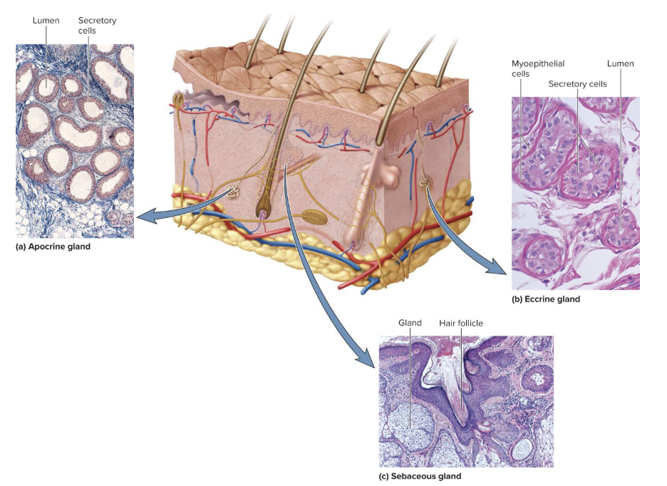
Apocrine sweat glands:
Groin, anal region, axilla, areola, beard area in men.
Ducts lead to nearby hair follicles; produce sweat that is milky and contains fatty acids.
Inactive until puberty; respond to stress and sexual stimulation. Believed to secrete sex pheromones—chemicals that can influence
the behavior of others. Can cause bromhydrosis with inadequate hygiene.
Eccrine (merocrine) sweat glands:
Simple tubular glands produce watery perspiration that
helps cool the body.Most numerous skin glands, 3-4 million in adult skin, especially dense on palms, soles, and forehead.

Bromhidrosis
disagreeable body odor; usually inadequate hygiene
Myoepithelial cells
Contract in response to stimulation by sympathetic nervous system and squeeze perspiration up the duct. Found in both apocrine and eccrine glands.
Sweat composition
99% water, pH range of 4 to 6. Acid mantle—inhibits bacterial growth.
Begins as a protein-free filtrate of blood plasma produced by deep secretory portion of gland. Some sodium chloride reabsorbed by duct, but some sodium chloride and other small solutes (potassium, urea, lactic acid, ammonia) remain in the sweat. Some drugs are excreted in sweat.
Insensible perspiration
not noticed; does not produce visible wetness of skin: 500 ml/day
Diaphoresis
Sweating with noticeable wetness of the skin, in response to heat, exercise, circulatory shock: 1 L or more of sweat per hour may be lost during exercise
Cutaneous tranpiration
Water loss from skin not due to sweating. Water diffuses between the keratinocytes and evaporates from the skin surface.
Sebaceous glands: describe the location, structure, and function.
Produce oily secreation called sebum. Gland structure includes flask-shaped, short ducts open into hair follicles.
Keeps skin and hair from becoming dry, brittle, and cracked.
Holocrine secretion style (where th whole cells disintergrates to discharge its sebum).
Ceruminous glands: describe the location, structure, and function.
Modified apocrine glands found only in the external ear canal.
Coiled, simple tubular glands produce cerumen (earwax): yellow, waxy secretion combined with sebum and dead epithelial cells.
Keeps eardrum pliable. Waterproofs the canal. Kills bacteria. Makes guard hairs of ear sticky to help block foreign particles from entering the auditory canal.
Mammary glands
Milk-producing glands that develop in female during pregnancy and lactation. Modified apocrine sweat glands. Rich secretion released through ducts opening at nipple.
Mammary ridges or milk lines—two rows of mammary glands in most mammals. Primates kept only two glands, but a few people have additional nipples along the milk line (polythelia).
Polythelia
additonal nipples along the milk line that cotnain mamary glands that produce the ruch aprocrine lactation./
Describe the three most common forms of skin cancer.
Most skin cancer is caused by exposure to UV rays of the sun. Most often on the head, neck, and hands. Most common in fair-skinned people and the elderly. One of the most common, easily treated cancers. Highest survival rates if detected and treated early.
Three types of skin cancer named for the epidermal cells in
which they originate:
• Basal cell carcinoma
• Squamous cell carcinoma
• Malignant melanoma
Basal cell carcinoma
Most common type, least dangerous because it seldom metastasizes. forms from cell sin the stratum basal.
Presentation: small, shiny bump with central depression and beaded edges.
Squamous cell carcinoma
Arrises from keratinocytes of stratum spinous.
Presentation: presents as lesions ussually on scar, ears, lower lip or back fo the hand. Raised, reddened, scaly apperance later forming a concave ulcer.
Chance of recovery is good with early detection and removal, but tends to metastasize to lymph nodes and may become lethal
Melanomia
Arises from melanocytes. Less than 5% of skin cancers. can be sucessfully removed if caught early, but usually fatal if metasizes. Greatest riskfactor is familial hisory. Highest incidence in men, redheads, and people who had sever sunburn as a child.
The reasons that Burns can result in death.
fluid lossm infection, and toxic effects of eschar—burned dead tissue
Describe the three classes of burns and the priorities in
burn treatment.
First-degree burn (superficial burns)—only involves the epidermis. Redness, slight edema, and pain. Heals within days.
Second-degree burn (partial-thickness burn)—involves part of dermis. May appear red, tan, or white; blistered and painful. Two weeks to several months to heal and may leave scars.
Third-degree burn (full-thickness burn)—involves all of dermis and often some deeper tissues. Often requires skin grafts; need fluid replacement, infection control, supplemental nutrition.

The evolution of skin color
Skin color variation due to evolutionary selection pressures as humans migrated from Africa:
Near equator and latitudes where UVR is strongest—benefit to have
dark, protective skin.Farther north or south where sunlight is weaker—benefit to have
lighter skin.
Melaninated skin protected itself from excess ultraviolet radiation (UVR). Brown and black skin today have SPF of 10 to 15, white skin only 2.5. UVR can be damaging (sunburn, cancer, degradation of folate) but also offers benefit of stimulating vitamin D synthesis.
Skin color in response to migration reflects compromise between need for folate (degraded by UVR) and vitamin D (promoted by UVR).
The skin is a barrier to all of the following except _________blank.
Metals such as mercury
Most of the skin is _________blank mm thick.
1-2
The narrow zone of dead skin overhanging the proximal end of a nail is called the _________blank.
Eponychium
Melanin, hemoglobin, and possibly carotene give color to the skin.
True
Blood vessels in the _________blank provide a hair with its sole source of nutrition.
Dermal papilla
The acidity of sweat contributes to the acid mantle that inhibits bacterial growth in the skin.
True
Which of the following is not found in the epidermis?
Multiple Choice
Melanocytes
Fibroblasts
Stem cells
Keratinocytes
Tactile cells
Fibroblast

Nail bed
Debridement is not necessary to infection control.
False
Which of the following cells stand guard against toxins, microbes, and other pathogens?
Dendritic cells
_________blank is/are formed partly from the secretions of glands in the external ear canal.
Cerumen
Excessive hairiness is called _________blank.
Hirtuism
Which layer of the epidermis consists of up to 30 layers of dead cells?
Stratum Corneum
The base where a hair follicle originates in the skin is known as the _________blank.
The hair bulb
A pilus is a _________blank.
The hair