Lymphatic system
 Call Kai
 Learn
 Practice Test
 Spaced Repetition
 Match
 Flashcards
 Knowt Play
1/62

Earn XP
Description and Tags
Dr. Delcambre
63 Terms
lymphatic system
•Open network of vessels, nodes, organs, tissues, and lymph that supports immunity and fluid balance
•Collects and returns excess interstitial fluid (ECF), removes waste and pathogens, produces and activates lymphocytes.
tertiary circulatory system member

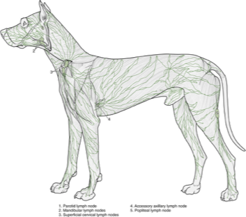
lymphocenters
Areas where several lymphatics come together. major sites of Lymph B and T
important for the proper functioning of immune system (like filter)

cisterna chyli
stopping point for all the fluid being returned to the heart

thoracic duct
last chance to dump (caudal to cranial to the heart). (dorsal to esophagus).
lymphadenopathy
enlarged, swollen, or changed lymph nodes
lymphedema
invrease in ECF in peripheral tissues (do not return and causes lymph buildup (edema)= lymphatic fluid stuck in peripheral tissue.

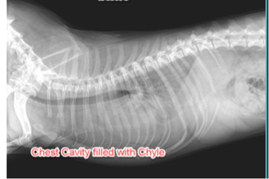
chyle
• fluid consisting of both fat and lymph, formed from small intestinal drainage into the lymphatic system – ex: in cisterna chyli and thoracic duct
found mostly in cysterna chyli and thoracic duct.
breed predisposition of spontaneous rupture of the thoracic duct (borzois).
fluid is going in the potential space between lung and = chylothorax

spleen
•Filter and storage for blood
•Generation of antibodies
•Removal of old blood cells
•Recycling of iron

thymus
maturation and differentiation of T-lymphocytes
Involutes (shrinks) with age in dogs

tonsilar tissue
no afferent vessels going to it, efferent. Palatine, pharyngeal (aka adenoids),
MALT
Mucosal associated lymphoid tissue - BALT and GALT
BALT
Bronchiole associated lymphoid tissue.
GALT
Gut associated lymphoid tissue

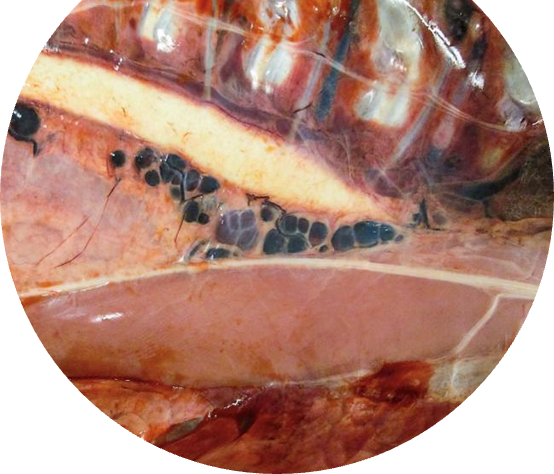
hemal nodes
collection of lympoid tissues. serve as mini spleens, filtering blood.
popliteal ln
drains lateral and distal hind limb (green)
mandibular ln
drain the external head
retropharyngeal ln
drain oral and nasal passageways, pharenges
superficial cervical ln
drain brachial region and cervical region
axillary ln
medial thoracic, cranial pectoral region (includes cranial aspects of the mammary chain)
mesenteric/jejunal ln
drains small intestine and proximal large intestine
superficial inguinal ln
caudal mammary chain + adductor region
iliosacral/sublumbar ln
distal large intestine / pelvic cavity /reproductive tract issues.
lymphatic system
Groups of cells, tissues, and organs that monitor body surfaces and internal fluid compartments and react to the presence of potentially harmful substances.

circulating lymphocytes
in blood or lymph - 70%

non-circulating lymphocytes
destined specific tissues - 30%
•(e.g. CT under epithelium of respiratory, GI, and urogenital tracts)

antigens
substances that can induce a specific immune response.
broken into small peptides to get a sufficient response
mononuclear phagocytic system
most APCs belong to this system, macrophages. they interact with T lymphocytes to facilitate immune response.

Primary lymphatic organs
bone marrow and thymus
this is where lymphocytes differentiate into immunocompetent cells (training) and to distinguish self/non-self.

secondary lymphatic organs
diffuse lymphatic tissue (GALT, BALT, MALT)
lymphatic nodules, lymph nodes, tonsils and spleen
where they activate
diffuse lymphatic tissue
Special type of LCT found under epithelium (i.e., in the Lamina propria) of the GI, respiratory, and genitourinary tracts
site of initial immune response
includes fibroblasts, plasma cells, eosinophils

lymphatic nodule
•Found in structures associated with GI tract such as ileum, tonsils, appendix.
•Concentration of lymphocytes in meshwork of reticular cells.
primary nodule
small lymphocytes, no germinal center. dark lymphocytes.

secondary nodule
has a germinal center (pale) and a mantle zone (darker), additional activation

Peyer Patches
aggregations of lymphatic nodules.
located in ileum, lamina propria
contain B and T lymphocytes
Tonsils
Immune surveillance of the mouth and nose.
•Stratified squamous epithelium (SSE) lines the tonsillar crypts= distinguishing feature of tonsils

palatine tonsil
located on either side of pharynx
pharyngeal tonsils
located at the roof of pharynx
lingual tonsils
located at the base of the tongue
lymph node
small bean shaped, encapsulated. located along a pathway of lymphatic vessels.
Filters lymph drained from peripheral tissues, screening it for pathogens.
•The principal site where T and B lymphocytes undergo antigen-dependent proliferation and differentiation into effector lymphocytes.

stroma of lymph node
capsule and trabeculae (dense. CT)
reticular tissue- fine mesh work

cortex of lymph node
secondary lymphoid nodules, composed mainly of B cells (proliferation and differentiation)

deep cortex
no nodules
mainly T cells (activation and differentiation)
High endothelial Venule

High Endothelial Venule
venules lined by cuboidal or low columnar endothelial cells
concentrate lymph
B and T ceclls leave blood through HEV and migrate into LN

lymph circulation
afferent lymph vessels
through the walls of post capillary (high endothelial) venules in deep cortex
lymph exits through the efferent lymph vessels

thymus
educates the T lymphocytes
Fully formed at birth, regresses after about 3-4 years in dogs (the lymphatic tissue is replaced by adipose tissue).
T cells are produced in bone marrow and become immunocompetent in thymus (primary)
no nodules
thymic lobules
subdivided into cortex and medulla.
only efferent vessels
surrounded by DICT

thymic cortex
dense population of small maturing T cells
•Macrophages reside in thymic cortex and are responsible for phagocytosis of T cells which do not fulfill thymic education requirement.

thymic medulla
•Mostly large lymphocytes (lighter staining).
•The center portion may be keratinized.

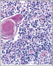
thymic corpuscles
isolated masses of closely packed, concentrically arranged epitheloreticular cells
blood thymic barrier
1.Capillary endothelium and its base membrane
2.Perivascular connective tissue space occupied by macrophages
3.Type I epithelioreticular cells
•Barrier between the T cells and the lumen of cortical blood vessels.
•Protects developing lymphocytes in thymus from antigens .
•Separates them from mature immuno-competent lymphocytes circulating in the blood stream.

spleen
largest lymphatic organ, filters blood and reacts to blood borne antigens
hematopoietic and immune functions

immune function of spleen
•Antigen presentation by APCs and initiation of immune response
•Activation and proliferation of B and T lymphocytes
•Removal of macromolecular antigens from blood
hematopoietic function of spleen
•Removal and destruction of aged RBCs
•Retrieval of iron from RBCs
•Formation of RBCs in fetal life
•RBC and platelet storage in some species
structure of spleen
encapsulated, trabeculae extend into parenchyma
•Parenchyma can be subdivided into: red pulp and White pulp


red pulp
vascular component (lots of RBCs)
consists of splenic sinuses separated by splenic cords

white pulp
lymphocyte aggregates, around central a, forming a PALS
contain central artery derived from splenic a.
nodules aooear as localizes expansions of PALS and displace a to the side (helps identify spleen)

splenic cords
loose meshwork of reticular cells and reticular fibers that contain large numbers of erythrocytes, macrophages, lympohocytes, plasma cells, and granulocytes.
splenic sinuses
special sinusoidal capillaries lined by rod-shaped endothelial cells (extremely long cells that run parallel to the direction of the vessel).
spaces allow blood cells to pass in and out, macrophages monitor lumen.


thymus


lymph node

spleen


tonsil

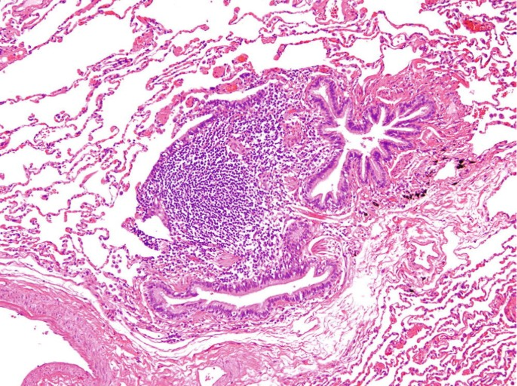
BALT
in lung