Lect 2 - Cell/Cell organization continued
 Call Kai
 Learn
 Practice Test
 Spaced Repetition
 Match
 Flashcards
 Knowt Play
1/180

There's no tags or description
Looks like no tags are added yet.
181 Terms
Q: What causes a 10-year-old child with progeria to develop the arteries of a 70-year-old (premature aging)?
A: Lamin A mutation → defective nuclear lamina.
Where does the Rb protein put the brake on the cell cycle?
G1 → S
Apoptosis differs from necrosis because it…
Uses energy, no inflammation
Which of the five compartments gives the cell its shape?
Cytoskeleton
Q: What is the lipid bilayer composed of?
A: Phospholipids and proteins.
Q: Where is the lipid bilayer found?
A: Forms the plasma membrane and the walls of every intracellular organelle.
Q: How do phospholipids self-assemble into a bilayer?
A: Water-seeking (hydrophilic) heads face outward toward water, while fatty acid (hydrophobic) chains face inward.

Q: What is the fluid mosaic model of the plasma membrane?
A: Proteins float in and across the phospholipid bilayer.

Q: What is the function of cholesterol in the plasma membrane?
A: Cholesterol is an important component of the lipid bilayer that regulates membrane fluidity.
Q: What is the difference between integral and transmembrane proteins?
A: Integral proteins are embedded in the membrane (some on one side), while transmembrane proteins span the entire lipid bilayer.
Q: What are the main structural components of the plasma membrane?
A: Phospholipids, cholesterol, integral/transmembrane proteins, and carbohydrate side chains (glycoproteins).
Q: What is the function of integral proteins in the plasma membrane?
A: Span the lipid bilayer and function as channels, transporters, and receptors.
Q: What is the function of peripheral proteins in the plasma membrane?
A: Sit on one face of the membrane, often linked to lipids.
Q: What is the sugar coat of the plasma membrane composed of?
A: Oligosaccharides attached to lipids (glycolipids) and proteins (glycoproteins).
What marks the “cells outside”?
the sugar coat (oligosaccharide)

Q: What causes cystic fibrosis?
A: A missing or misfolded CFTR chloride channel in the plasma membrane.
Q: What happens to secretions in cystic fibrosis?
A: They become thick and sticky.
Q: What organs and systems are affected by cystic fibrosis?
A: Lungs, pancreas, intestine, liver, and reproductive tract.
Q: What is the inheritance pattern of cystic fibrosis?
A: Autosomal recessive.
Q: What causes familial hypercholesterolemia?
A: Defective LDL receptors, preventing LDL from entering cells.
Q: What causes retinitis pigmentosa?
A: Defects in photoreceptor membrane proteins.
Q: What are the characteristic symptoms of retinitis pigmentosa?
A: Progressive loss of night and peripheral vision.
Q: What do cystic fibrosis, familial hypercholesterolemia, and retinitis pigmentosa have in common?
A: All three are associated with plasma membrane defects.
Q: What causes Marfan syndrome?
A: A defect in the extracellular matrix caused by an FBN1 mutation → defective fibrillin in elastic fibers.
Q: What are the characteristic features of Marfan syndrome?
A: Tall stature, lens dislocation, and aortic disease.
Q: What is the inheritance pattern of Marfan syndrome?
A: Autosomal dominant.
Q: What causes Duchenne muscular dystrophy?
A: Loss of dystrophin, which normally anchors actin (cytoskeleton) to the cell membrane → muscle fibers tear and waste.
Q: What is the inheritance pattern of Duchenne muscular dystrophy?
A: X-linked.
Q: What causes epidermolysis bullosa?
A: Failure of cell adhesion proteins that anchor skin layers together (keratins, laminin, collagen VII).
Q: What is the characteristic symptom of epidermolysis bullosa?
A: Blisters after minor trauma.
Q: What causes Bardet-Biedl syndrome?
A: Defects in primary cilia due to failure of basal body/cilia proteins.
Q: What are the characteristic features of Bardet-Biedl syndrome?
A: Polydactyly, retinal degeneration, obesity, and kidney failure.
Q: What is the inheritance pattern of Bardet-Biedl syndrome?
A: Autosomal recessive.
Q: What are the main functions of the nucleus?
A: Storage, replication, and transcription of DNA.
Q: What is the nuclear envelope?
A: A double membrane that surrounds the nucleus.
Q: What supports the nuclear envelope?
A: Lamins (part of the nuclear cytoskeleton).

Q: What is the function of nuclear pores?
A: Nuclear pore complexes regulate traffic, allowing substances to move in and out of the nucleus.
Q: What organelle is closely associated with the nuclear envelope?
A: The endoplasmic reticulum (ER).
Q: What is the nuclear pore complex (NPC)?
A: A huge protein channel that passes through both nuclear membranes and controls what enters and exits the nucleus.
Q: What substances exit the nucleus through nuclear pore complexes?
A: mRNA and ribosome subunits.
Q: What substances enter the nucleus through nuclear pore complexes?
A: Proteins with nuclear localization signals.
Q: What has cryo-electron microscopy (cryo-EM) revealed about nuclear pore complexes?
A: Cryo-EM can resolve nuclear pores at molecular resolution and has revealed different inner-ring channel widths.
Q: Are nuclear pore complexes fixed openings or dynamic structures?
A: Dynamic structures — nuclear pores can dilate (change their channel width).
Q: What is the nuclear lamina?
A: A meshwork of lamin intermediate filaments lining the inner nuclear membrane.
Q: What are the main functions of lamins?
A: Give the nucleus its shape and organize chromatin.
Q: What happens when lamins are defective?
A: They can cause a laminopathy.
Q: What disease is associated with a defect in the nuclear lamina?
A: Progeria (caused by a lamin A mutation).
Q: What is Hutchinson-Gilford progeria?
A: A rare disorder characterized by premature aging.
Q: What is the typical life expectancy of individuals with Hutchinson-Gilford progeria?
A: Individuals usually die in their teens.
Q: What are the characteristic clinical features of progeria?
A: Abnormal growth, skin changes, loss of hair, small fragile bodies, wrinkled skin, atherosclerosis, and cardiovascular problems.
Q: What cellular structure is defective in progeria?
A: The nuclear lamina (lamin A mutation).
Q: Why does progeria affect multiple tissues throughout the body?
A: The nuclear lamina is important for maintaining nuclear structure in cells throughout the body, so defects can affect many tissues.
Q: What specific mutation in the LMNA gene causes progeria?
A: A single C → T change in LMNA exon 11 (G608G), which is a "silent" mutation.
Q: Why does a "silent" mutation in LMNA cause progeria?
A: It changes RNA splicing, not the codon, by activating a cryptic splice site.
Q: What happens when the cryptic splice site is activated in progeria?
A: Abnormal mRNA splicing causes a deletion of 50 amino acids.
Q: What is progerin?
A: A truncated, unprocessed prelamin A protein that retains a toxic farnesyl group.
Q: What is the dominant-negative effect of progerin on the nuclear lamina?
A: Misshapen nuclei, disorganized chromatin, and defective DNA repair.
Q: What happens to genomic stability in cells affected by progeria?
A: Defective DNA repair leads to increased genomic instability.
Q: What is the overall result of the LMNA mutation in progeria?
A: Premature aging in every tissue due to defective nuclear lamina function (laminopathy).
In cystic fibrosis, the CFTR chloride channel is missing from the
Plasma membrane
mRNA leaves the nucleus through
Nuclear pore complexes
Progeria results from a defect in
Lamin A
Q: What is the secretory pathway of proteins?
A: Ribosome → Rough ER → Golgi → Vesicles → Destination (outside the cell, plasma membrane, or lysosome).
Q: What is the function of ribosomes in the secretory pathway?
A: Translate mRNA into proteins. ER-bound ribosomes make secreted, membrane, and lysosomal proteins.
Q: What is the function of the rough ER in the secretory pathway?
A: Proteins enter the rough ER, where they fold and undergo post-translational modifications.
Q: What is the function of the Golgi apparatus in the secretory pathway?
A: Adds sugars to proteins and sorts them for delivery to their destinations.
Q: What is the order of protein movement through the Golgi apparatus?
A: Cis → Medial → Trans.
Q: What is the function of vesicles in the secretory pathway?
A: Carry protein cargo to its designated destination.
Q: What are the three possible destinations of proteins in the secretory pathway?
A: Secreted outside the cell, delivered to the plasma membrane, or sent to the lysosome.
Q: What is the primary function of ribosomes?
A: Ribosomes are the sites of protein synthesis, where RNA is translated into protein.
Q: What are the two locations of ribosomes in the cell?
A: Free in the cytoplasm or bound to the rough ER.
Q: What types of proteins are produced by free ribosomes in the cytoplasm?
A: Cytosolic, nuclear, mitochondrial, and peroxisomal proteins.
Q: What types of proteins are produced by ribosomes bound to the rough ER?
A: Secreted, membrane, and lysosomal proteins.
Q: What determines whether a ribosome is free in the cytoplasm or bound to the rough ER?
A: The protein's signal sequence determines its destination. Both locations use the same type of ribosome.
Q: What is the main structural difference between rough ER and smooth ER?
A: Rough ER is studded with ribosomes, while smooth ER has no ribosomes.

Q: What are the main functions of the rough ER?
A: Synthesis of secreted and membrane proteins and post-translational modification.
Q: In what types of cells is the rough ER especially abundant?
A: Secretory cells, such as plasma cells and pancreatic cells.
Q: What are the main functions of the smooth ER?
A: Steroidogenesis (steroid production) and detoxification reactions.
Q: In what organs is the smooth ER especially prominent?
A: Liver and adrenal cortex.
Q: How does the ER help with wound healing?
A: The ER reorganizes based on cell and tissue curvature, helping with gap repair during wound healing.
Q: What new role does the ER play during wound healing?
A: It acts as a central mechanotransducer, sensing changes in cell shape and helping the cell respond.
Q: Is the ER only responsible for protein synthesis and post-translational modification?
A: No, the ER can also sense changes in the cytoskeleton and help respond to them.
Q: What cellular compartment does the ER work with during wound healing?
A: The cytoskeleton, including actin bundles and focal adhesions.
Q: What is the main function of the Golgi apparatus?
A: Adds oligosaccharides to proteins and lipids, and sorts them for transport to their destinations.
Q: In what types of cells is the Golgi apparatus highly developed?
A: Secretory cells and cells exposed to cytotoxins.
Q: What is the order of transport through the Golgi apparatus?
A: Cis → Medial → Trans.

Q: What is the function of the cis face of the Golgi apparatus?
A: Receives incoming transport vesicles from the ER.
Q: What is the function of the trans face of the Golgi apparatus?
A: Ships out newly formed vesicles to their destinations.
Q: What are the two main pathways that the Golgi apparatus sorts proteins into?
A: Lysosomal pathway or exocytosis pathway.
Q: What is the main function of exocytosis?
A: Release of secretory products and renewal of membrane components.
Q: What are the two types of exocytosis?
A: Constitutive and regulated exocytosis.
Q: What is constitutive exocytosis?
A: Continuous secretion that is always active and requires no signal. It also renews membrane components.
Immunoglobulin secretion is an example of what?
constitutive exocytosis

Q: What is regulated exocytosis?
A: Secretory products are stored in granules until a chemical or electrical signal (like Ca²⁺) triggers their release.
Insulin and neurotransmitter release is an example of what?
regulated exocytosis
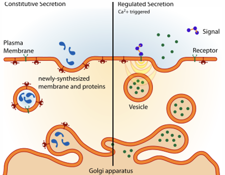
Q: What triggers regulated exocytosis?
A: A chemical or electrical signal involving Ca²⁺.
Q: What is the final step shared by constitutive and regulated exocytosis?
A: Vesicle docking and membrane fusion.
Q: What is the main function of endocytosis?
A: Bringing cargo into the cell through vesicles.
Q: What is the first step of receptor-mediated endocytosis?
A: A receptor recognizes and binds its ligand.
Q: What happens after a receptor binds its ligand during endocytosis?
A: The receptor-ligand complex gathers in a clathrin-coated pit.