Heart Anatomy Structures
 Call Kai
 Learn
 Practice Test
 Spaced Repetition
 Match
 Flashcards
 Knowt Play
1/30

There's no tags or description
Looks like no tags are added yet.
31 Terms
Layers of the heart
visceral pericardium (epicardium), myocardium, endocardium

Left and right auricles
ear-shaped muscular pouches attached to the outside of each atrium
expandable blood reservoirs and hormone-producing sensors attached to the upper chambers of the heart

atrium
collect incoming blood and pump it into the lower pumping chambers (ventricles)
right atrium
Function (Deoxygenated Blood):
-Receives Body Blood. It collects oxygen-poor blood returning from the rest of the body through three main veins: the superior vena cava (from the upper body), the inferior vena cava (from the lower body), and the coronary sinus (from the heart muscle itself).
-Delivers Blood to the Right Ventricle. When the heart relaxes between beats, the right atrium opens the tricuspid valve to let blood flow into the right ventricle, which then pumps it to the lungs for oxygen.
Left atrium
Function (Oxygenated Blood):
Receives Lung Blood. It collects freshly oxygenated blood returning from the lungs through four pulmonary veins (two from the left lung and two from the right lung).
Delivers Blood to the Left Ventricle. When the heart relaxes, the left atrium opens the mitral (bicuspid) valve to fill the left ventricle, which then pumps that oxygen-rich blood out to the entire body.

Right Ventricle
Deoxygenated (oxygen-poor) blood received from the right atrium.
Pumps blood through the pulmonary valve into the pulmonary trunk toward the lungs.

Left Ventricle
Oxygenated (oxygen-rich) blood received from the left atrium.
Pumps blood through the aortic valve into the aorta out to the entire body.
Right Coronary Artery
Feeds the right atrium, the right ventricle, and the bottom (inferior) wall of both ventricles.

Left Main Coronary Artery
Does not feed much heart tissue directly. Instead, it acts as the main trunk pipeline for the entire left side of the heart before splitting (bifurcating) into the LAD and LCX.

Left Anterior Descending (LAD) Artery
Supplies the front (anterior) wall of the left ventricle, the front two-thirds of the interventricular septum (the muscular wall separating the ventricles), and the apex of the heart.
Supplies roughly 45% to 50% of the left ventricle's muscle mass. A blockage near its origin is commonly called a "widowmaker" heart attack because it cuts off blood to the heart's primary pumping wall.


Left Circumflex (LCX) Artery
Feeds the left atrium and the outer side (lateral) and back (posterior) walls of the left ventricle.
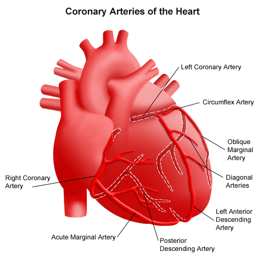

Ascending Aorta
Gives off only two branches right above the aortic valve: the left coronary artery and the right coronary artery,
!!Feed oxygen-rich blood directly to the heart muscle.

Aortic Arch
Its walls contain specialized stretch sensors called baroreceptors. They continuously monitor blood pressure and send rapid signals to the brainstem to adjust heart rate and blood vessel tightness if pressure rises or falls too quickly.

Descending Aorta
-Thoracic Aorta (Above the Diaphragm). Supplies blood to the chest wall, ribs (intercostal arteries), esophagus, and bronchi.
-Abdominal Aorta (Below the Diaphragm). Gives off major branches to the digestive organs (celiac trunk, superior and inferior mesenteric arteries) and kidneys (renal arteries) before splitting to feed the lower body.

Brachiocephalic trunk
the first and largest branch of the aortic arch, supplying oxygenated blood to the right side of the head, neck, and right upper limb

Left common carotid artery
the second and longest major branch of the aortic arch, supplying oxygenated blood to the left side of the head, neck, and brain

Left subclavian artery
the third and final major branch of the aortic arch, supplying oxygenated blood to the left upper limb, parts of the posterior brain, neck, and anterior chest wall.

Pulmonary trunk
the primary outflow vessel of the right ventricle, carrying deoxygenated blood from the heart toward the lungs for gas exchange. Along with the umbilical arteries in a fetus, it is one of the only arteries in the human body that transports oxygen-poor blood

left and right pulmonary arteries
transport deoxygenated blood from the right side of the heart to the lungs
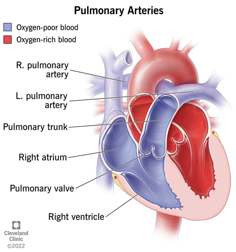

left and right pulmonary veins
a set of four large blood vessels (two from each lung) that carry oxygenated blood from the lungs directly into the left atrium of the heart.

Semilunar valves- pulmonary and Aortic valves
one-way valves located at the bases of the pulmonary trunk and aorta that prevent blood from flowing backward into the ventricles after each heartbeat

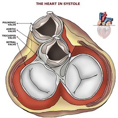
antrioventricular valves
one-way valves situated between the upper atria and lower ventricles that
!!ensure blood flows forward into the ventricles and never leaks backward during heart contractions
2 folds= bicuspid
3 folds- tricuspid

interventricular septum
the thick, triangular wall of cardiac tissue that separates the right and left ventricles of the heart

anterior interventricular sulcus
a shallow vertical groove on the front surface of the heart that marks the external boundary between the right and left ventricles
Superior Vena Cava
Drains upper body. It collects all deoxygenated blood from structures above the diaphragm, including the brain, head, neck, both upper limbs, and the chest wall.
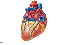
coronary sinus
the main venous collection vessel of the heart muscle, gathering deoxygenated blood from the myocardium and emptying it directly into the right atrium.
Right opening of the coronary sinus
When the right atrium contracts (atrial systole), the Thebesian valve helps prevent blood from surging backward into the coronary sinus.

Fossa ovalis
a shallow, oval-shaped depression on the interatrial septum inside the right atrium of the heart

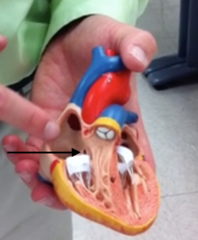
Chordae tendineae
strong, inelastic cords of fibrous connective tissue inside the ventricles that connect the papillary muscles to the cusps of the atrioventricular (tricuspid and mitral) valves.

papillary muscles
cone-shaped projections of cardiac muscle located inside the right and left ventricles of the heart

moderator band
a specialized, bridge-like band of cardiac muscle located exclusively in the right ventricle of the heart