SA commonly occuring hernias
 Call Kai
 Learn
 Practice Test
 Spaced Repetition
 Match
 Flashcards
 Knowt Play
1/33

There's no tags or description
Looks like no tags are added yet.
34 Terms
When do incisional hernias usually occur?
Following abdo surgery —> usually due to surgeon error
Can be acute (within 2 week of surgery) or chronic (weeks to years aftery surgery)
How does an incisional hernia present? How would you diagnose it?
Swelling/mass at surgical site
Carefully palpate wound for any defects —> confirm by US, rads, CT or surgical exploration (last resort)
How are acute incisional hernias treated?
Hospitalise & support w/ bandage until surgery —> risk of evisceration
Reopen & surgically explore
Remove ineffectual sutures
Debride devitalised fat/ tissue between wound edges then close primarily (PDS suitable)
How are chronic incisional hernias treated?
Less risk of evisceration because skin healed
Conservative management
if asymptomatic, no incarceration, owner can closely monitor
Surgical repair
Approach over original incision
Identify ring and excise the edge
Close the defect primarily / as for chronic traumatic hernias
How do you manage evisceration?
Surgical emergency
Protect viscera using large sterile dressing and place collar
Stabilise with fluid and AB
Ex Lap, resect damaged viscera, debride wound edges, lavage and close abdo
Close wound
How do body wall ruptures (traumatic ruptures) occur?
As result of significant trauma e.g. RTA, fall, kick
multiple inuries & rupture may be overlooked initially —> check body wall ruptures, diaphragmatic ruptures, injuries to urinary tract
What are the most common body wall ruptures (traumatic rupture)
Paracostal —> avulsion of muscles from last rib
Rupture / avulsion of pre pubic tendon —> body wall ruptured off caudal attachement
How do acute body wall ruptures present?
Marked bruising, swelling, pain @ site of rupture
Patient may have concurrent life threatening injuries
(Ability to palpate herniated organs subcutaneously is unlikely in acute injury)
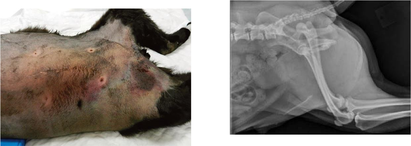
^^^ prepubic tendon rupture —> muscle not attached dorsally (follow from ventral m.) —> do cd coeliotomy
intramuscular & intra abdominal haemorrhage —> high risk anaesthesia
local inflam, subcut tissue injury
Referral!
How are body wall ruptures (acute presentation) diagnosed?
Imaging —> plain (± contrast) rads, U/S, CT
large clip
What should you do for cd abdo ruptures / concern for the urinary tract? (diagnosis)
retrograde urethrogram (RUG) & +ve contrast cystogram to assess integrity of bladder & urethra
How do you treat an acute body wall rupture?
Delay surgery 1-5 days to stabilise
Emergency surgery if cannot stabilise, penetrating abdo wound present or strangulation present
Explain the surgical approach to a body wall rupture repair
Prepare large area
Debride devascularised / necrotic tissue
Repair m. layers individually
Use appositional synthetic monofilament absorbable sutures
Include strong tissues in repair (fascia / ligament / bone)
Consider referral for multidisciplinary approach (imaging, anesthesia, critical care, surgery)
How would a chronic body wall rupture present?
Patient presents w/ mass/swelling (= herniated tissue, may not be @ location of the hernia)
Hernial ring may be palpable
Herniated structures may be felt subcut
Differentiate from seroma, neoplasia, cyst, abscess etc. (imaging)
How is a chronic body wall rupture treated?
Surgery once diagnosis is confirmed via imaging
Incise directly over rupture
Enlarge hernial ring if needed to reduce herniated structures
Repair as for acute hernias
Consider referral
Explain how a diaphragmatic rupture happens
Usually due to blunt trauma w/ glottis open
Incr pleuroperitoneal pressure gradient (due to air moving out of lungs through open glottis) causes tearing of m. of diaphragm
Commonly ass. w/ RTAs, kicks, falls
What are the main presenting signs of diaphragmatic rupture?
Dyspnoea (Incr RR and shallow, decr lung sounds)
Lethargy
Reduced appetite
Borborygmi in thorax due to decr lung sounds
Reduced/ abnormally positioned heart sounds
displaced apex beat due to herniated abdo contents
Tucked up abdomen
Can exist w/ other significant injuries e.g. long bone fracture (may be overlooked on initial presentation)
Cats can present weeks/ months after injury
How is diaphragmatic rupture diagnosed?
Care re-positioning / handling —> risk of decompensation / death
U/S —> do conscious in sternal recum
Thoracic rads —> might have to do w/ single DV view
CT (usually unnecessary)
What is being shown in this radiograph?
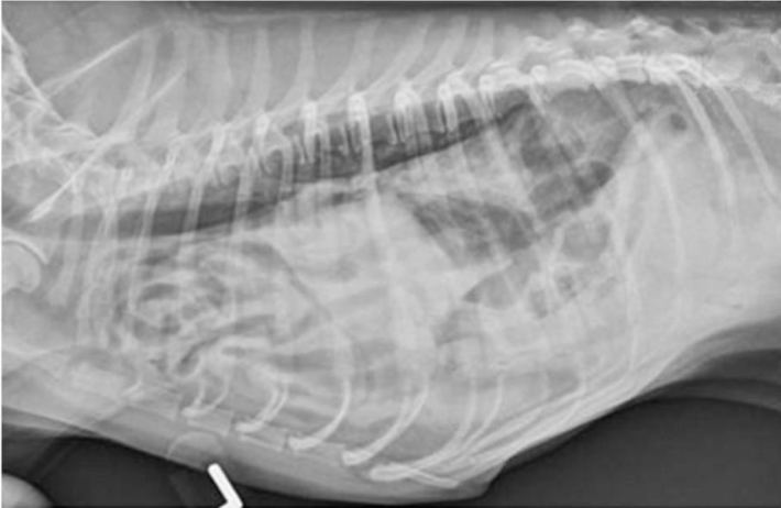
Diaphragmatic hernia
How do you treat an acute diaphragmatic rupture?
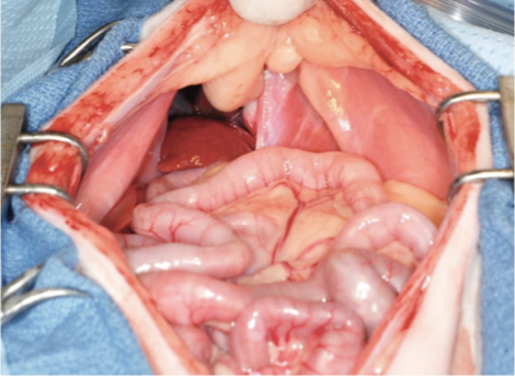
Try to stabilise prior to surgery & GA
intra op risk of decompensation / death
induce → GA = short time as poss
Tilt table so head / thorax slightly elevated
Can decompress —> prepare for intra-op manual ventilation
Perform cr midline coeliotomy
ID diaphragmatic tear (check for more), reduce herniated organs, assess viability → resect devitalised tissues
Close tear (simple int / cont / cruciate mattress), drain thorax to resolve intra-op pneumothorax (chest drain?), check abdo organs for concurrent trauma
Routine closure of abdo
How do you treat a chronic diaphragmatic rupture? How will it differ in acute presentation
Surgical
More likely to be stable for GA (not suffered recent trauma)
May be adhesions of herniated structures making surgery more challenging
be prepared to extend incision to caudal sternum / abdo if required
Post- op greater risk of re-inflation pulmonary oedema (life threatening)
lungs squashed into smaller vol for long period of time → correction → more space in pleural cavity → ventilation → overinflated lungs too quickly → fibrotic lesions in lungs tear
place chest drain, empty air and fluid q.4-6hrs over a couple days
What causes a peritoneopericardial diaphragmatic hernia (PPDH)?
Congenital —> due to failure of fusion of transverse septum → midline communication between abdo & pericardium
often incidental finding
What does PPDH often commonly coexist with?
Other defects e.g. split caudal sternal, cr abdo wall defect etc.
What are the clinical signs of a PPDH?
Often asymptomatic and is an incidental finding on thoracic radiographs
Respiratory signs —> reduced volume of thorax for lungs to expand
Elevated respiratory rate, cough, exercise intolerance
GI signs
Various including V+, D+, inapp, polyphagia, etc
Cardiac signs
Sudden collapse
R-HF (tamponade)
What is this radiograph showing?
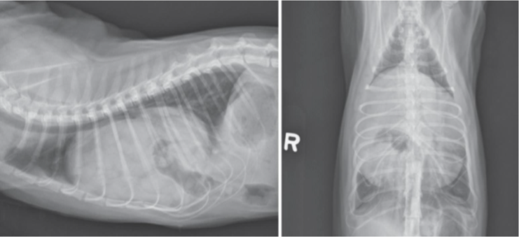
PPDH
Massive cardiac sil w/ gas filled opacity superimposed and cannot differentiate from diaphragm
diaphragm fused with pericardium
Dorsally displaced trachea
V. clear silhouette because no movement as heart is just somewhere within sac
How do you treat a PPDH?
Asymptomatic patients —> conservative management
Symptomatic patients —> surgical repair
How is PPDH surgically repaired (symptomatic patients)?
(clip higher up sternum)
Ventral midline coeliotomy
Reduce viscera (may need to extend defect)
Resect any devitalised/strangulated tissues (unusual)
Close defect with interrupted / cont. monofilament absorbable suture (cruciate mattress if under tension)
For large defect —> harvest pericardium from attachment to diaphragm & use to close the defect → pneumothorax
Drain air from pericardial sac (& pleural cavity using needle & syringe if caused pneumothorax)
Routine closure of abdomen
What causes a perineal hernia?
Weakness of pelvic diaphragm = internal obturator muscles, levator ani & coccygeus muscles (either side of rectum)
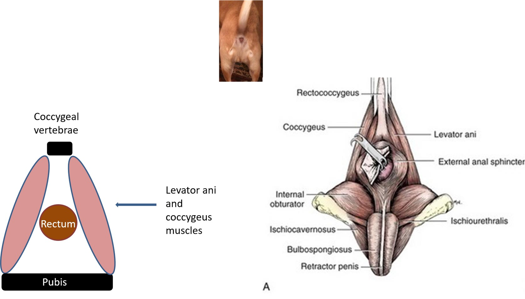
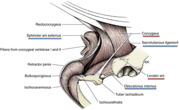
dilation of rectum & accumulation of faeces
→ tenesemus & constipation
What is the typical presentation of a perineal hernia?
Common
Almost exclusively seen in middle aged/older male entire dogs (/ male dogs castrated in later life)
Muscles of perineal diaphragm weaken & atrophy
What is being shown in this radiograph?
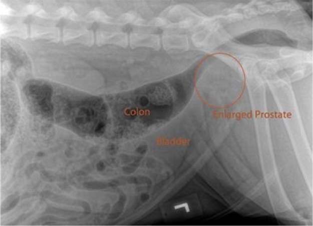
Perineal hernia can be seen @ same time as enlarged prostrate gland but not causative (cause most likely hormonal)
distal colon gets squeazed → faecal material in rectum → defecatory tenesemus
What happens due to a perineal hernia?
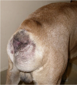
Loss of muscular support of rectum
Sacculation (expansion of rectum on one side i.e. unilateral perineal hernia), deviation (into hernia) or dilation of rectum (bilateral perineal hernia)
Accum of faeces in rectum
Constipation, straining to defaecate
Swelling/bulge of perineum on affected side(s)
Perineal bulge can disappear after defaecation
Owner may push on bulge to aid defaecation
faeces witin descended rectum = bulge
Bladder can retroflex into the hernia
What is being shown here? What clinical signs are associated with this?
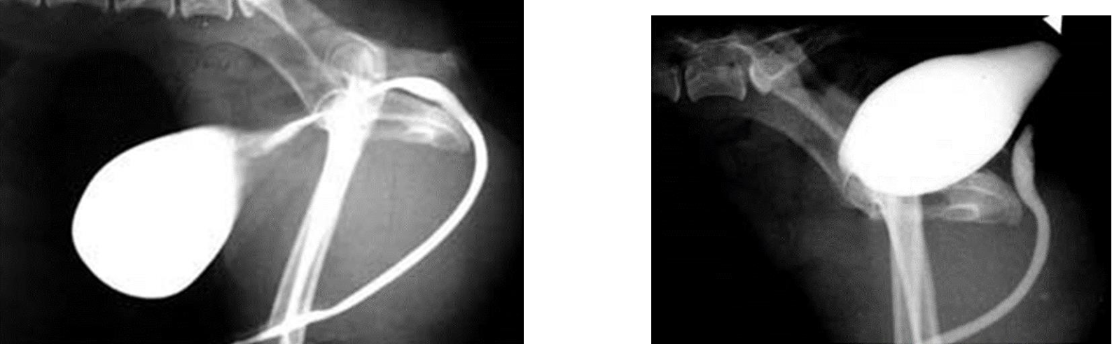
Retroflexion of the bladder → dysuria
How is a perineal hernia diagnosed?
Rectal exam —> palpate laterally and there will not be lateral support muscles (+/- imaging)
feel laterally to feel pelvic diaphragm —> can feel bands of tissue while muscles being lost → finger can be pushed towards skin & seen through subcut tissue = diagnosis of perineal hernia!

What other investigations can be done for perineal hernia?
Abdo rads & or U/S to assess other potential causes of defaecatory tenesmus
often concurrent benign prostatic hypertrophy (concurrent, not causative)
CT rarely indicated
+ve contrast retrograde urethrogram to locate bladder in dysuric patients —> bladder herniation?
How is a perineal hernia treated?
Conservative = Not reccommended unless GA contraindicaton
add fibre, laxative, castration (medical/surgical)
muscles cont. to deteriorate (delayed sx may be more challenging) & rectal dilation gets worse
risk of bladder herniation → life threatening urethral obstruction
Surgical (recommended)
castration plus hernia repair (concurrent) —> inclusion of adjacent int. obturator m. & / or superficial gluteal muscles
consider referral
good prog (relatively minor complications but possibility of recurrence)