R5 - lung structure,function and physiology
 Call Kai
 Learn
Learn Practice Test
 Spaced Repetition
 Match
Match Flashcards
 Knowt Play
1/39

There's no tags or description
Looks like no tags are added yet.
Name | Mastery | Learn | Test | Matching | Spaced | Call with Kai | Chat |
|---|
No analytics yet
Send a link to your students to track their progress
40 Terms
What is the gross structure of the lungs ?
Gross structure:
• Right lung: three lobes
• Left lung: two: the heart takes up the space of teh third lobe
• Each lung is in a pleural sac
o Visceral and parietal layers
o Thin fluid film between - allows movement
• Hilum: airways, arteries and veins enter together
• Pleural pressure is negative - keeps the lungs expanmded against their natural tedecy to recoil

What are the two types of circulation in the lungs?
Two circulations, two jobs
Pulmonary - carries the whole cardiac output, at a low pressure for gas exchange
Bronchial circulation - comes from systematic circulation ,supplies the airway wall
What are the two functional zones of the lungs?
Conducting zone: moves air; no gas exchange.- tube
Respiratory zone: where gas exchange occurs.- sac
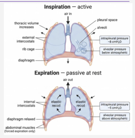
What happens during inspiration ( active) ?
The diaphragm contracts and flattens, external intercostal muscles contract,lift ribs upwards and outwards
Thoracic volume increases
intrapleural pressure decreases
alveolar pressure falls below atmospheric pressure
air flows into the lungs.
What happens during expiration (passive)?
External intercostal respiratory muscles relax
Elastic recoil reduces lung volume
Alveolar pressure rises above atmospheric pressure
Airflow out of the lungs
Note: air flows down the pressure gradient
What equation is used to calculate air flow ?
Flow = ΔP ÷ R
Flow- volume of air moved per second
ΔP = pressure difference ( alveolar pressure—atmospheric)
R = airway resistance
Air therefore flows down a pressure gradient.
What is the Wiebels idealised model for the structure of the trachea to the alveolus?
23 divisions from trachea to alveolar sac,at each division the number double
trachea= 218 mm
bronchiole = o.5 mm

How does the wall change as you go down from the trachea to the alveolus?
cartilage rings change to plates , none below 1mm
bronchiole
patency - muscle tone and alveolar tethering
What are the conducting and respiratory airway generations?
Conducting zone: generations 0–16
Respiratory zone: generations 17–23
What is a bronchiole?
A small airway with no cartilage. Cartilage disappears from the airway wall below approximately 1 mm diameter.
Where is resistance the highest?
In the medium-sized bronchi, not the smallest airways.
Why does the smallest airway have relatively low total resistance?
Because airway branching creates a very large total cross-sectional area, reducing overall resistance.
What is the structure of the airway wall in cross-section?
1. Mucus
2. Periciliary layer
3. Ciliated epithelium
• Goblet cells, sealed by tight junctions
4. Basement membrane - Reticular lamina
5. Lamina propria
• Mast cells, eosinophils, macrophages ,Microvessels,Sensory nerves
6. Airway smooth muscle - Only layer that actively contracts
7. Submucosal gland - Duct to the lumen

What is the role of teh ciliated epithelium?
It forms a protective barrier and provides mucociliary clearance, moving mucus and trapped material out of the airway.
What is the role of the goblet cells and submucosal glands?
They produce mucus. Excess mucus can fill and narrow the airway lumen
What are the changes to the structure of the airway wall in asthma and COPD?
• Reticular basement membrane thickens (subepithelial fibrosis)
• Smooth muscle mass increases
• Microvessels leak — this is the wall oedema (Slide 11)
Key concept: Every asthma and COPD drug acts somewhere on this diagram
What are the cells in the airway walls

Which inflammatory cells are important in asthma and COPD?
Asthma: eosinophils are characteristic; IL-5 driven.
COPD: neutrophils are characteristic.
Macrophages: contribute to chronic inflammation.
What is the dominant autonomic nerve supply to the airway, and how it work?
The parasympathetic nervous system, mainly through the vagus nerve.
ACh activates the M3 receptors ( Gq), which leads to smooth muscle contraction and increased gland secretion
M2 ( Gi) autoreceptor on the nerve terminal further limits ACh release; resting bronchomotor tone is vagal
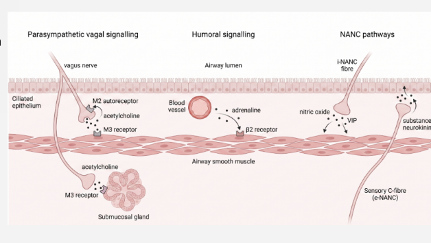
Does the smooth muscle have functional sympathetic innervation ( connected nerves) /
No. Airway β₂ receptors respond to circulating adrenaline rather than to direct sympathetic nerves. β₂ agonists can also activate these receptors as drugs.
What are the differences between airflow obstruction in asthma and COPD?
Asthma: variable and substantially reversible.
COPD: persistent, progressive and only limited/barely reversible.

What are some characteristics of asthma and COPD?

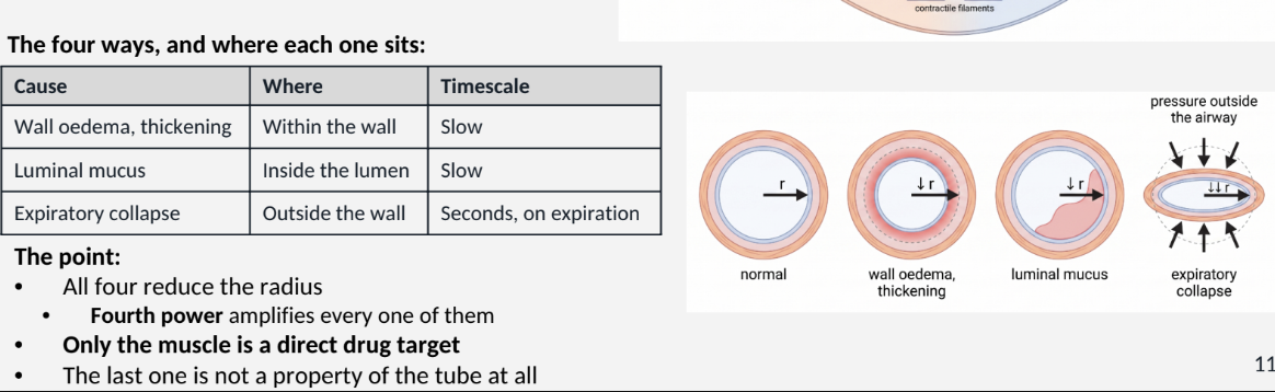
What causes airway narrowing within the airway ?
Smooth muscle contraction - the wall moves in
Airway-wall oedema/thickening
Mucus in the lumen
What is the result of narrowed airways?
lumen radius decrease
airflow decreases, lungs work harder,pateints wheeze
no drugs can affect the length and the viscosity
radius is the only way to control asthma and copd
How does airway radius affect airway resistance?
Resistance is related to the fourth power of the radius. Therefore, even a small decrease in radius produces a large increase in airway resistance.
eg.radius is halved = 2^4 = 16 , so resistance increase 16 fold
How does M3 receptor activation cause airway smooth- muscle contraction normally? ( ans 19) AIRWAY CONSTRICTION
Acetylcholine binds to M3 receptors on airway smooth muscle cells.
Gq Protein: Activates the internal Gq messenger protein.
↑ Ca²⁺ (Calcium): Triggers a sudden flood of calcium inside the cell.
MLCK Activation: High calcium activates Myosin Light Chain Kinase (the muscle "ignition switch").
Contraction: The muscle fibres slide together, clamping down and narrowing the airways.
How do β2 receptors cause Bronchodilation normally ? - AIRWAY DIALATION
β₂ Receptor: Adrenaline binds to beta-2 receptors on airway smooth muscle cells.
Gs Protein: Activates the internal Gs (stimulatory) messenger protein.
↑ cAMP: Gs activates adenylyl cyclase, causing a surge of cyclic AMP (cAMP) inside the cell.
PKA Activation: High cAMP turns on Protein Kinase A (PKA).
↓ Ca²⁺ (Calcium): PKA forces calcium levels inside the cell to decrease
Relaxation: Without calcium, the muscle fibres let go, opening up the airways.
What are four factors that can reduce airway radius during obstruction ?
Increased smooth-muscle tone
Airway-wall thickening/oedema
Luminal mucus
Expiratory airway collapse
What factors of the alveolus hold the airway open ?
the structure
radial traction
the scaffold
destroy the septa
What is the structure of the alveolus
1 The structure:
• Septum — the wall shared by two alveoli
• Elastin · collagen — the scaffold
• Alveolar attachments — septa insert into the airway wall
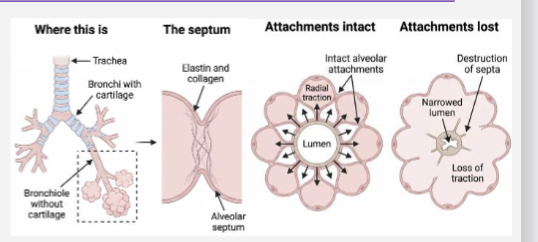
What is radial traction?
Alveolar septa attach to the airway wall and pull the airway outwards, helping keep small airways open.
What does the scaffold give you ?
Elastic recoil — elastin, collagen, surface tension
• Compliance — ease of inflation · emphysema high, fibrosis low
• High compliance is not good lungs
• Surfactant — type II pneumocytes
• Stops surface tension shutting small alveoli
What happens when alveolar septa are destroyed in emphysema?
Alveolar attachments and radial traction are lost → elastic recoil decreases → small airways collapse more easily during expiration → air trapping occurs.
note - no drugs repair the septum
How do emphysema and fibrosis affect lung compliance?
Emphysema: ↑ compliance but ↓ elastic recoil.
Fibrosis: ↓ compliance because the lungs are stiff.

How do we see or measure obstruction?
Spirometry — one manoeuvre:
Biggest breath in, then blow out as hard and as long as you can

What do FEV₁, FVC and PEF measure?
FEV₁: forced expiratory volume in 1 second.- SPIROMETER
FVC: forced vital capacity exhaled.-everything you can blow out
PEF: peak expiratory flow — the fastest expiratory flow achieved.- PEAK FLOW METER
When do these values fall ?
Narrow tube → high resistance → slow emptying — FEV1 falls (slide 10)
• Blowing hard compresses the airway — that is slide 13
• Lost recoil traps air behind shut airways — FVC falls (slide 12
What does an FEV₁/FVC ratio below 0.7 indicate?
Airflow obstruction. FEV₁ can fall in both obstruction and restriction, so the ratio is important for distinguishing them. In asthma, variability/reversibility is particularly important.
the airway wall target map

key concepts
Think of the lung as “the tube and the sac”:
Tube = airway
→ Determines resistance
→ Narrower radius = much greater resistance
→ Asthma/COPD drugs target things such as β₂, M3, glucocorticoid, H1 and CysLT1 receptors
Sac = alveoli
→ Determines compliance + elastic recoil
→ Helps tether small airways open through radial traction
→ Alveolar destruction in COPD contributes to airway collapse and air trapping
→ The lecture emphasises that the drugs discussed do not repair destroyed alveolar septa.