Benign Tumors
 Call Kai
 Learn
Learn Practice Test
 Spaced Repetition
 Match
Match Flashcards
 Knowt Play
1/250

There's no tags or description
Looks like no tags are added yet.
Name | Mastery | Learn | Test | Matching | Spaced | Call with Kai | Chat |
|---|
No analytics yet
Send a link to your students to track their progress
251 Terms
What is the 3d most common site of spread of cancers
Skeleton
Majority of bone metastases are from what cancers
Breast lung and prostate
Neoplasm that originates from non bone forming supportive connective tissue of marrow
Giant cell tumor
Does a giant cell tumor have pain
YES
Age for giant cell tumor
20-40
Another name for giant cell tumor
Osteoclastoma
Location for giant cell tumor
Mostly long bones
Sacrum newar SIJ
Giant cell tumors that are distal to the radius mean
Higher chance of being malignant
Most common benign tumor of the sacrum
Giant cell tumor
Clinical presentation of giant cell tumor
Localized pain
Restricted ROM
Radiographic features of giant cell tumor
Geographic
Eccentric
Metaphyseal-epiphyseal
Subarticular (doesn't enter joint space)
Cortical thinning
Soap bubbly

Giant cell tumor

Another giant cell tumor

Giant cell tumor in the distal ulna

Giant cell tumor in the distal radius

Giant cell tumor in the sacrum

Treatment of giant cell tumors
Surgical cureetage- recurrence of 12-50%
Radiation of spinal lesions
5 yr survival for malignant about 10%
Large geographic tumor, eccentric location, distal part of the radius but the X ray has open growth plates... is it a giant cell tumor
NO- because it is a kid and doesn't go to the end of the bone

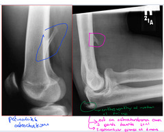
Pedunculated Osteochondroma

Bony exostosis on the external surface of a bone and usually lined by hyaline cartilage cap
Solitary Osteochondroma
What is the most common benign osseous tumor
Solitary Osteochondroma
Age of solitary osteochondroma
Below age 20 (before growth plates close)
Clinical features of solitary Osteochondroma
Asymptomatic
Painless hard mass around a joint
Pain from fractured stalks or cord compression
Obstructive uropathy from pelvic tumor
If there is pain with a solitary Osteochondroma then what does this mean
Pain and new growth may indicate malignant transformation
-it should stop growing once the growth plates close
-usually turns into a Chondrosarcoma or osteosarcoma
What are the 2 types of solitary osteochondroma
Sessile
Pedunculated
What type of solitary Osteochondroma has a broad base and no stalk, common in the humerus and scapula
Sessile
What type of solitary Osteochondroma has a long stalk with cartilage cap, common in knee, hip and ankle
Pedunculated
Ways to describe a Pedunculated solitary Osteochondroma
Coat hanger exostosis
Cauliflower cap
Osseous location of solitary Osteochondroma
Any bone formed by enchondral ossification
Relationship of solitary Osteochondroma and growth plates
When growth plate stops growing some does the growth of the solitary osteochondroma
Radiographic features of solitary Osteochondroma
Bony exostosis
Pedunculated and sessile
Pedunculated base with elongated bony stalk
Tip may have dense cortical rime
Sessile may cause metaphyseal widening
If there is a single solitary Osteochondroma on a single bone than it is
Solitary Osteochondroma
If there are 2 or 3 bones with a solitary Osteochondroma then and no family history then
It's just multiple Osteochondromas
Widespread solitary osteochondromas with a family history then what is it called
Hereditary multiple exostosis (HME)
Pedunculated solitary osteochondroma

Sessile osteochondroma

Inherited automal dominant condion characterized by multiple Osteochondroma often causing metaphyseal overgrowth
Hereditary multiple exostosis (HM)
Age of occurrence for hereditary multiple exostosis
2-10 yrs
Difference between Pedunculated Osteochondroma and a supracondylar process slide
If it points away then Pedunculated Osteochondroma, if ti points towards the joint space then is a supracondylar process
What is it when there are multiple osteochondroma`s around multiple joints
Hereditary multiple exostosis
Does hereditary multiple exostosis hurt
No, its multiple painless hard masses
Radiographic features of hereditary multiple exostosis
Shortening of 4th and 5th metacarpals
Supernumerary fingers and toes
Madelung or bayonet deformity of forearm
Disproportionate shortening of extremity
Non-neoplastic fluid filled cystic cavity lined with fibrous tissue
Simple/solitary/unicarmeral bone cyst
-not really a tumor
Age for simple/solitary bone cyst
3-14 yrs
Hereditary multiple exostosis
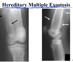
Example of madelungs deformity and hereditary multiple exostosis

Clinical symptoms of solitary bone cyst
Asymptomatic unless fractured
Where do most solitary bone cysts occur
Proximal humerus (MAINLY THIS)
Femur, fibula, calcaneal neck, talus
Where on the bone does a solitary bone cysts occur
Metaphyseal adjacent to growth palte
Central, long axis parallel to host bone
What does solitary bone cysts look like
Loculatd with Endosteal scalloping
Truncated cone appearance (wider at metaphysics and narrower near diaphysis)
Fallen fragment sign if fractured
Most effective treatment for solitary bone cyst
Injection of steroids
Non-neoplastic Expansile lesion containing thin-walled blood-filled cystic cavities
Aneurysmal bone cyst
What do you look for to see if a solitary bone cysts fractured
Fallen fragment sign

Solitary bone cyst up the proximal humerus

Solitary/simple bone cyst

Solitary bone cyst in the calcaneus

Age for aneurysmal bone cyst and mainly occurs in who
10-30 yrs in females
Most aneurysmal bone cysts are primary or secondary? If they're secondary what are they from
Mainly primary
Secondary dare from another lesion (Giant cell tumor, osteosarcoma, simple bone cyst)
Clinical features of aneurysmal bone cyst
ACUTE ONSET OF PAIN WIT RAPID INCREASE IN SEVERITY
Often people with an aneurysmal bone cyst also have
History of trauma
Most aneurysmal bone cyst occur when
Spine, especially the posterior elements of the spine
Radiographic features of aneurysmal bone cyst
Purely lytic and eccentric, metaphysis
Soap bubble appearance
Aneurysmal bone cyst is hard to see on what imaging
CT
-cause how thin the cortex is
What do you see on CT with an aneurysmal bone cyst
FLUID FLUID LEVELS
-due to blood layering
What do you see on bone scan with aneurysmal bone cyst
Increased uptake
What do you see on MRI with aneurysmal bone cyst
Multiple different signal intensities representing different stages of blood by-products
-also can see fluid fluid levels
What part of the spine does an aneurysmal bone cyst like
Neural arch part in the spine
Treatment for aneurysmal bone cyst
Surgical curettage
Radiation therapy especially for when its in the spine
Aneurysmal bone cyst and why would it not be a giant cell tumor
Giant cell tumors are age 20-40 and the patient here is a 13 yr old kid, ant the tumor is not subarticular

Fluid fluid levels on an MRI from an aneurysmal bone cyst

Aneurysmal bone cyst

Difference between aneurysmal bone cyst and giant cell tumor in the distal radius slide

A rare benign tumor with unlimited growth potential
Osteoblastoma
Aneurysmal bone cyst causing a missing pedicle

Another aneurysmal bone cyst in the spine

Age for osteoblastoma
10-20 years
What resembles an Osteoid osteoma but is large
Osteoblastoma
Symptoms of osteoblastoma
Dull pain of insidious onset, worse at night and NOT RELIEVED BY ASPRIN
Swelling and tenderness
Locations of osteoblastoma
Spine (especially the posterior elements)
Long bones
Radiographic features of an osteoblastoma
Spinal lesions radiolucent and Expansile
Long bones show progressive Expansile lesion that may rapidly increase in metaphyseal to diaphyseal direction
Treatment for osteoblastoma
Small legions are by excision or curettage
Spinal lesion respond well to radiation
Osteoblastoma in the spine
(Also read slide here)

Osteoblastoma of the spine in lumbars and on CT

Another sample of an osteoblastoma in the cervical spine

Giant osteoid osteoma

Benign skeletal neoplasm composed of osteoid and woven bone that is less than 1.5 cm in diameter
Osteoid osteoma
Age for Osteoid osteoma
10-25 yrs of age
Clinical features of Osteoid osteoma
Local pain weeks to years in duration, decreased by activity
PAIN RELIEVERS HELP WITHIN 20-30 minutes
What occurs in the spine with an osteoid osteoma
Painful scoliosis (concave)
-effects the posterior elements of lower thoracic and upper lumbars
Other than the spine where else does Osteoid osteoma like
Metaphysis and into the diaphysis of long bones show progressive
Radiographic features of osteoid osteoma
Lesion is the radiolucent nidus (central part of radiolucent where the tumor lives) with REACTIVE SCLEROSIS
CORTICAL: NIDUS IN CORTEX
Example of that ovoid radiolucency (the nidus) with surrounding reactive sclerosis that comes along with osteoid osteoama

Osteoid osteoma in the femur

What may occur with an osteoid osteoma when they occur in Intracapsular areas
Limited amount of reactive sclerosis

Another Osteoid osteoma in the femur

One more osteoid osteoma in the femur

What do you see on a bone scan that indicates is it an osteoid osteoma
Double density sign
-super dense in the middle and lighter color on the outer parts of it

List the possible causes of increased density of pedicle/lamina area on a vertebrae
Missing pedicle
tumor (osteoblastoma- primary tumor or osteoid osteoma)
Blastic Mets
Facet arthrosis
Pars defect on opposite side
Osteoid osteoma effecting the pedicle in the spine

What the nidus looks like for an osteoid osteoma when looking vertically though it on a CT
