Thorax 2 - Coronary Arteries & Veins/Pericardium/Heart Walls
 Call Kai
 Learn
Learn Practice Test
 Spaced Repetition
 Match
Match Flashcards
 Knowt Play
1/96

There's no tags or description
Looks like no tags are added yet.
Name | Mastery | Learn | Test | Matching | Spaced | Call with Kai | Chat |
|---|
No analytics yet
Send a link to your students to track their progress
97 Terms
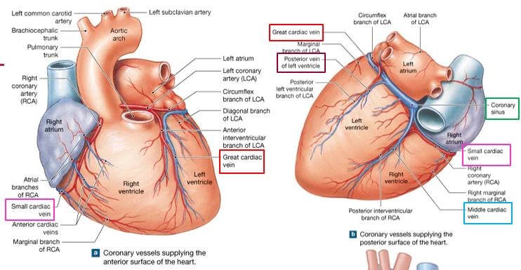
Why is coronary circulation necessary?
The heart wall is too thick to get enough O₂/nutrients from blood inside its chambers. The heart muscle needs its own blood supply.
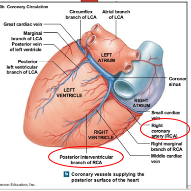
What are the two main coronary arteries?
Right coronary artery (RCA) and left coronary artery (LCA).

Where are the openings of the coronary arteries?
Just outside the aortic semilunar valve.

Where does the RCA travel?
Toward the right side in the coronary sulcus.

What are the atrial branches of the RCA?
RCA branches that travel toward/supply the atrial region.

What are the anterior ventricular branches of the RCA?
RCA branches that travel toward the anterior ventricular region.

Where does the right marginal branch travel?
Along the right margin toward the apex.

What is another name for the posterior interventricular branch?
R-PDA, or right posterior descending artery.

What determines right vs. left coronary artery dominance?
Whether the right or left coronary circulation gives rise to the PDA.
Where does the LCA travel?
To the left of the aorta along the coronary sulcus.

How does the diameter of the LCA usually compare with the RCA?
The LCA is usually larger.

What is another name for the anterior interventricular branch?
LAD — left anterior descending artery.
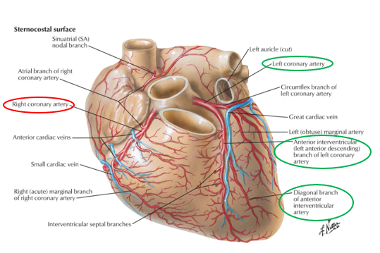
What artery does the LAD anastomose with?
The R-PDA….rare

What is the diagonal branch a branch of?
The LAD.
Why is the diagonal branch important?
It contributes to collateral circulation to areas of the ventricle.

What major artery branches from the LCA and wraps around the heart?
Circumflex artery.

What are the left atrial branches of the circumflex artery?
Branches of the circumflex that travel toward the left atrium.

What is the left marginal branch?
A branch associated with the LCA/circumflex that travels along the left margin.

What is the posterior ventricular branch of the LCA?
A branch of the left coronary circulation supplying the posterior ventricular region.

Heart attack
A complete blockage of a coronary vessel that cuts off the blood supply to cardiac tissue that vessel normally supplies….tissue dies…impaired ventricle function….depends on collateral circulation
Angioplasty
open narrowed or blocked blood vessels and restore healthy blood flow

Coronary artery bypass graft (CABG)
“cabbage”
Fat (plaque) build up, restricts blood to heart…take a healthy blood vessel (graft) from body…attach above and below the blocked area = new "detour"

Where do the majority of cardiac veins drain?
Into the coronary sinus.

Where does the coronary sinus drain?
Into the right atrium.

Which cardiac vein runs with the LAD?
Great cardiac vein.

Which artery is associated with the great cardiac vein?
LAD.

Which cardiac vein runs with the R-PDA?
Middle cardiac vein.

Which artery is associated with the middle cardiac vein?
R-PDA.

Which cardiac vein runs with the right marginal artery?
Small cardiac vein.
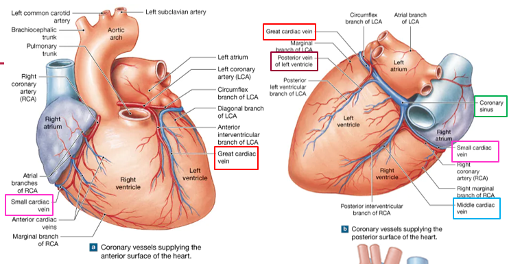
Which artery accompanies the small cardiac vein?
Right marginal artery.

What artery accompanies the posterior vein of the left ventricle?
Left posterior ventricular artery.

Where do the anterior cardiac veins empty?
Directly into the right atrium….NOT coronary sinus
What is the endocardium?
Thin innermost heart layer in contact with blood inside the atria and ventricles.

What is the myocardium?
The contractile cardiac muscle layer.

What is the epicardium?
The outer covering of the heart wall….Visceral pericardium.
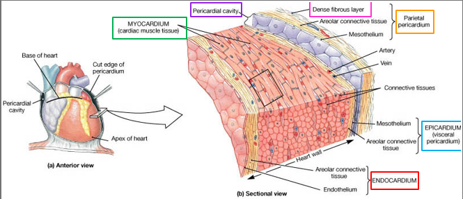
What is another name for the epicardium?
Visceral pericardium.

What is the parietal pericardium?
The outer serous pericardial layer separated from visceral pericardium by the cavity.

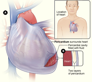
What is the pericardial cavity?
The space between visceral and parietal pericardium.

What is the fibrous pericardium?
The most superficial outer fibrous layer surrounding the heart.

Put the heart/pericardial layers in order from inside outward.
Endocardium → myocardium → epicardium/visceral pericardium → pericardial cavity → parietal pericardium → fibrous pericardium

What is the relationship between visceral and parietal pericardium?
They are continuous with one another.
Where does visceral pericardium fold over to form parietal pericardium?
Near the great vessels.
What does visceral pericardium become when it folds over near the great vessels?
Parietal pericardium.
What do the visceral and parietal pericardium create?
A closed pericardial sac.
What is the space inside the pericardial sac called?
Pericardial cavity.
What is mesothelium?
A single layer of epithelial cells lining the pericardial cavity….this is a serous membrane, secretes pericardial fluid
What type of membrane lines the pericardial cavity?
The mesothelium…a serous (smooth slippery, stop friction) membrane
What does the mesothelium (serous membrane) secrete?
Pericardial fluid.
What is the function of pericardial fluid?
It lubricates the surfaces and reduces friction as the heart beats.
What type of tissue makes up the fibrous pericardium?
Dense connective tissue.
What is the fibrous pericardium fused to?
Parietal pericardium.
What is the pericardial sac?
The pericardium that encloses the heart.

How does the pericardial sac help maintain heart position?
It attaches to connective tissue between the lungs.
What nearby source of infection does the pericardium help protect the heart from?
Lungs

What fluid is held within the pericardial sac?
Pericardial fluid

Why is pericardial fluid important?
Reduces friction as heart beats

What type of membrane is the pericardium?
Serous membrane

Pericarditis
Inflammation of the pericardium…viral, bacterial, lupus, cancer, injury

What physical finding can result from pericarditis?
A friction rub
What can severe pericarditis lead to if more fluid accumulates?
Cardiac tamponade….fluid inside pericardial sac…fibrous pericardium not stretchy to expand…pressure around heart…muffled heart sounds
What is cardiac tamponade?
Increased pressure/compression of the heart from fluid within the pericardial sac.
What procedure is associated with cardiac tamponade?
Pericardiocentesis with ultrasound imaging.
Internal thoracic/mammary artery
branches off the subclavian artery

What artery gives rise to the internal thoracic artery?
Subclavian artery.

The internal thoracic artery lies between which two branches?
Thyrocervical trunk and vertebral artery.

What vessel gives rise to anterior intercostal arteries of ribs 1-7?
Internal thoracic artery

Where do posterior intercostal arteries arise?
Descending thoracic aorta.

Anterior vs. posterior intercostal artery origin?
Anterior = internal thoracic; posterior = descending thoracic aorta.

What supplies the anterior intercostal arteries of ribs 8–10?
Musculophrenic artery.
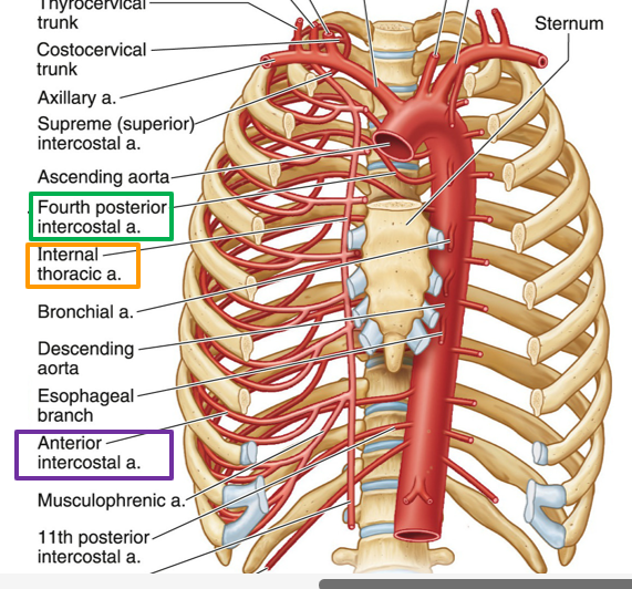
The musculophrenic artery is a continuation of what vessel?
Internal thoracic artery.

Trace the supply to anterior intercostal arteries of ribs 8–10.
Internal thoracic → musculophrenic → anterior intercostal arteries 8–10.

Where do anterior intercostal veins drain?
Into the internal thoracic vein.

Where does the internal thoracic vein drain?
Into the brachiocephalic vein.

Trace anterior intercostal venous drainage.
Anterior intercostal → internal thoracic → brachiocephalic vein.
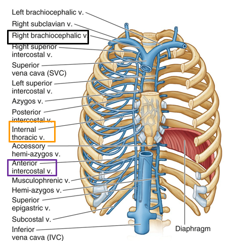
Are right and left posterior intercostal veins drained identically?
No. Their drainage differs.

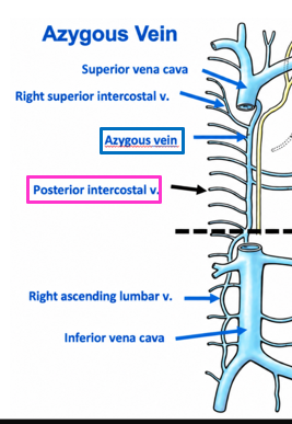
Where do right posterior intercostal veins drain?
Into the azygos vein.

Where does the azygos vein then drain?
Toward the SVC.

Where do inferior left posterior intercostal veins 9–11 drain?
Into the hemiazygos vein.

Where does the hemiazygos vein drain?
Into the azygos vein.
Where do superior left posterior intercostal veins 4–8 drain?
Into the accessory hemiazygos vein.

Where does the accessory hemiazygos vein drain?
Into the azygos vein.

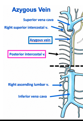
According to the slide, what veins also drain into the azygos/hemiazygos system?
Lumbar veins and femoral veins.

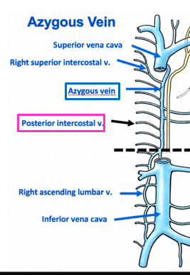
What major function does the azygos system provide?
A collateral pathway between the SVC and IVC.

Azygos vs. hemiazygos vs. accessory hemiazygos?
Right = azygos; lower left = hemi; upper left = accessory hemi.
Where is the intercostal VAN located relative to each rib?
Inferior to the rib.

Where does the anterior intercostal artery come from?
Internal thoracic artery.

Where does the posterior intercostal artery usually come from?
Descending aorta.

What relationship exists between anterior and posterior intercostal arteries?
They anastomose with each other.

Which posterior intercostal arteries are NOT supplied by the aorta?
Those associated with ribs 1 and 2.

What else is different about ribs 1 and 2 besides arterial supply?
Their venous drainage differs.

Are posterior intercostal arteries 1 and 2 supplied by the descending aorta?
No

What supplies posterior intercostal arteries 1 and 2?
Superior/Supreme intercostal artery.

The superior/supreme intercostal artery is a branch of what?
Costocervical trunk.

What is the costocervical trunk?
A branch of the subclavian artery that gives rise to the superior intercostal artery.

Where does the costocervical trunk originate?
From the subclavian artery.

Where is the costocervical trunk relative to the thyrocervical trunk?
It arises distal to the thyrocervical trunk.

Trace the arterial supply to posterior intercostal arteries 1 and 2.
Subclavian → costocervical trunk → superior/supreme intercostal artery → ribs 1–2.
