Lab 8 Thoracic Anatomy
 Call Kai
 Learn
 Practice Test
 Spaced Repetition
 Match
 Flashcards
 Knowt Play
1/59

There's no tags or description
Looks like no tags are added yet.
60 Terms
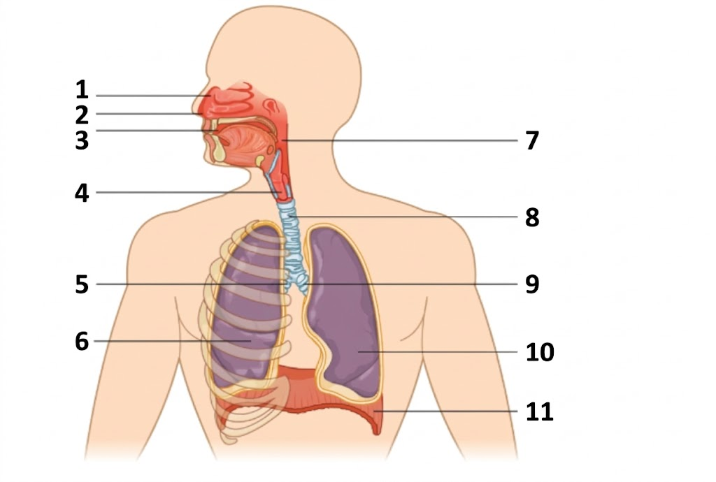
1
nasal cavity

2
nostril

3
oral cavity

4
larynx

5
right main bronchus

6
right lung

7
pharynx
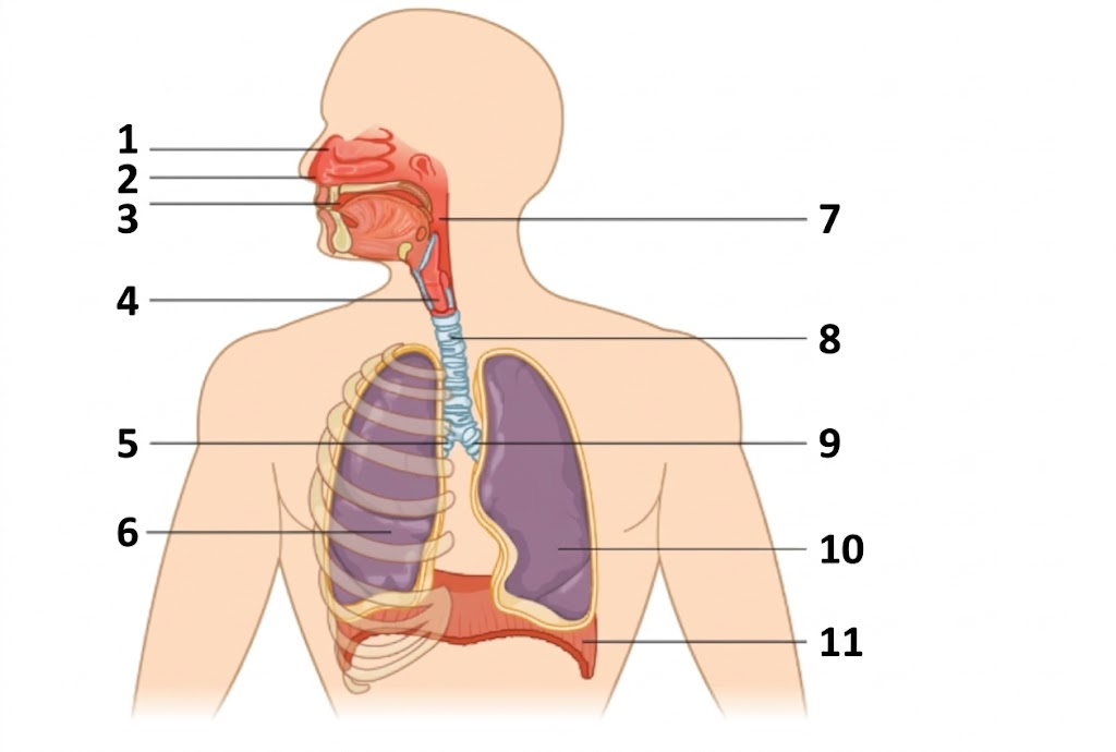
8
trachea

9
left main bronchus

10
left lung

11
diaphragm

1
main bronchus

2
segmental bronchus

3
trachea

4
lobar bronchus

1
cricoid cartilage
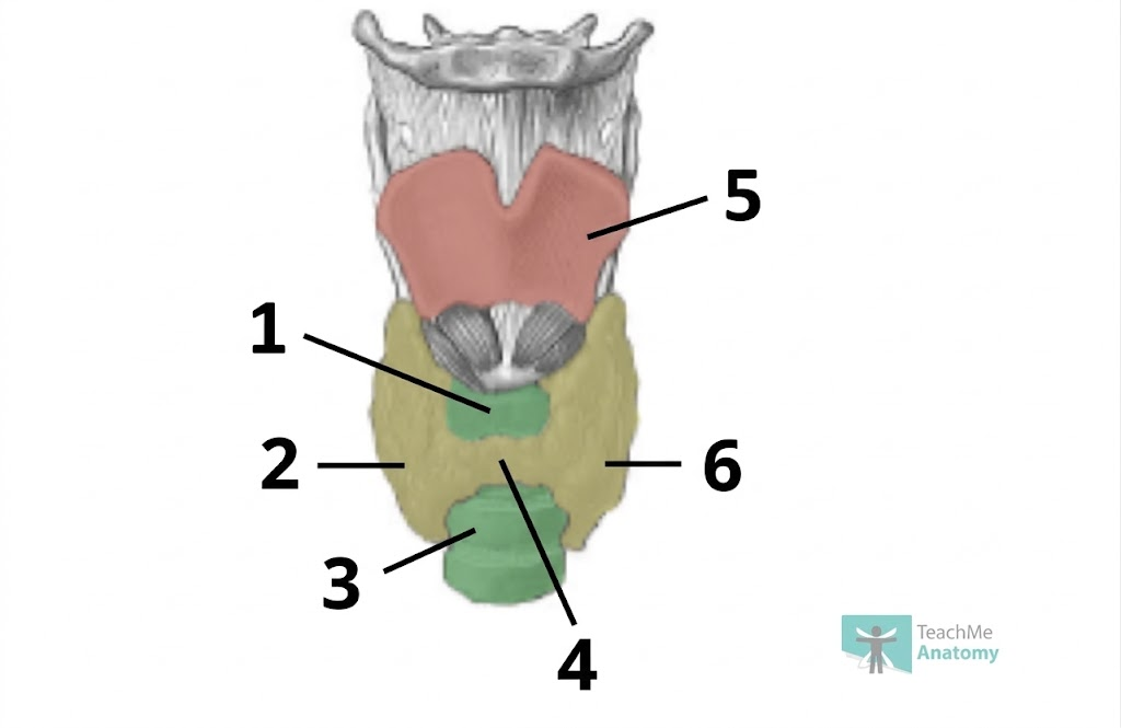
2
right lobe of thyroid gland
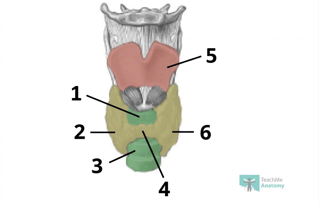
3
trachea
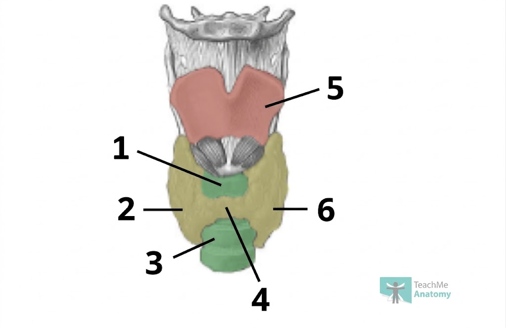
4
isthmus

5
thyroid cartilage

6
left lobe of the thyroid gland

1
glands

2
trachealis muscle
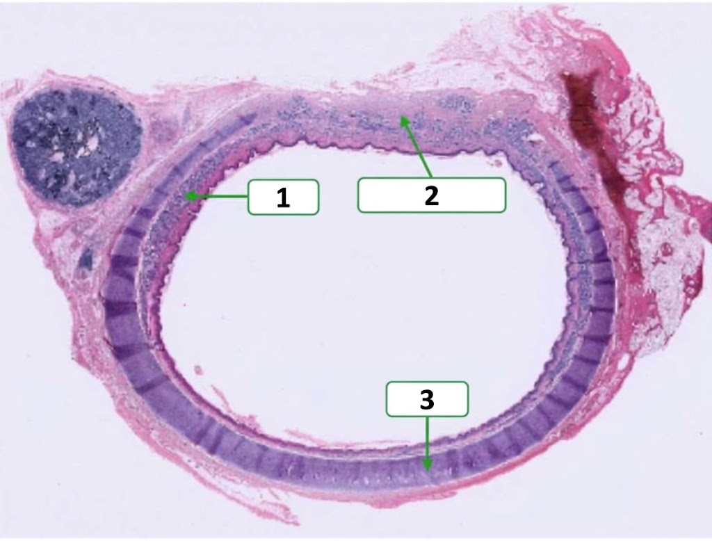
3
cartilage

1
ciliated columnar epithelium
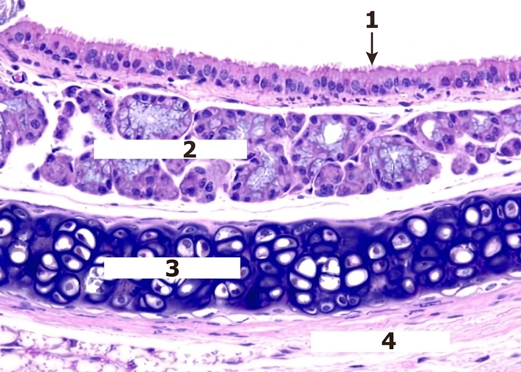
2
seromucinous cells

3
hyaline cartilage

4
fibroelastic tissue
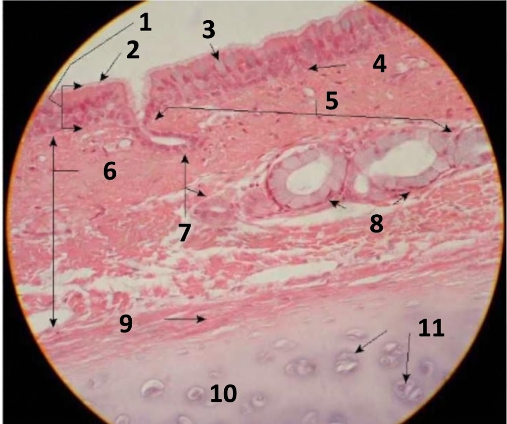
1
pseudostratified ciliated columnar epithelium

2
cilia

3
goblet cell

4
basement membrane

5
tracheal glands secrete mucus

6
Lamina propria

7
ducts of tracheal glands (lined with simple cuboid)

8
mucous alveoli: nuclei at periphery

9
perichondrium
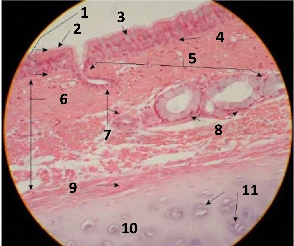
10
hyaline cartilage

11
chondrocytes

1
bronchi
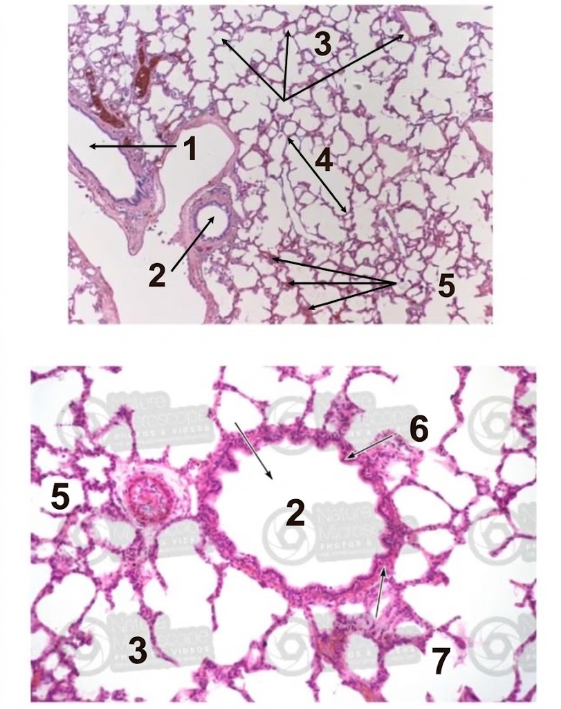
2
bronchiole

3
alveoli

4
alveolar sac

5
capillaries
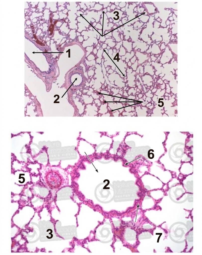
6
columnar epithelium

1
capillary endothelium

2
type 1 pneumocyte

3
pneumocyte (type II)

4
alveolar air space

5
red blood cells
type 1 AEC
large flat cells minimize the distance between alveolar gas and blood to facilitate efficient gas exchange
type 2 AEC
cuboidal cells produce surfactant which lowers alveolar surface tension. also, maintain alveolar epithelium and can differentiate into type 1 AEC if needed
macrophage
engulfs debris and particles that reach the lungs
typical diameter of alveolar sac
Alveolar sac is typically 200 μm in diameter.
Density: 31.6 ±3.4 alveoli/mm3 (can convert to cm3
how close is alveolar exterior surface to capillaries
very close to the surface (easier to find by looking for erythrocytes then looking for thin endothelial cells)

1
type 1 pneumocytes

2
capillaries

3
type II pneumocyte

4
air
Name for the collection of connective tissues between the pulmonary vascular endothelium and the alveolar epithelium
alveolar septum