Topic 5 and 6
 Call Kai
 Learn
 Practice Test
 Spaced Repetition
 Match
 Flashcards
 Knowt Play
1/14

There's no tags or description
Looks like no tags are added yet.
15 Terms
Describe the structure of DNA
DNA consists of four nucleotides that are covalently linked
2 DNA strands, held together by hydrogen bonds, run antiparallel to each other
twisted into a double helix
DNA nucleotides in one strand are held together by covalent bonds
Phosphodiester bonds linking the 5’ end of one sugar with 3’ end of the next
Linkages on either side of the bond are different; 5’ phosphate, 3’ hydroxyl → polarity • DNA strands therefore have a 5’ and a 3’ end

Describe the role and structure of the nucleosome
Nucleosome: DNA wrapped around histone proteins (histones)
Basic unit of eukaryotic chromosome structure
• Individual nucleosomes are linked
• Histone octamer: 2x each of H2A, H2B, H3, H4
• Histone proteins have high proportion of positively charged amino acids (lysine & arginine) → helps binding of histones to DNA (negatively charged due to sugar-phosphate backbone)
Histone tails (extends out of nucleosome core) important for regulation
Binding of Histone H1 helps pull adjacent nucleosomes together → more condensed chromatin fiber

Describe the structure and function of SMC ring complex in chromatin packing
During interphase, converts linear chromatin fiber into series of chromatin loops; ‘bunny ears’ shape • Structural Maintenance of Chromosome (SMC) form ring complexes that push out loops of DNA using ATP hydrolysis
Cohesin (= SMC ring complex that organizes the structure of interphase chromosomes) works together with sequence specific clamp proteins to produce an extended series of chromosome loops
Condensins (= another set of SMC ring proteins) replace cohesins during cell division
• Using ATP hydrolysis they form even tighter loops (incl loops within loops). This winding and tightening produce the structure of mitotic chromosomes

Summarise how DNA is condensed into chromosomes.
DNA double helix: DNA consists of two strands twisted into a double helix.
Nucleosomes: DNA wraps around histone proteins, forming structures that resemble beads on a string.
Chromatin: Nucleosomes interact and fold into more compact structures, forming chromatin. (Interphase)
Chromatin loops: Chromatin forms loops that attach to structural proteins, allowing further organisation and condensation. (Interphase)
Condensed chromosomes: During cell division, chromatin becomes highly compacted into visible chromosomes, helping ensure DNA is distributed correctly to daughter cells. (mitosis)

How does changes in Nucleosome Structure Allow Access to DNA
• Chromatin-remodeling complexes alter accessibility of DNA (either more or less accessible depending on cell cycle stage)
• Interact with histones and DNA; ATP-dependent process
Histone tails can be chemically modified (reversible) • Histone-modifying enzymes can add/remove acetyl, methyl or phosphate groups
Acetylation of lysine residues (on histone tails) reduces affinity for neighboring nucleosomes → chromatin structure is loosened
Methylation usually results in compacting chromatin (dense heterochromatin) → gene expression is reduced
Describe how Interphase Chromosomes Contain both Highly Condensed and More Extended Forms of Chromatin
Heterochromatin (highly condensed) vs euchromatin (less condensed)
• During interphase, chromosomes show regions of hetero- and euchromatin
• Gene expression usually occurs within euchromatin (DNA more accessible)
Heterochromatin can ‘spread’ along the DNA
• Modifications of histone tails (ie methylation of lysine in tail of H3) attracts heterochromatin specific proteins adding modifications to adjacent nucleosomes
X-chromosome inactivation:
• Example of the use of dense heterochromatin to shut down/silence gene expression
• Random condensation/inactivation of either maternal or paternal X chromosomes in females
• Pattern of inactivation inherited to all daughter cells
In cats, X-chromosomes encode genes responsible for coat color
• Skin cells with inactivated X chromosomes: if black is inactivated, coat color is orange and vice versa
• Size of color patches depends on number of skin cells that descended from the embryonic cells
Inheritance of condensed chromatin structure (heterochromatin) to silence genes = cell memory BMS143 Topic 5 • Histone proteins in daughter cells during cell division contain the covalent modifications of parent histone proteins

Describe the initiation of DNA Synthesis
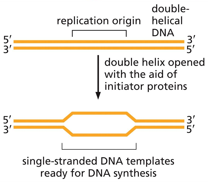
To begin the process of replication, initiator proteins bind replication origins → DNA strands are opened (breaking the hydrogen bonds)
Exposed single strands can then be used as template to copy the DNA
• Y-shaped junctions are formed at replication origins → replication forks
• DNA replication in both prokaryotes and eukaryotes is bidirectional. At each fork replication machinery moves away from each other
New strand grows from 5’ to 3’. Hydrolysis of incoming phosphate bond provides the necessary energy
Describe the direction of synthesis of the replication fork
• Parental DNA strands have opposite orientation (polarization). Newly synthesized strands move in opposite direction
in eukaryotes “backstitching” method solves the problem of the asymmetrical replication fork
Lagging strand is synthesized in 5’ 3’ direction as a series of short DNA strands → Okazaki fragments • After synthesizing a fragment, DNA polymerase moves back along parental DNA template strand to create the next fragment

Explain the role of proofreading enzymes in correcting replication errors.
DNA replication: DNA polymerase adds complementary nucleotides to the growing DNA strand.
Error detection: DNA polymerase detects when an incorrect nucleotide has been added, causing improper base pairing.
Error removal: The enzyme uses its 3′ → 5′ exonuclease activity to remove the incorrectly added nucleotide.
Correction: DNA polymerase inserts the correct nucleotide and continues replication.
What proteins for the replication machine
Protein/enzyme | Function |
|---|---|
Helicase | Unwinds the DNA double helix by breaking hydrogen bonds between complementary bases. |
Topoisomerase | Relieves twisting and tension ahead of the replication fork. |
Single-strand binding proteins (SSBs) | Keep separated DNA strands apart and prevent them from rejoining. |
Primase | Synthesises short RNA primers to provide a starting point for DNA synthesis. |
DNA polymerase | Adds complementary DNA nucleotides to the growing strand and proofreads for errors. |
Sliding clamp | Holds DNA polymerase onto the DNA, allowing it to synthesise DNA efficiently. |
Clamp loader | Loads the sliding clamp onto DNA using energy from ATP. |
RNase H and/or other primer-removal enzymes | Help remove RNA primers so they can be replaced with DNA. |
DNA ligase | Seals gaps in the sugar-phosphate backbone, joining DNA fragments together. |
Telomerase | Extends chromosome ends in cells that require it (replicates ends of chromosomes), helping prevent loss of DNA during replication. |

Describe damage that occurs in DNA
• Depurination (removal of purine bases from sugar-phosphate backbone)
• Deamination (removal of amino group from base → most common: C-to-U (read as T by DNA polymerase))
Thymine Dimers:
UV-light radiation can induce the formation of thymine dimers: covalent linkage between two adjacent pyrimidine bases
‘bulky’ DNA damage such as thymine dimers can stall the DNA replication machinery at the site of the damage

Explain how single-strand DNA damage is repaired in cells
If one strand becomes damaged, the intact strand can serve as template for DNA repair
• The cell has different ways to repair DNA; most involve three basic steps:
Step 1 – Excise damaged section of DNA
Step 2 – Repair missing DNA by using intact strand as template
Step 3 – Seal the gap
• Mismatch repair proteins only recognize and repair the newly synthesized strand • Humans with mutations in genes coding for mismatch repair proteins are predisposed for cancer (Lynch-Syndrome)
Describe how the cell repairs double strand DNA damage
The cell has two major pathways to repair a double strand break:
1. NHEJ (Non-homologous End Joining)
Simply fusing both ends back together
Quick. But: error-prone
2. Homologous Recombination (HR)
Precise, error-free repair mechanism
Needs a template to guide the repair (usually found during DNA replication before cell division)
Template and broken strand have nearly identical sequence (outside the broken region) = homologous sequences
Can be ‘hijacked’ for gene editing purposes
1. Removing 5’ ends of broken strands
2. Strand invasion: one of the broken strands (chewed back 3’ end) ‘invades’ the intact homologous DNA double-strand duplex and searches for complementary sequences
3. Once match is found, invading strand is elongated using the intact strand as template
4. After ‘passing’ the original breakpoint, the elongated invading strand is released and rejoins with its original strand
5. Additional DNA synthesis at the 3’ ends of the broken DNA double strands, followed by ligation completes the repair.
