Learn: BMS 307 exam 1
 Call Kai
 Learn
Learn Practice Test
 Spaced Repetition
 Match
Match Flashcards
 Knowt Play
1/180

There's no tags or description
Looks like no tags are added yet.
Name | Mastery | Learn | Test | Matching | Spaced | Call with Kai | Chat |
|---|
No analytics yet
Send a link to your students to track their progress
181 Terms
simple squamous epithelium
Single layer of flat cells
Allows for exchange of oxygen, nutrients, and gases
simple squamous cells
Alveoli, bowmans capsule, capillaries, endothelium of vasculature
stratified squamous epithelium
Multiple layers of squamous cells
Can be keratinized or non-keratinized

Non-Keratinized stratified squamous
Has nucleus, protects
found in mouth, anus, vagina

Keratinized stratified squamous
No nucleus, protects from water
found on skin

Simple cuboidal
One layer, cube shaped
Secretion and absorption
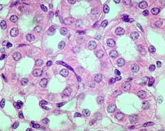
simple cuboidal cells
Kidney tubules, common bile duct
Simple columnar
Single layer of column cells
Aids in absorption and secretions

simple columnar cells
Lines the intestinal tract (stomach, small intestines, large intestine)
Microvilli
Increases surface ares for absorption

pseudostratified columnar epithelium
One layer of cells, but appears as multiple
Goblet cells secrete mucus to fight bacteria, and cillia structure collects particles to protect

pseudostratified columnar cells
Upper respiratory tract, trachea
Cillia
Structure on apical surface of columnar epithelium to catch bacteria and foreign particles and secrete mucus
Transitional
Multiple layer to allow for stretching
Stretches as volume within bladder changes

Transitional cells
bladder
Anatomy
the study of the relationship between body parts
Cytology
study of cells
Histology
study of tissues
Surface anatomy
refers to the superficial anatomical markings
Regional anatomy
refers to all structures in a specific area of the body, whether they are superficial or deep
Systemic anatomy
The study of the organ systems of the body
anatomical position
body erect
head, eyes, palms, and toes face forward
Arms at side
Back
posterior (dorsal)
front
anterior (ventral)
above
superior
below
inferior
head
cranial
rear
caudal
to the side of the body
lateral
middle
medial
proximal
above
distal
below
Median plane
vertical direction
intersects anterior and posterior midline
divides body into left and right halves

frontal plane
vertical direction
divides body into anterior and posterior parts

Transverse plane
horizontal direction
divides body into superior and interior parts

lying on back
supine
lying on stomach
prone
lateral rotation
rotation away from the midline
medial rotation
Rotation toward the midline
circumduction
circular movement of a limb
dorsiflexion and plantarflexion
up and down movement of the foot
Inversion
turning inward
eversion
turning outward
Elevation Movement
superior movement of body part (raising shoulders)
Depression Movement
inferior movement of body (mouth opening)
Protrusion
moving jaw forward
Retrusion
moving jaw back
protraction
moving a part forward (pushing forward with shoulders)
Retraction
moving a part backward (pushing shoulder back)
3 types of connective tissue
skeletal
Cardiac
Smooth
Skeletal muscle
striated and voluntary
moves or stabilizes the position of the skeleton

skeletal muscle
combined with connective tissues and neural tissue in skeleton
Smooth muscle
has striations and intercalated discs, involuntary
Moves food , urine, and reproductive tract secretions

smooth muscle
in the walls of blood vessels and throughout internal organs
cardiac muscle
end of cells are pointy
circulates blood; maintains blood pressure
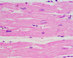
cardiac muscle
heart
neurons
maintain charge across membrane
communication via specialized chemicals
unipolar, bipolar, multipolar

Glial Cells
supporting cells
Schwann cells
Supporting cells of the peripheral nervous system responsible for the formation of myelin.
layers of skin
epidermis, dermis, hypodermis
epidermis
Outer layer of skin
Keratinized epithelium
5 layers of skin
move superficially as growth occurs
avascular
Epidermis Layers
Stratum Corneum
Stratum lucidum
Stratum granlosum
Straum spiosum
Stratum Germinativum
Stratum germinativum (basale)
bottom of epidermis (base)
cells are cuboidal/columnar shape
Keratinocytes/melanocytes
Stratum Spinosum
several layers thick
Keratinocytes
Distinct intercellular attachment
Responsible for strength of skin
stratum granulosum
3-5 layers thick
Keratohyalin granules present
cells are flattened/dying
stratum lucidum
present only in thick skin
high concentration of immature keratin
cellular organelles disrupted
ex:sole of foot
Stratum corneum
cells are "bags" of keratin
Keratin is combined with glycolipid to add water resistance
ex: dandruff
Dermis
Connective tissue under epidermis
Hypodermis (subcutaneous layer)
Loose connective tissue
Immune system, blood cells, adipocytes
appendage
hair, sweat glands, nails
Hair
Embedded in epithelial pockets (follicles)
Hair root
portion within follicle, forms hair bulb
Hair growth process
Active- mitosis occuring, hair is growing (longest portion of cycle)
Resting- Mitosis stopped, club hair occupies follicle (cuts hair from blood supply)
Hair types
terminal- typical hair of scalp
vellus- "peach fuzz" present on infant
Intermediate- growth associated with sex hormones
Sebaceous Glands
oil glands
not present at nipple, glands, labia
merocrine sweat glands
tubular shape with myoeplithelial cells
Merocrine type secretion
Nails
plates of hard keratin
act to protect skin
prevent distortion of finger and toe tips
Nail Root
Matrix- hard keratin, germinative zone
nail bed
Eponychium- Cutical
Hyponychium- thick skin under "free edge" of nail
growth of Integumentary System
Epidermis becomes thinner
Dermis becomes thinner
Vitamin D production declines
Melanocyte activity declines
Glandular activity declines
hair follicles stop functioning
Skin repair slows down
cervical vertebrae
C3-C7
Capable of wide range of movement
Extension, flexion, lateral flexions

thoracic vertebrae
T1-T12
Some extension, flexion, lateral flexion and rotation

lumbar vertebrae
L1-L5 fused
Some extension, flexion, little lateral flexion, very limited rotation

coccygeal vertebrae
3-5 fused
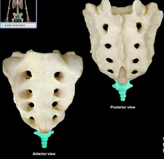
Scoliosis
excessive lateral curvature of the spine

kyphosis
excessive thoracic curvature (humpback)

lordosis
excessive lumbar curvature (sway-back)

Spinous process of vertebra
muscle and ligament attachment sites

Lamina
part of the vertebral arch

transverse process
two lateral projections from the vertebral arch

pedicle

intervertebral discs
contributes to 25% of total length of spine
-Annulus fibrosus
-Nucleus pulposus
annulus fibrosus
Fribrocartilage

Neucleus pulposus
-jelly-like material that holds water
-Swells during periods of recumbency(lying down)
-May herniate
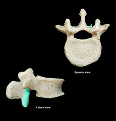
Inferior articular process
Serratus Posterior Superior
Attachment: Nuchal ligament, C7-T3
Insertion: 2nd-4th ribs
Innervation: Spinal, T2-T5
Action- Elevates ribs
Blood supply: Superior intercostal artery via the dorsal rami of the upper two posterior intercostal arteries
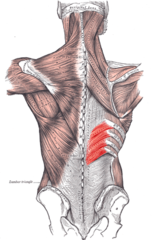
Serratus Posterior inferior
Attachment: T11-L2
Insertion: 8th and 12th rib
Innervation: Spinal, T9-T12
Action: Depresses ribs
Blood supply: lowest posterior intercostal artery, subcostal artery, first 2 lumbar arteries

Spinal cord
has 31 pairs of spinal nerves
Cauda equina- L2 to coccyx
pia mater
thin, delicate inner membrane of the meninges
arachnoid mater
weblike middle layer of the three meninges
subarachnoid space
contains cerebrospinal fluid, between pia and arachnoid matter