Lec. 11 - Serological Tests
 Call Kai
 Learn
Learn Practice Test
 Spaced Repetition
 Match
Match Flashcards
 Knowt Play
1/26

Earn XP
Description and Tags
Exam 2
Name | Mastery | Learn | Test | Matching | Spaced | Call with Kai | Chat |
|---|
No analytics yet
Send a link to your students to track their progress
27 Terms
Antigen-Antibody binding relies on affinity. What is affinity? What 2 things effect the strength of attraction?
The attraction force between the Ab (Fab region) and the Ag determinant site (epitope).
Strength of attraction depends on the specificity between the Ab & Ag
Binds together better if the Ag fits all the binding sites on the Ab
Cross-reacting Ag’s usually have low affinity, but if it closely resembles the original Ag it will bind to the Ab well (high affinity)
Antigen-Antibody binding also involves avidity. What is avidity? What strengthens avidity binding?
The sum of the attractive forces between Ag and Ab that keep them held together.
Higher valency on the Ab increases it’s avidity (IgM has highest avidity)
What Ab’s have high affinity but low avidity and why? What Ab(s) have high avidity but low affinity and why?
High affinity and low avidity: IgG, IgE, IgA → b/c affinity is formed from B cell maturation (creating specificity for Ag), only having a valency of 2 gives them lower avidity.
High avidity and low affinity: IgM → high avidity due to high valency (10) but less specificity (affinity) due to being a first response Ab
What do immuno-assays test/detect for?
Detects antigens or antibodies via Antigen-Antibody binding
Immuno-assays can be labeled or unlabeled. What are the two different categories of unlabeled immuno-assays? Describe what type of antigens or antibodies are used.
Precipitation: soluble antigen and antibody to produce insoluble complexes
Agglutination: particulate antigen and soluble antibody form large complexes
What does the unlabeled precipitation immuno-assay require antibodies and antigens to have? (hint *3 things)
Multivalency: multiple binding sites for Ab-Ag
Equal concentration relative to each other
Affinity & Avidity
Precipitation tests are usually seen as a curve. What are the three zones (in order of curve progression) and briefly describe them.
Prozone: excess Ab → prevents Ag from binding enough Ab’s (no cross-linkages)
Zone of Equivalence: Ab-Ag multivalent sites are equally bound → precipitation detectable
Postzone: excess Ag → small amount of Ab w/ multiple Ag around it (no lattice)
Where can false-negatives occur in precipitation reactions? How can these false-negatives be fixed?
Prozone false-negative occurs due to high [Ab] → Fix by diluting Ab and performing test again
Postzone false-negative occurs due to high [Ag] → Fix by testing patient sample after 1 week has passed ( [Ab] may have increased)
What are the 2 liquid precipitation tests that can be used to test for Ab-Ag interactions?
What are the 4 gel precipitation tests that can be used to test for Ab-Ag interactions?
Liquid precipitation tests: Measure via light
Turbidimetry
Nephelometry
Gel precipitation tests:
Radial immunodiffusion (RID) (passive techn.)
Ouchterlony double diffusion (passive techn.)
Serum protein electrophoresis (SPE)
Immunifixation electrophoresis (IFE)
One of the liquid precipitation tests is turbidimetry. How does it measure Ab-Ag interactions?
Via the absorbance units as light decreases while passing through the liquid.
One of the liquid precipitation tests is Nephelometry. How does it measure Ab-Ag interactions? What does it specifically measure regarding Ag-Ab interaction? What all can it test for (hint* what proteins)?
Scatters light at an angle as it passes through the liquid sample.
Ag-Ab complex formation over time
Tests for: Ig’s (Ab), complement proteins, C reactive protein
One of the gel precipitation tests is Radial Immunodiffusion (RID). What kind of diffusion is it? Explain how RID works and describe the two types of “end-points”/conclusions of the reaction. What all can it test for (hint* what proteins)?
Single-diffusion
Ab is in gel, Ag is added into wells in gel → Ag diffuses out until equivalence is reached → larger radius, the greater the amount of Ag present
End-point (Mancini): rxn goes to completion (takes time)
Kinetic (Fahey): msrmnts taken before completion (estimate) → faster than end-point
Tests for: Ig’s (Ab) & complement proteins
One of the gel precipitation tests is Ouchterlony diffusion. What does this test determine? What kind of diffusion is it? Describe how this test works and the 3 different end-points that can occur. What all can it test for (hint* what proteins)?
Determines relatedness of Ag to an Ab
Double-diffusion
→ Gel has wells in it and known Ab or known Ag (lab type) are added in along with patient serum (that contains either an unknown Ab or Ag)
→ incubated to allow for diffusion out
→ Line of precipitate forms when equivalency between Ag and Ab is met
End-point possibilities:
Identity (forms an arc/V shape)
Nonidentity (forms an X)
Partial identity (forms partial X)
Tests for: complex Ag’s (ex. fungal Ag)

One of the gel precipitation tests (really a immunoglobulin eval test) is Serum Protein electrophoresis (SPE). What does this test determine? What test should you reflex to if an abnormality is found in this one?
Determines where the immunoglobulins (Ig) are located. Indicates presence of disease if gamma has a monoclonal (M) spike.
Reflex to Immunofixation electrophoresis (IFE)

One of the gel precipitation tests (really a immunoglobulin eval test) is Immunofixation electrophoresis (IFE). How does this test work? What does it determine?
Unknown Ag (patient serum) is placed in gel → All Ab’s is added into gel → Precipitate forms where Ag-Ab combines and creates a band in the gel
Determines: over/under production of Ab types and differentiates monoclonal/polyclonal gammopathies

What is agglutination? What are the antibodies called that cause agglutination? What kind of process is agglutination and describe the steps.
Agglutination: visible clumping of particles (RBCs, bacteria, latex-beads) that combine with a specific Ab.
Ab’s: Agglutinins
2-step process:
Sensitization (initial binding): Ab-Ag bind - this stage is quick, but reversible
Lattice formation (creates large aggregates): multiple Ag and Ab rearrange bonds to form stable lattice

What are the two predominant Ab that agglutinate? What effects agglutination and is IgM affected by ionic strength? Know the Tm that the two main Ab work best at.
Ab: IgM & IgG
Factors effecting agglutination:
lower ionic strength leads to high agglutination (in lab use LISS to allow for increased agglutination of small Ab)
IgM does not need LISS b/c it’s large enough to overcome electrostatic repulsion
pH: 7.0-7.4 is best for agglutination (<4.0 & >9.0 bad)
TM for IgG best at body Tm (30-37 C)
IgM best at Rt (4-27 C)
Does agglutination also have a curve it follows? If so, what happens in prozone, equivalence, and postzone?
Prozone: excess Ab
Equivalence: Balanced - lattice forms
Postzone: excess Ag
What are the 5 types of agglutination reactions? Briefly describe them.
Direct (Hemagglutination): Ag markers found on the particle (RBC) to catch lab Ab
Passive (indirect): Particle (ex. latex-bead) is bound with Ag marker to catch patient Ab
Reverse passive: Particle (ex. latex-bead) is bound with Ab marker to catch patient Ag
Agglutination inhibition: Positive test is no agglutination of particles
Hemagglutination inhibition: Positive test is no agglutination of RBCs
How does direct and hemagglutination work? What is considered a positive test? What is it used for?
Direct agglutination:
Known Ag (from known RBC type - ex. Type A ag markers on RBCs) are mixed with patient serum to test for their Ab’s (unknown) → ex. if patient Ab binds to RBCs that are known to have A ag’s → they have anti-A Ab, indicates they may have Type B or Type O blood.
Agglutination indicates prescence of patient Ab (positive test)
Clumping indicates what kind of blood type they don’t have.
Hemagglutination:
Patient RBC is mixed with made lab Ab to determine blood type.
If it clumps, that’s the patients blood type (positive test)

Explain how Passive (indirect) agglutination works. What is this test usually used to check for?
Lab carrier particles (ex. lab RBCs, latex-beads, silicate beads) are bound to a known Ag → Patient serum is added to check for patient Ab (unknown) binding to lab Ag (known)
Positive test: agglutination
Checks for: virus immunity (patient should have Ab for it), rheumatoid factor (autoimmune response if binds), Group A strep immunity
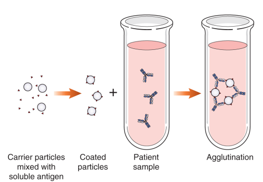
Explain how reverse passive agglutination works. What does it detect for?
Known Ab is attached to carrier particle → mixed with patient serum to check for patient Ag (unknown) → patient is positive for Ag if known Ab bind and causes agglutination
Detects: Infectious agents (rapidly, ex. Strep), soluble Ag’s present in urine, spinal fluid, and serum

Another form of agglutination is the agglutination inhibition. What is this test based on and governed by? What is considered a positive test? What is it used to detect and what would a positive test mean?
Based on: competition between lab particulate (Ag) and soluble patient Ag to bind limited Ab (lab bound Ab)
Governed by: Avidity of Ab → how well it binds to Ag
Positive test: no agglutination
Detects: hapten Ag’s (ex. drugs) → positive test indicates they have drugs in bloodstream b/c patient Ag bound to lab Ab before lab Ag could bind (patient Ag out-competed)
Negative would mean that there would be agglutination b/c lab Ag would have bound with lab Ab due to lack of prescence of patient Ag binding prior

Hemagglutination inhibition is the last test for agglutination. Explain how it works and what it detects. What is a positive test and what does that mean?
Competition between Ag (known/lab) and reagent RBC (RBC coated with known Ag markers) to bind to patient Ab (unknown).
Detects: Patient immunity to certain viruses → checks Ab binding to Ag of viruses (ex. Rubella, mumps, measles, flu, HBV, HSV, RSV, and adenovirus)
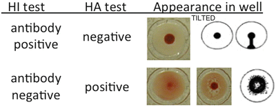
Positive test: no agglutination (looks like a button) → indicates patient has Ab to Ag (lab)

How is antibody titer checked for hemagglutination and what does it mean if there is a high titer?
High titer: more agglutination is inhibited means more Ab is present
Done by doing serial dilutions of patient serum and adding same amount of known Ag into each dilution
1:10 dilution shows a button (no agglutination) up until 1:1280 (shows button) then 1:2560 shows a “mat” of red meaning agglutination happened → Ab is strong until dilution of 1280 (last effective dilution)
How do you avoid cross-reactivity?
Use a monoclonal Ab against an antigenic determinant unique to Ag
Store reagent properly & check expiration dates (also follow instructions)
Account for sensitivity & specificity of kits used
Does a negative test result rule out presence of disease or antigen?
No.