Antomy quiz 10 &11
 Call Kai
 Learn
 Practice Test
 Spaced Repetition
 Match
 Flashcards
 Knowt Play
1/42

There's no tags or description
Looks like no tags are added yet.
43 Terms
Apex of the heart
The pointed inferior tip of the heart, oriented toward the left hip.

Parietal pericardium
The outer sac-like membrane that anchors and protects the heart within the mediastinum.

Visceral pericardium (Epicardium)
The innermost layer of the pericardium that sits directly on the outer surface of the heart muscle.

Myocardium
The thick middle layer of the heart composed entirely of cardiac muscle tissue responsible for pumping.

Endocardium
The smooth, thin inner lining of the heart chambers and valves.

Atria (Right and Left)
The two superior receiving chambers of the heart.
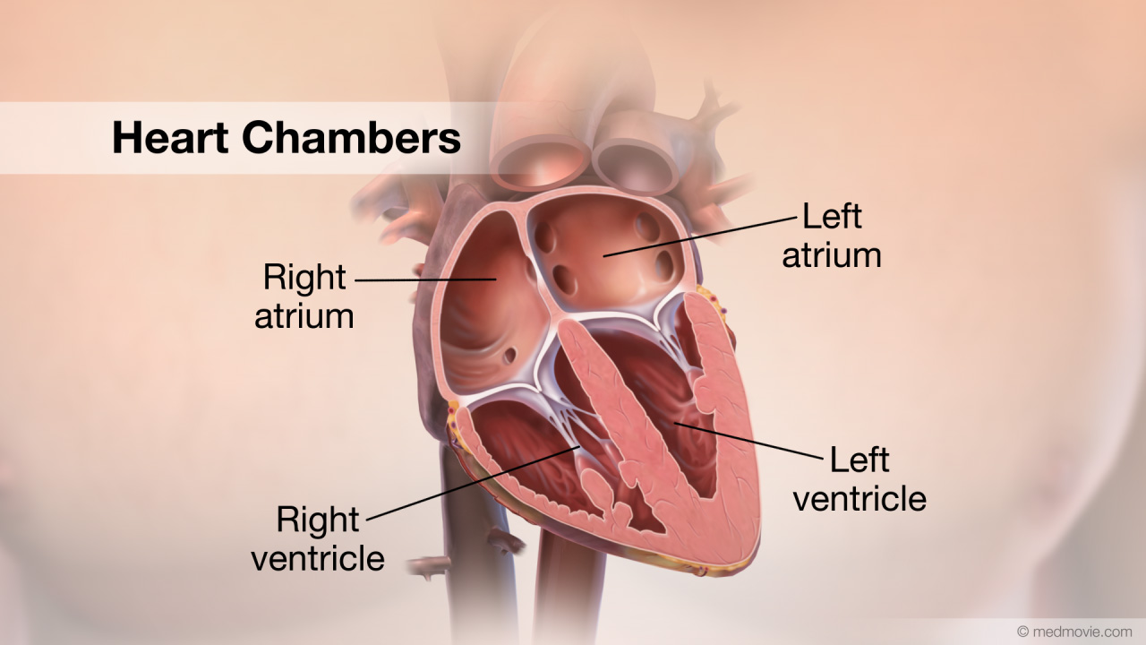
Auricles
Ear-like flap extensions on the exterior of the atria that increase total atrial volume.

Ventricles (Right and Left)
The two inferior pumping chambers of the heart.

Interventricular septum
The thick muscular wall that separates the right and left ventricles.

Fossa ovalis
A depression in the right atrium that represents the remnant of the fetal foramen ovale.
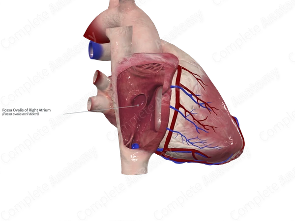
Foramen ovale
The fetal shunt between the right and left atria that closes at birth to become the fossa ovalis.

Sinoatrial (SA) Node
The natural pacemaker of the heart, located in the superior wall of the right atrium.

Tricuspid valve
The right AV valve, consisting of three cusps.

Bicuspid (Mitral) valve
The left AV valve, consisting of two cusps.

Chordae tendineae
Tendon-like strings ("heart strings") that connect AV valve cusps to papillary muscles.

Papillary muscles
Structural muscles located in the ventricles attached to chordae tendineae to anchor AV valves during systole.

Semilunar valves
Valves located at the exits of the ventricles that prevent backflow from the great arteries.

Pulmonary semilunar valve
Valve located between the right ventricle and the pulmonary trunk.

Aortic semilunar valve
Valve located between the left ventricle and the aorta.

Superior vena cava
Great vein bringing deoxygenated blood from the upper body to the right atrium.

Inferior vena cava
Great vein bringing deoxygenated blood from the lower body to the right atrium.

Pulmonary trunk
The major vessel carrying deoxygenated blood out of the right ventricle toward the lungs.

Pulmonary arteries
Left and right branches of the pulmonary trunk carrying low-oxygen blood to the lungs.
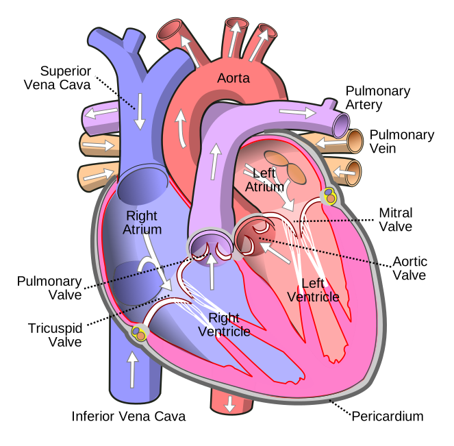
Pulmonary veins
Four vessels carrying oxygen-rich blood back from the lungs into the left atrium.
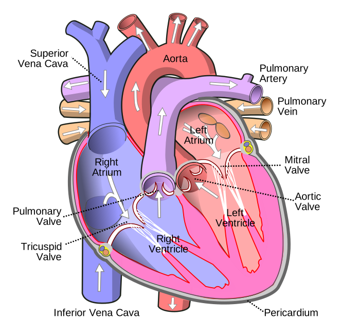
Aorta
The largest artery in the body, carrying oxygen-rich blood out of the left ventricle to systemic circulation.

Right and left coronary arteries
Vessels originating at the proximal end of the aorta that supply oxygenated blood to the myocardium.

Cardiac (coronary) veins
Vessels that drain deoxygenated blood from the heart muscle tissue.

Coronary sinus
A large collecting vein on the posterior heart surface that receives blood from cardiac veins and empties into the right atrium.

Pulmonary circuit
The path of blood flow from the right side of the heart to the lungs for gas exchange, and back to the left side.

Systemic circuit
The path of blood flow from the left side of the heart to all body tissues for oxygen delivery, and back to the right side.

Deoxygenated blood pathway
Superior/Inferior Vena Cava & Coronary Sinus -> Right Atrium -> Tricuspid Valve -> Right Ventricle -> Pulmonary Valve -> Pulmonary Trunk & Arteries.

Oxygenated blood pathway
Pulmonary Veins -> Left Atrium -> Bicuspid/Mitral Valve -> Left Ventricle -> Aortic Valve -> Aorta & Systemic Arteries.

Systole
The phase of contraction of the heart muscle that produces a pulse of blood.
Diastole
The phase of relaxation of the heart muscle that allows for the filling of the ventricles.
Blood Pressure (BP)
The physical force that blood exerts against a vessel wall, typically measured at the brachial artery.
Systolic blood pressure
The peak arterial blood pressure attained during ventricular contraction (systole).
Diastolic blood pressure
The minimum arterial blood pressure occurring during ventricular relaxation (diastole).
Normal young adult BP
120/75mm Hg
Pulse pressure
The difference between systolic and diastolic pressure; an important measure of stress exerted on small arteries.
Hypertension
A chronic state of elevated resting blood pressure above 130 systolic or 80 diastolic.
Complications of hypertension
Destroys arterioles, damages kidneys and heart, causes blindness, and leads to heart attacks or aneurysms.
Systolic reading technique
The pressure value noted on the gauge when you first hear the rhythmic pulse sound through the stethoscope.
Diastolic reading technique
The pressure value noted on the gauge when the pulse sounds completely disappear.