OMFPR 12 - Radiographic Appearance Of Dental Anomalies (Dr. Campos)
 Call Kai
 Learn
 Practice Test
 Spaced Repetition
 Match
 Flashcards
 Knowt Play
1/152

There's no tags or description
Looks like no tags are added yet.
153 Terms
cementum
what structure on a tooth is NOT visible under normal conditions?
- Variation in number
- Variation in size
- Variation in morphology
What are the three types of developmental dental anomalies?
hyperdontia/supernumerary teeth
Define the following:
Presence of additional erupted or unerupted teeth
mesiodens
Define the following:
Extra teeth located in the maxillary central incisor region
distodens
Define the following:
Extra teeth located distal to the 3rd molar
peridens
Define the following:
Extra teeth located in the premolar region
maxillary central incisor region
Patient has mesiodens. What is the location of this supernumerary tooth?
distal to the 3rd molar
Patient has distodens. What is the location of this supernumerary tooth?
premolar region
Patient has peridens. What is the location of this supernumerary tooth?
mesiodens
ID the anatomical anomaly:
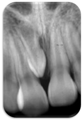
mesiodens
ID the anatomical anomaly:

peridens
ID the anatomical anomaly:

mesiodens
ID the anatomical anomaly:

peridens
ID the anatomical anomaly:

peridens
ID the anatomical anomly

Developing supernumerary teeth
ID the anatomical anomaly:

Cleidocranial dysplasia
Patient presents with the following:
- Multiple supernumerary teeth
- Does not have any clavicles on radiograph (aplasia/hypoplasia of clavicles)
- Brachycephalic skull with open fontanelles.
What is the syndrome?
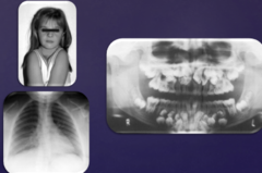
Gardner's syndrome
Patient presents with the following:
- Multiple supernumerary teeth as well as some retained primary teeth
- Osteomas of the jaws and facial bones
- Colon polyps
What is the syndrome?

colon polyps (most likely Gardner's Syndrome)
Patient presents with multiple supernumerary teeth as well as some retained primary teeth. Radiograph also shows osteomas of the jaws. Given all the signs, what else should the patient be cautious about?
Hypodontia
Define the following:
Lack of development of one or few teeth (1-4)
Anodontia
Define the following:
Total lack of development of teeth
Oligodontia
Define the following:
Lack of development of numerous teeth (>5)
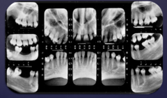
hypodontia
ID the anatomical anomaly:

oligodontia
ID the anatomical anomaly:

Ectodermal dysplasia
Patient presents with many missing teeth that never developed (oligodontia). Patient also has abnormal hair growth pattern and nails, and missing sweat glands. What is the syndrome?

- Abnormal hair
- Abnormal nails
- Abnormal or missing sweat glands
(needs at least 2 of above)
For a patient to be diagnosed with Ectodermal dysplasia, along with oligodontia, what other features would lead to the diagnosis?
ectodermal dysplasia
What is the diagnosis?

Macrodontia
Define the following:
- Teeth larger than normal
- Single/group of teeth
Macrodontia
ID the anatomical anomaly:

Macrodontia
ID the anatomical anomaly:

Macrodontia
ID the anatomical anomaly:

Microdontia
Define the following:
- Teeth smaller than normal
- Single/group of teeth
Microdontia
ID the anatomical anomaly:

Microdontia
ID the anatomical anomaly:

Peg lateral
ID the anatomical anomaly:

maxillary lateral incisor
What tooth most commonly experiences microdontia?
generalized microdontia
Patient presents to your office. All of their teeth are very small. Patients father has similar presentation. What is this called?

generalized microdontia
What condition?
fusion
Define the condition:
The union of two adjacent teeth via the dentin
dentin
In teeth that experience fusion, what part of the tooth is confluent?
Count the teeth - In fusion there are fewer than normal number of teeth present in arch
How do you determine if a tooth fusion has occured vs. macrodontia?
fusion
Patient presents with a larger than normal lateral incisor. All permanent teeth have erupted, but you count only 31 teeth. What event has occured?

fusion
ID the anatomical anomaly:

fusion
ID the anatomical anomaly:

Gemination
Define the condition:
The attempt for partial division of a single tooth germ will result in a normal number of teeth in the mouth via invagination of crown
Twinning
Define the condition:
The attempt for complete division of a single tooth germ will result in hyperdontia via normal tooth & supernumerary tooth
32 (normal number)
In partial division germination how many teeth are there in the arch?
Hyperdontia
In complete division germination/twinning how many teeth are there in the arch?
Gemination
ID the anatomical anomaly:

Twinning
ID the anatomical anomaly:

cementum
In teeth that experience concrescence, what part of the tooth is confluent?
Concrescence
Patient present for an extraction. But once you mobilize the 1st tooth, the adjacent one mobilizes just the same. You look back at the radiograph and know that the dentin isn't fused. What event has occurred?

Concrescence
when two adjacent roots of teeth are fused by cementum and it complicates the extraction, this is known as _________
- Space restriction during development
- Local trauma
What causes concresence?
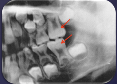
Taurodontism
What is the condition?
- Body of teeth appear elongated with short roots
- Elongated pulp chamber, apically positioned pulpal floor
Taurodontism
ID the anatomical anomaly:

Taurodontism
ID the anatomical anomaly:

Taurodontism
ID the anatomical anomaly:
Down Syndrome
what patient population is prone to developing teeth with taurodontism?
Dens invaginatus (dens in dente)
Define the condition:
Infolding of the enamel into the interior of the tooth
Dens invaginatus (dens in dente)
ID the anatomical anomaly:

Dens invaginatus (dens in dente)
ID the anatomical anomaly:

Dens invaginatus (dens in dente)
ID the anatomical anomaly:

Dens invaginatus (dens in dente)
ID the anatomical anomaly (red arrows):

Dens invaginatus (dens in dente)
ID the anatomical anomaly:

Dens in dente is incisal edge and dens invaginatus is in the cingulum
In clinic, what is the difference between dens invaginatus and dens in dente?
Dens evaginatus
Define the condition:
Outpouching/outfolding of enamel organ resulting in enamel-covered tubercle on the occlusal surface
Dens evaginatus
ID the anatomical anomaly:

Dens evaginatus
ID the anatomical anomaly:

Dilaceration
ID the anatomical anomaly:
Sharp bend or curve in the tooth
Dilaceration
ID the anatomical anomaly:
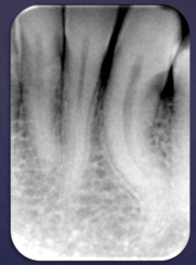
Dilaceration
ID the anatomical anomaly:

Amelogenesis imperfecta
Patient presents with small square shaped crowns on all of the teeth. The enamel layer appears to be extremely thin and cusps are difficult to distinguish on molar teeth (low/absent cusps). There are multiple open contacts between all teeth, giving a "picket fence" appearance. What do you suspect?

- Hypoplastic
- Hypomaturation
- Hypocalcification
- Combination of Hypomaturation/Hypocalcification
What are the four general types of amelogenesis imperfecta?
amelogenesis imperfecta
ID the condition:
- Square crown
- Relatively thin layer or absence of enamel
- Multiple open contacts between teeth
- Anterior teeth: "picket fence" appearance
Amelogenesis imperfecta
ID the anatomical anomaly:

Amelogenesis imperfecta
ID the anatomical anomaly:

Amelogenesis imperfecta
ID the anatomical anomaly:

Amelogenesis imperfecta
ID the anatomical anomaly:
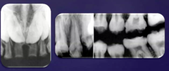
true
T/F: Amelogenesis imperfecta is a genetic anomaly that has marked changes in the enamel involving all teeth
dentin
Dentinogenesis imperfecta is a genetic anomaly that involves what?

Osteogenesis imperfecta (type I)
Dentinogenesis imperfecta Type 1 (Radicular Dentin Dysplasia - Griffins update) is associated with what condition?
type 2
Dentinogenesis imperfecta without any associated skeletal defects is what type?
Medical history (type 1 associated with osteogenesis imperfecta)
What do you need to identify type 1 vs type 2 dentinogenesis imperfecta?
Dentinogenesis imperfecta
ID the anomaly:
- Bulbous crowns
- Cervical portion of tooth is constricted
- Slender roots
- Partial/complete obliteration of pulp chamber and root canals
- Areas of apical rarefying osteitis
Dentinogenesis imperfecta
Patient presents with teeth that have a brownish hue to them. Crowns are bulbous with the cervical portion of tooth constricted. Pulp chambers on most teeth appear completely obliterated and roots are calcified. What do you suspect?

Dentinogenesis imperfecta
ID the anatomical anomaly:

Dentinogenesis imperfecta
ID the anatomical anomaly:

Dentinogenesis imperfecta
ID the anatomical anomaly:

Dentin Dysplasia Type I (Radicular Dentin Dysplasia)
ID the anomaly:
- Roots are short or abnormally shaped
- Spontaneous exfoliation
- Obliteration of pulp chambers (before eruption)
- Rarefying osteitis-with non-carious teeth
Dentin Dysplasia Type I (Radicular Dentin Dysplasia)
Patient presents complaining of spontaneous exfoliation of permanent teeth. Radiograph shows all roots that are extremely short and abnormal and pulp chambers of all teeth appear obliterated. What do you suspect?

Dentin Dysplasia Type I (Radicular Dentin Dysplasia)
Rarefying osteitis-with non-carious teeth is a feature of what condition?
Dentin Dysplasia Type 1a (Radicular Dentin Dysplasia)
ID the anatomical anomaly:
- Complete pulp obliteration
- Minimal root development

Dentin Dysplasia Type 1b (Radicular Dentin Dysplasia)
ID the anatomical anomaly:
- Crescent shaped pulp chamber
- Short conical roots
- Rarefying osteitis

Dentin Dysplasia Type 1c (Radicular Dentin Dysplasia)
ID the anatomical anomaly:
- 2 crescent shaped lines (pulp) concave to each other at the level of CEJ
- Roots are half the normal length
- Molar roots "W" shaped

Dentin Dysplasia Type 2 (Coronal Dentin Dysplasia)
New patient presents to your office. Radiograph shows all teeth have normal appearing roots but have what look to be pulp stones in the root canals. Most pulp chambers are obliterated after eruption or are flame/thistle shaped. What do you suspect?

Dentin Dysplasia Type 2 (Coronal Dentin Dysplasia)
ID the anatomical anomaly:
- Obliteration of pulp chamber and root canals (after eruption)
- Pulp chambers are flame or thistle shaped
- Multiple pulp stones
- Roots appear normal

Regional odontodysplasia
New patient presents to your office. Most of their teeth clinically look completely fine except for the right mandibular region is currently edentulous. Radiograph shows teeth that have failed to erupt, both enamel and dentin is hypoplastic and hypoclacified. The roots and pulp chambers on those teeth are large. What do you suspect?

Regional odontodysplasia
ID the anatomical anomaly:
- Enamel and dentin hypoplastic and hypocalcified
- Large pulp chamber/root canal
- Aka "Ghost teeth"
- Affects few adjacent teeth in a quadrant
regional odontodysplasia
_________ is also known as "ghost teeth"