Mitochondrial Dynamics II: Fission, Fusion, and Architecture
 Call Kai
 Learn
 Practice Test
 Spaced Repetition
 Match
 Flashcards
 Knowt Play
1/59

Earn XP
Description and Tags
Vocabulary practice flashcards covering the key proteins, regulatory mechanisms, division steps, fusion models, and clinical pathologies associated with mitochondrial dynamics.
60 Terms
MICOS
The mitochondrial contact site and cristae organizing system; a protein complex in the inner mitochondrial membrane that interacts with Mic60 and connects to the outer membrane sorting and assembly machinery (SAM) complex to establish membrane contact sites.
Miro1
A mitochondrial Rho GTPase that connects the MICOS complex to regulatory signaling networks, cytoskeletal transport machinery, and cellular redox control mechanisms.
Dynamin Superfamily Proteins (DSPs)
A group of four large GTPases that regulate mitochondrial dynamics and share structural similarities with bacterial dynamin-like proteins due to common evolutionary ancestry.
DRP1 (Dynamin-Related Protein 1)
A cytosolic dynamin-related GTPase that functions as the master regulator of mitochondrial division in mammals by translocating to the outer mitochondrial membrane to assemble into constricting rings.
MFN1 and MFN2 (Mitofusins 1 and 2)
Dynamin-related GTPases anchored in the outer mitochondrial membrane with cytosol-exposed functional domains that mediate outer membrane fusion
OPA1 (Optic Atrophy 1)
A dynamin-related GTPase anchored in the inner mitochondrial membrane that drives inner mitochondrial membrane fusion and maintains cristae architecture.
fission and fusion regulation by post-translational modifications
phosphorylation
Ubiquination
SUMOylation
Acetylation
DRP1 Ser 637 Phosphorylation
Phosphorylation of DRP1 at Ser 637 by PKA inhibits fission, whereas dephosphorylation by calcineurin promotes mitochondrial division.
does phosphorylation by PKA inhibit fission
YES it phosphorylates DRP1 and Ser 637
SUMO modifies what?
modifies DRP1 and MFNs modulates stability, localization
Ubiquitination Modification
E3 ubiquitin ligases (such as Parkin and MARCH5) regulate the turnover and activity of mitochondrial fusion and fission factors,
Lysine Acetylation
affects protein-protein interactions and enzymatic activities
Mitochondrial marking the division sites (what are the 2 key factors marking future division sites)
mitochondrial DNA replication
ER contact formation
Mitochondria-ER Contact Sites (MERCS)
Specialized contact sites where the endoplasmic reticulum wraps around mitochondria, marking future division sites, coordinating mtDNA replication, and facilitating phospholipid transfer to remodel membranes.
mtDNA replication
mitochondrial DNA replication occurs at sites that will becomes division sites
what serves as the MERCs facilitate
MERCS facilitate transfer of lipids essential for membrane remodelling during division.
Overview of mitochondrial division
requires events to recruit DRP1 to the OMM where it drives membrane constriction.
Actin-Mediated Pre-Constriction
An early division event at MERCS where actin polymerization generates mechanical force to narrow mitochondrial diameter from 300–500nm to approximately 150nm before DRP1 ring assembly.
What are 3 key players in actin-mediated pre-constriction
INF2
Spire1C
Myosin IIA
These generate actin filaments tethering the ER to mitochondria, creating forces needed for initial constriction.
INF2
An ER-resident protein that nucleates actin filaments
Spire1C
A mitochondrial outer membrane-associated actin nucleation factor
Myosin IIA
A motor protein that generates the contractile force required to narrow the mitochondrion during actin-mediated pre-constriction.
what is the role of DRP1 and is it cytosolic?
YES- its cytosolic
The primary mediator of mitochondrial division in mammals.
DRP1 structure
Contains G domain, BSE, stalk domain and variable domain for membrane binding
DRP1 GTPase activity and Oligomerization
The ability of DRP1 to hydrolyze GTP is crucial for its function in mitochondrial fission.
Oligomerization of DRP1 facilitates its recruitment to the mitochondria,
forms helical structures around mitochondria
Where does DRP1 recruitment of the adaptor proteins occur
requires adpator proteins at the outer mitochondrial membrane for proper recruitment
3 adaptor proteins in DRP1 recruitment
MFF
MiD49
mId51
MFF (Mitochondrial Fission Factor)
An outer mitochondrial membrane receptor that directly recruits cytosolic DRP1 to mediate mitochondrial fission.
MiD49 and MiD51
Outer mitochondrial membrane dynamic proteins that bind DRP1 and regulate its recruitment, linear copolymer assembly, and GTPase activity.
miD51
Stabilize linear filaments that curl into constricting rings through GTP hydrolysis
is DRP1 ATP or GTP dependent
GTP dependent
the constriction mechnasims (5 steps)
Recruitment
DRP1 are recruited to pre-constricted sites by adaptor proteins at MERCs
Linear assembly
DRP1 adpator form linear filaments along mitochondria
GTP binding
stabilize conformational chnages DRP1
Ring Formation
GTP hydrolysis triggers curling of linear filaments into constricting rings
Membrane Scission
Midzone Fission
Mitochondrial division occurring at central regions
marked by MFF, mtDNA replication, ER tubules, and actin polymerization,
serving the purpose of mitochondrial biogenesis to produce new functional mitochondria.
Peripheral Fission
Mitochondrial division occurring at organelle tips and edges
marked by FIS1, lysosomal contacts, and autophagic machinery,
serving the purpose of quality control by segregating damaged regions and degradation.
Why fusion matters
stress production → protect againt autophagic degradation
content mixing → distrribution of mtDNA
cell fate → t cell and stem cell determination
what 2 proteins that mediate outer membrane fusion
MFN1 and MFN2
both are GTPases
key features of MFN1 and MFN2
66% identical amino acid sequences
both required fro embryonic development
form homo and heterotypipc complexes
MFN1 has higher GTPase activity
MFN2 also tethers ER to mitochondria
how do the mitofusins in mitochondria dymerize?
GTP-bound MFNs from 2 mitochondria in close proximity interact and dimerize in trans through domains, which tethers mitochindria togther .
Trans-Dimerization of Mitofusins
The GTP-dependent interaction and dimerization of G domains between MFN proteins anchored on opposing outer mitochondrial membranes, effectively tethering the two organelles together.
The fusion mechanism: step by step (6 steps)
Autoinhibited step → in the cytososl, MFNs are folded with the G domain locked by HB2 domain preventing premature activation.
GTP binding → triggers MFN folded state to “stand-up state” exposing dimerization surface
Trans-Dimerization → the GTP-bound MFNs from opposing mitochondria interact with their G domains, forming a stable dimer and tethering the organelles.
GTP hydrolysis → open to closed confirmation, bringinf mitohcindria closer togther
Membrane docking→ HB2 domains dock membrane creating flattened contact area
membrane fusion
MIM structure and function
regions of high curvature make ATP and controlled transport of lipids
compartmentalization of the MIM allows for controlled reactions
specific location of cytochrome C in the cristae and its controlled release into the cytosol triggers programmed cell death
what is OPA1?
controls cristae and MIM fusion
its a dynamin related GTPase
has a N-terminal mitochondrial targeting sequence that is cleaved upon import
the bulk of the protein is exposed to the inter membrane space
OPA1 overexression promotes tight crista junctions while OPA1 knockdown causes widened cristae.
exhibits characteristics of both intergral and peripheral membrane proteins
L-OPA1 and S-OPA1 - proteolytic processing
The long (membrane-anchored) and short (soluble) forms of OPA1 produced by proteolytic cleavage at three sites by YME1L and OMA1 proteases; a balanced ratio of both forms is required for efficient fusion.
OPA1 key structural elements
G Domain → GTPase domain
BSE → Bundle signaling element of 3 alpha-helices
Stalk →antiparallel bundle
Paddle domain
Paddle Domain
A structural membrane-binding region in OPA1 containing a membrane-inserting loop (MIL) that directly interacts with lipid bilayers to facilitate membrane curvature and fusion.
Models of OPA-1 mediated Membrane Fusion
Bud Model
Crista Rim Model
Bud Model of MIM Fusion (OPA1)
A mechanism of inner mitochondrial membrane fusion where OPA1 oligomerizes on both sides of the inner membrane into curved filaments to form membrane buds that undergo GTPase-dependent structural changes and fuse at their tips.
in depth bud model of MIM fusion
(L-OPA1) generates membrane buds in the septum by oligomerizing on each membrane leaflet in a ring-like fashion
the bud tips fuse to connect the two matrix spaces
recruitment of F1Fo-ATP synthase and MIM proteins generate new cristae

what does the septa rim have?
high membrane curvature
Crista (Septa Rim) Model of MIM Fusion
Membrane fusion occurs at the septa rim, which has high membrane curvature. OPA1 bridges opposing MIMs and possibly creates a contact site to the outer membrane. By inserting hydrophobic residues from the paddle into the membrane,
OPA1 promotes curvature at the rim and facilitates fusion.
picture crista rim model of mIM fusion
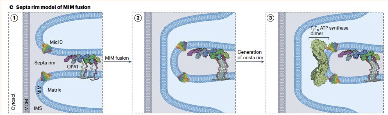
OPA1 MIM fusion steps
MOMs are tethered with help of dynamin-like mitofusins
MIMs are separated before they align to form a septum between the two intermembrane space
MIM fusion generate a connecting membrane tubule which may be converted into cristae
MIM architecture in disease
OPA1 mutations → affect GTPase domain
MICOS disorders → can cause Parkisons disease
ATP synthase disease → mutations in F1Fo-ATP lead to cardiomyopathies
Autosomal Dominant Optic Atrophy (ADOA)
A disease resulting in degeneration of the optic nerve caused by more than 200 point mutations in OPA1, the majority of which affect the GTPase domain and impair nucleotide binding or GTP hydrolysis.
Behr Syndrome
A severe neurodegenerative condition occurring when both OPA1 alleles carry loss-of-function mutations, causing severe optic atrophy, myopathy, peripheral neuropathy, ataxia, and hearing loss.
Heteroplasmy
The presence of both wild-type and mutant mitochondrial DNA (mtDNA) within a single cell or organism, whose variable distribution among offspring is caused by a genetic bottleneck during oogenesis.
Reproductive options for women with mitochondrial DNA mutations
in vitro fertilization and fertilizing all oocytes
pronuclear transfer
metaphase II spindle transfer
Metaphase II Spindle Transfer
A mitochondrial donation technique performed prior to fertilization in which the nuclear spindle is removed from an unaffected donor oocyte and replaced with the nuclear spindle of the patient oocyte before in vitro fertilization.
Pronuclear Transfer
A mitochondrial donation technique performed after fertilization in which the pronuclei from a patient zygote containing pathogenic mtDNA are transferred into an enucleated donor zygote.
Mic60 - disease mechanisms
altered Mic60 expression has been associated with down syndrome, parkisons disease and neurodegeneration