Chapter 13 - The Spinal Cord and Spinal Nerves (Matt)
 Call Kai
 Learn
Learn Practice Test
 Spaced Repetition
 Match
Match Flashcards
 Knowt Play
1/53

There's no tags or description
Looks like no tags are added yet.
Name | Mastery | Learn | Test | Matching | Spaced | Call with Kai | Chat |
|---|
No analytics yet
Send a link to your students to track their progress
54 Terms
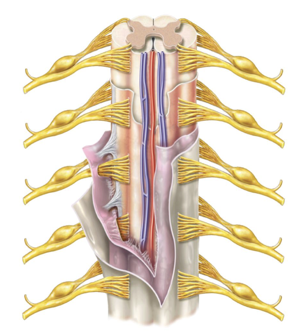
Functions of the spinal cord
responsible for reflexes, integrating sensory information, and conducts sensory/motor impulses to effectors from the brain

Vertebral Column
a bony structure that houses and protects the spinal cord
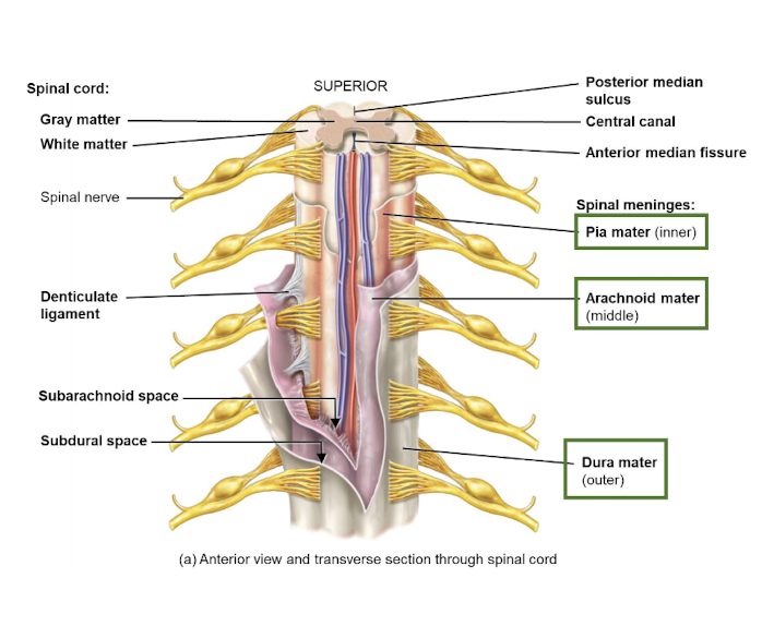
Meninges
the three protective membranes that cover the brain and spinal cord, consisting of the dura mater, arachnoid mater, and pia mater.
Epidural space
the space filled with fat and connective tissue

Dura mater
the outermost layer of the meninges, made of dense, irregular connective tissue (tough mother)

Subdural space
the space filled with interstitial fluid

Arachnoid mater
the layer of the meninges made of avascular, collagen and elastin fibres (spider-like)

Subarachnoid space
the space filled with cerebrospinal fluid

Pia mater
the layer of the meninges made of thin, vascular layer of connective tissue; stuck to spinal cord and brain (delicate)
Cerebrospinal fluid (CSF)
the fluid that cushions the CNS, maintains a stable environment, and removes wastes

Spinal taps
procedures used to collect cerebrospinal fluid from the subarachnoid space for diagnostic, therapeutic purposes, and meningitis

Conas medullaris
the cone-shaped end of the spinal cord between L1 and L2

Filum terminale
the extension of pia mater that fuses with the arachnoid and dura mater; anchoring the spinal cord to the coccyx

Caudae equina
the roots of the lowest spinal nerves
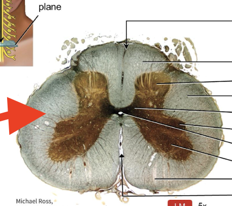
White matter
the outer region of spinal cord, made of myelinated axons to conduct signals to and from brain
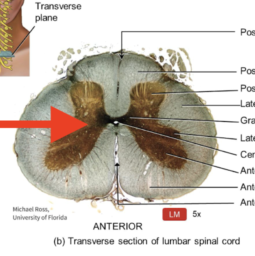
Grey matter
the inner region of spinal cord, made of dendrites, cell bodies, unmyelinated axons, and neuroglia to integrate IPSPs and EPSPs

Grey commissure
the region of grey matter that cross the midline, allowing communication between the two halves

Central canal
the space in the center of spinal cord that is filled with cerebrospinal fluid acting as cushioning

Posterior grey horns
region of grey matter that contains axons from incoming sensory neurons and interneurons

Anterior grey horns
region of grey matter that contains somatic motor nuclei (sends signals to skeletal muscles for movement)
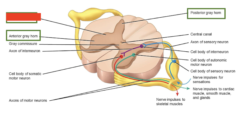
Lateral grey horns
region of grey matter that contains autonomic motor nuclei (regulates activity of cardiac muscle, smooth muscle, and glands.

Sensory tracts
axons that conduct nerve impulses to the brain (ascending/afferent)

Motor tracts
axons that conduct nerve impulses away from the brain (descending/efferent)
Nerves
bundles of axons that transmit signals between the spinal cord and body.

Posterior root
the root of a spinal nerve that contains sensory axons

Spinal ganglion
located in the posterior root of spinal nerves containing sensory cell bodies

Anterior Root
the root of a spinal nerve that contains motor axons for controlling muscle movements.
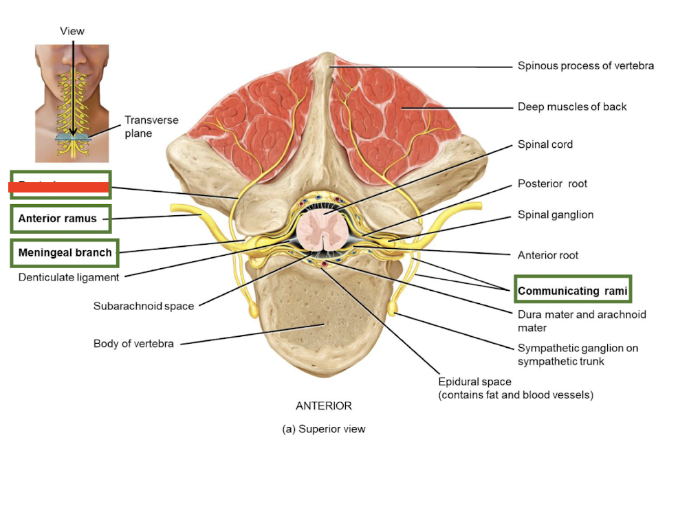
Posterior rami
the branch of a spinal nerve that serves the posterior trunk
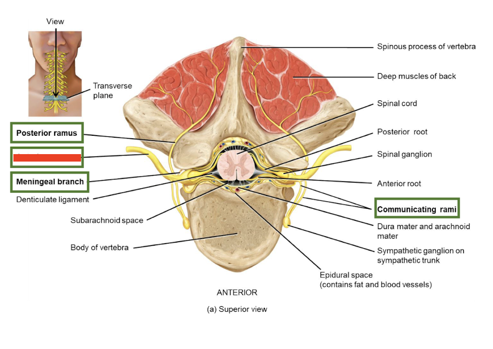
Anterior rami
the branch of a spinal nerve that serves the limbs and anterolateral trunk

Meningeal branches
the branches of spinal nerves that re-enter the vertebral canal to innervate the meninges and blood vessels.

Communicating rami
the branches of spinal nerves that are a part of the autonomic nervous system

Cervical plexus (C1-C4)
supplies the neck, upper shoulder, and diaphragm

Brachial plexus (C5-T1)
supplies shoulder and upper limbs

Lumbar plexus (L1-L4)
supplies the abdomen, external genitals, and lower limbs

Sacral plexus (L4-S4)
supplies the buttocks, perineum, and lower limbs (contains sciatic nerve)

Intercostal nerves (T2-T12)
formed when the anterior rami of the spinal nerves innervate intercostal spaces

Dermatome
an area of skin whose sensory information is carried to the CNS by one pair of spinal nerves
Reflex
a fast, involuntary, unplanned response to a particular stimulus

Babinski sign
a reflex when lateral fanning of toes in response to firmly stroking the sole of the foot (<1.5 years)

Plantar grasp reflex
a reflex when infants curl their toes when pressure is applied directly beneath them (<1 year)
Spinal reflexes
a reflex that is integrated in the spinal cord
Cranial reflexes
a reflex that is integrated in the brainstem
Somatic reflexes
a reflex that contracts skeletal muscle
Autonomic reflexes
a reflex that contracts smooth muscle or glands
Reflex arc
the pathway followed by nerve impulses to produce a reflex (contains: sensory receptor, sensory neuron, integrating centre, motor neuron, effector)
Monosynaptic
reflex arc that involves only one synapse between a sensory and a motor neuron, resulting in a quick response.
Polysynaptic
reflex arc that involves multiple synapses between sensory, interneurons, and motor neurons, resulting in a more complex and slower response.
Ipsilateral Reflex
type of reflex where sensory neurons and motor neurons leave from the same side

Stretch reflex
a somatic reflex that causes contraction of a skeletal muscle in response to stretching of the muscle (ex. patellar reflex occurs in response to the tapping on the tendon of the knee, quadriceps femoris contracts)
Reciprocal innervation
the simultaneous contraction of one muscle and relaxation of its antagonist during a reflex action.
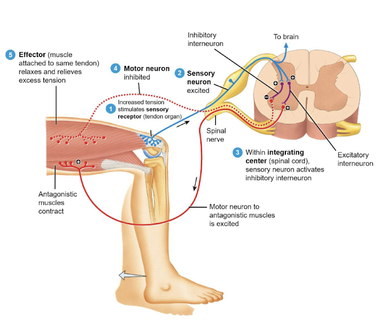
Tendon reflex
A reflex action that causes muscle relaxation when the force on a tendon is so great that tendons might be torn
Tendon organs
sensory receptors in the tendon that detects stretching

Flexor reflex
A reflex action that causes withdrawal of a limb to avoid injury or pain

Crossed Extensor reflex
A reflex action that stabilizes the body by extending the opposite limb when one limb is withdrawn, often in response to pain.