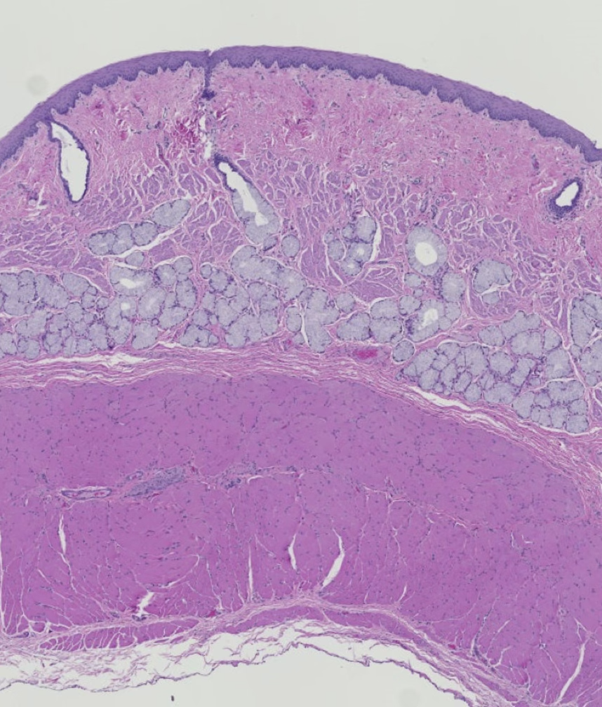
histo self-quiz- teeth, tongue and esophagus

0.0(0)
Studied by 1 person
call kaiCall Kai
learnLearn
examPractice Test
spaced repetitionSpaced Repetition
heart puzzleMatch
flashcardsFlashcards
GameKnowt Play
Card Sorting

1/24

encourage image

There's no tags or description

Looks like no tags are added yet.

25 Terms

1
New cards
<p>ID the labeled regions of this tooth (region 1 and region 2)</p>

ID the labeled regions of this tooth (region 1 and region 2)

1- crown, 2- root

2
New cards
<p>ID the labeled regions of this tooth (region 3 and region 4)</p>

ID the labeled regions of this tooth (region 3 and region 4)

region 3- alveolus/alveolar bone, region 4- pulp cavity

3
New cards
<p>ID the labeled regions of this tooth (regions 5 and 6)</p>

ID the labeled regions of this tooth (regions 5 and 6)

5- gingiva, 6- gingival sulcus

4
New cards
<p>which cells extend their cytoplasmic processes through the dentinal tubules (analogous to canaliculi in bone) this maintaining this living mineralized tissue?</p>

which cells extend their cytoplasmic processes through the dentinal tubules (analogous to canaliculi in bone) this maintaining this living mineralized tissue?

odontoblasts (nucleated cells on the far right image)

5
New cards
<p>the dentin crown of the brachydont tooth is normally covered in a highly mineralized tissue, but this tissue dissolves during demineralization. which mineralized tissue of the tooth covers the dentin crown?</p>

the dentin crown of the brachydont tooth is normally covered in a highly mineralized tissue, but this tissue dissolves during demineralization. which mineralized tissue of the tooth covers the dentin crown?

enamel

6
New cards
<p>the simple columnar cells enclosed within the black rectangle in this image produce tooth enamel. ID these cells</p>

the simple columnar cells enclosed within the black rectangle in this image produce tooth enamel. ID these cells

ameloblasts

7
New cards
<p>the dentin root of the brachydont tooth is covered in a mineralized, bone-like tissue. <br></p><p>what is this tissue?</p><p>which cells produce this tissue?</p><p>what do we call the spaces where these cells reside?</p>

the dentin root of the brachydont tooth is covered in a mineralized, bone-like tissue.

what is this tissue?

which cells produce this tissue?

what do we call the spaces where these cells reside?

cementum

cementocytes

lacunae

8
New cards
<p>the periodontal ligament of the brachydont tooth inserts its collagen and elastic fiber into both the adjacent cementum and alveolar bone. what do we call these fibers of insertion that are utilized to anchor tendons and ligaments to the bone?</p>

the periodontal ligament of the brachydont tooth inserts its collagen and elastic fiber into both the adjacent cementum and alveolar bone. what do we call these fibers of insertion that are utilized to anchor tendons and ligaments to the bone?

sharpeys fibers
9
New cards
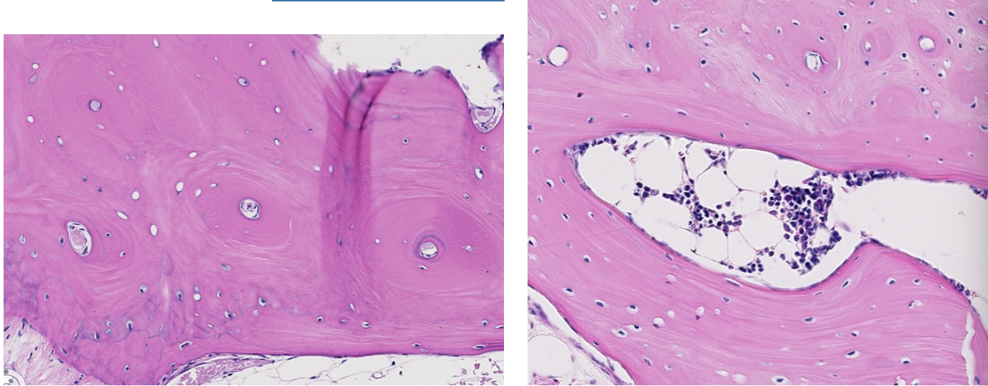
<p>review: is this alveolar bone composed of lamellar ot woven bone?</p>

review: is this alveolar bone composed of lamellar ot woven bone?

lamellar (both osteons ans circumferential lamellae are visible

10
New cards
<p>the slender, elongated cells seen in this image produce predentin, which is mineralized to become dentin. ID the circled cells</p>

the slender, elongated cells seen in this image produce predentin, which is mineralized to become dentin. ID the circled cells

odontoblasts

11
New cards
<p>ID the surface epithelium of the tongue</p>

ID the surface epithelium of the tongue

stratified squamous

12
New cards
<p>the surface epithelium of the tongue varies as to whether it is cornified or non-cornified. which of the 2 indicated regions is cornified?</p>

the surface epithelium of the tongue varies as to whether it is cornified or non-cornified. which of the 2 indicated regions is cornified?

green (thinly cornified)

13
New cards
<p>tongue review:</p><ol><li><p>ID the type of muscle in the tongue (be specific)</p></li><li><p>what do we call the fine CT that surrounds individual myofibers? (one is indicated by the yellow line)</p></li><li><p>the RBC is inside which type of blood vessel that is encircled in green?</p></li></ol><p></p>

tongue review:

  1. ID the type of muscle in the tongue (be specific)

  2. what do we call the fine CT that surrounds individual myofibers? (one is indicated by the yellow line)

  3. the RBC is inside which type of blood vessel that is encircled in green?


  1. skeletal

  2. endomysium

  3. capillary


14
New cards
<p>tongue review: what do we call the CT (indicated by asterisks) that surrounds skeletal muscle fascicles and through which small arteries and veins travel?</p>

tongue review: what do we call the CT (indicated by asterisks) that surrounds skeletal muscle fascicles and through which small arteries and veins travel?

perimysium

15
New cards
<p>ID the type of tongue papilla. be specific</p>

ID the type of tongue papilla. be specific

vallate (circumvallate)

16
New cards
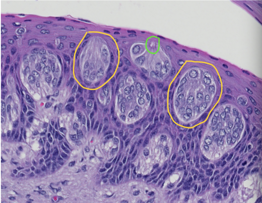
<p>is this surface epithelium from a tongue papilla gustatory or nongustatory?</p>

is this surface epithelium from a tongue papilla gustatory or nongustatory?

gustatory
17
New cards
<ol><li><p>ID the structures encircled in yellow</p></li><li><p>ID the specific region of these structures that is encircled in green</p></li></ol><p></p>
  1. ID the structures encircled in yellow

  2. ID the specific region of these structures that is encircled in green


  1. taste buds

  2. taste pore


18
New cards
<p>ID the tubular digestive organ</p>

ID the tubular digestive organ

esophagus

19
New cards
<p>ID this surface epithelium from the esophagus</p>

ID this surface epithelium from the esophagus

stratified squamous (non-cornified)

20
New cards
<p>does this esophagus have a complete or incomplete tunica mucosa? on what did you base your decision?</p>

does this esophagus have a complete or incomplete tunica mucosa? on what did you base your decision?

complete, has lamina muscularis

21
New cards
<ol><li><p>what type of muscle makes up the tunica muscularis in this portion of the esophagus?</p></li><li><p>what do we call the nerve tissue in this image betweent he 2 muscle layers of this tunic? (fascicle or autonomic ganglion)</p></li><li><p>which specific plexus does this nerve tissue belong to? (myenteric or submucosal)</p></li></ol><p></p>
  1. what type of muscle makes up the tunica muscularis in this portion of the esophagus?

  2. what do we call the nerve tissue in this image betweent he 2 muscle layers of this tunic? (fascicle or autonomic ganglion)

  3. which specific plexus does this nerve tissue belong to? (myenteric or submucosal)


  1. skeletal

  2. autonomic ganglion (has nerve cell bodies)

  3. myenteric plexus


22
New cards
<p>ID the type(s) of muscle in this portion of the tunica muscularis of the esophagus</p>

ID the type(s) of muscle in this portion of the tunica muscularis of the esophagus

both skeletal and smooth

23
New cards
<ol><li><p>these submucosal glands are passing though the lamina muscularis; which type of muscle is this?</p></li><li><p>which type of glands are these submucosal glands?</p></li></ol><p></p>
  1. these submucosal glands are passing though the lamina muscularis; which type of muscle is this?

  2. which type of glands are these submucosal glands?


  1. smooth

  2. mucous


24
New cards
<p>does this section of esophagus have a tunica adventitia or a tunia serosa? why?</p>

does this section of esophagus have a tunica adventitia or a tunia serosa? why?

serosa, lined by mesothelium

25
New cards
<p>review: ID this structure located in the serosa of the esophagus</p>

review: ID this structure located in the serosa of the esophagus

nerve fascicle