AE 1.2 - Scope of Practice, Cardiac Anatomy, Optimization
 Call Kai
 Learn
Learn Practice Test
 Spaced Repetition
 Match
Match Flashcards
 Knowt Play
1/31

There's no tags or description
Looks like no tags are added yet.
Name | Mastery | Learn | Test | Matching | Spaced | Call with Kai | Chat |
|---|
No analytics yet
Send a link to your students to track their progress
32 Terms
- functions as a delegated agent of the physician
- does not practice independently
- sonographers CAN not provide interpretation of exam to patient
-- interpretation comes from physician '
- recognize, evaluate, quantify
as Low as Reasonably Achievable
- lowest output power & the shortest scan time
-- consistent with acquiring the required diagnostic information
Name & Date of Birth
ask the patient the date of birth instead of saying it and having them agree
Intersocietal Accreditation Commission
– the minimum standards and guidelines for echocardiography accreditation

- sound beam cannot travel through air inspiration
- brings lung artifacts into view expiration
- brings lungs out of view
Patient Positioning
Patient Breathing
AdJust image/window
Ergonomics
Highest possible transducer frequency
Focus centered at ROI (region of interest)
Don’t rely on auto-optimization
Every image should be re-evaluated for quality and optimized
Low frequency = better penetration
High frequency = better resolution
- positioning, cannot see anything if patient is not in right position
- your goal is to image structures on-axis whenever possible
- on-axis imaging gives the most accurate representation of size, shape, motion, and relationships
- don't accept an off-axis image just because you can see the structure
- go off-axis with purpose, not by accident
- ability of the ultrasound machine to accurately show the position of moving structures at a particular instant in time
- ↑ Depth = ↑ pulse time
- ↑ Sector size = ↑ the # of scan lines
- narrow sector
- shallow imaging
- above 30 fps in 2D images
- above 18 fps in color images
pulmonic circuit
- right atrium (smallest, thinnest, lowest pressure)
- right ventricle (most anterior)
systemic circuit
- left atrium (most posterior) (conduit, reservoir, pump)
- left ventricle (largest, thickest, highest pressure)

RA
- anterior
- low pressure
- 3 inlets
- thin walls; coarse pectinate muscles
- chiari network, eustachian valve
LA
- posterior
- high pressure
- 4 inlets
- thick walls; smooth pectinate muscles
- no fetal remnants
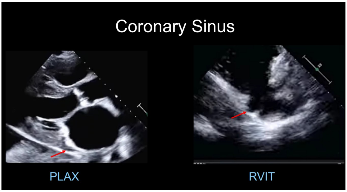
- lies within a sulcus called the AV sulcus or AV groove (epicardium) and empties into RA
- drains the coronary veins into the RA
- Thebesian valve sits at the opening
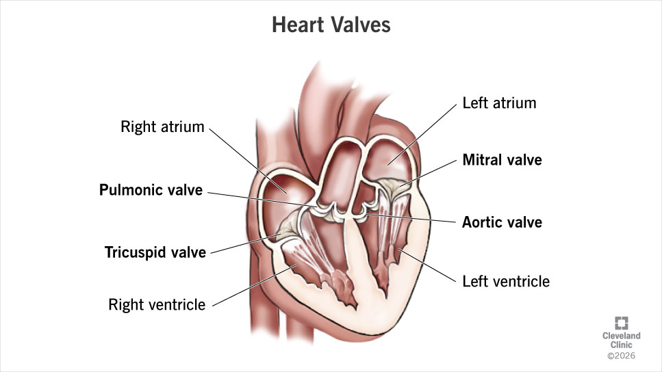
tricuspid valve
pulmonic valve (most anterior & superior)
mitral valve (2 leaflets)
aortic valve
leaflets
1) anterior
2) posterior
3) septal

- leaflets
1) anterior
- anchored to aortic root
- larger
- no scallops
2) posterior
- wider attachment to annulus
- divided into 3 scallops

TV
- 3 leaflets, thinner
- expandable annulus
- sits lower in heart
- 3 commissures
- ant, post, sept = papillary muscles
MV
- 2 leaflets
- D shaped, rigid annulus
- 2 commissures
- AL, PM papillary muscles
- semilunar
- cusps
1) anterior
2) left
3) right
- most anterior & superior valve

- semilunar
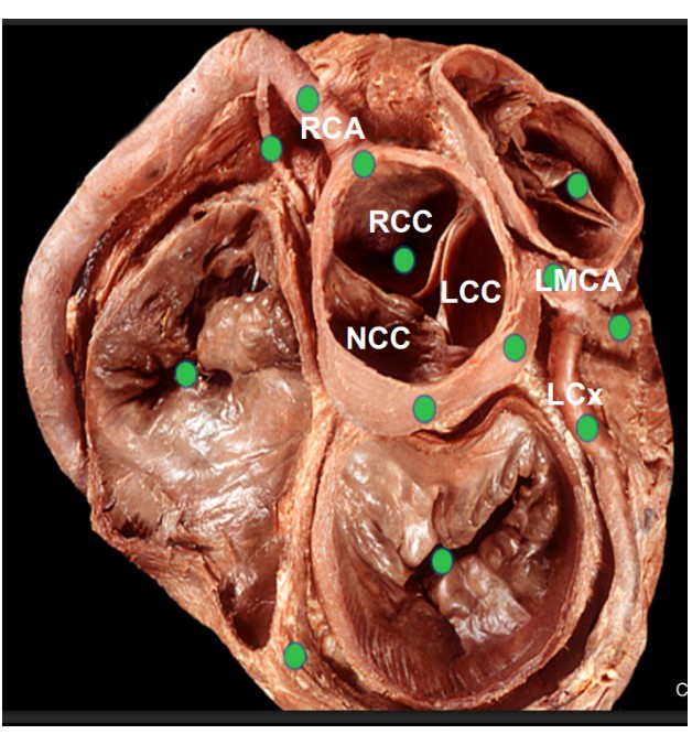
- based on where coronary arteries originate
1. Left coronary cusp (LCC)
2. Right coronary cusp (RCC)
3. Non coronary cusp (NCC)

"waist”

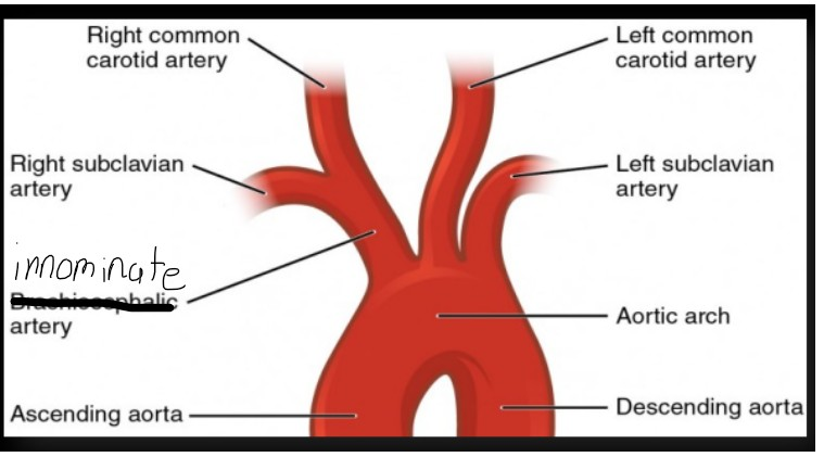
- Innominate Artery
-- splits into right common carotid Artery & right subclavian Artery
- Left common carotid
- Left subclavian
- muscle column that courses from IVS to the base of anterior papillary muscle
- electrical conduction path
- CAN also act as a protective mechanism to resist overdistention of the RV

- annulus
- leaflets
- chordae tendineae
- papillary muscle
- commissures

- small, string-like structures
- connect the leaflets of the valve with the papillary muscles
- anchor and support the leaflets
- prevent leaflet prolapse during ventricular systole

- contract, pulling the leaflets toward one another, helping valve closure
- connect to the chordae tendineae

- long and narrow, “finger like”
- decompresses LA when atrial pressure is high
- frequent location for blood clots

- thin walls
- heavily trabeculated
- moderator band
- smaller
- crescent shape
- rough interior
- thick walls
- no moderator band
- fine trabeculations at apex
- ellipsoid shape
- larger
- smooth interior
right heart - lower pressure
left heart - higher pressure
right heart = 75%
left heart = 98%