A+P Chapter 18 Cardiovascular System
 Call Kai
 Learn
Learn Practice Test
 Spaced Repetition
 Match
Match Flashcards
 Knowt Play
1/74

There's no tags or description
Looks like no tags are added yet.
Name | Mastery | Learn | Test | Matching | Spaced | Call with Kai | Chat |
|---|
No analytics yet
Send a link to your students to track their progress
75 Terms
Function of the cardiovascular system
Continuously circulate blood, oxygenate tissues, remove waste products (co2), circulate hormones, and deliver nutrients to tissues.
Pulmonary Circulation
Blood flow through the right side of the heart that sends deoxygenated blood to the lungs then returns that blood to the left side of the heart for distribution.
Systemic Circulation
Blood flow through the left side of the heart that sends oxygenated blood to the body, delivering nutrients and oxygen to tissues, picking up co2, and returning deoxygenated blood to the heart.
Heart
4 chambered (2 atria, 2 ventricles) muscular organ made from cardiac muscle that contract to generate pressure to push blood through the body.
Where is the heart located in the body?
In the pericardial cavity behind the sternum, between the lungs. The base (top) is between the 2nd and 3rd ribs. The apex (bottom/point) is between the 5th and 6th ribs towards the left side of the chest.
Where do major vessels attach at the heart?
The base
Pericardium
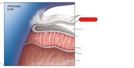
Protective sac that covers the heart. Includes three layers. Fibrous pericardium, parietal pericardium, visceral pericardium. Contains a fluid between the parietal and visceral layers called the pericardial fluid. This helps lubricate the heart as it beats.

Fibrous Pericardium
Outermost, and toughest layer of the pericardium.


Parietal Pericardium
Middle layer of the pericardium.

Visceral Pericardium
Innermost layer of the pericardium, against the heart wall. Also known as the epicardium.

What are the layers of the heart wall?
Epicardium (Outer layer, also known as visceral pericardium), myocardium (middle layer, cardiac muscle), and the endocardium (inner layer).

Identify the epicardium (visceral pericardium)


Identify the myocardium


Identify the endocardium

Interventricular Septum
Muscular wall in the heart that separates the right and left ventricles.
Congestive heart failure
Progressive disease where the heart becomes inefficient at pumping blood. Commonly associated with an enlarged heart. This leads to poor return of blood to the heart causing blood to backup in the venous system, specifically in the lungs. This causes pulmonary venous hypertension (increase in lung vein blood pressure) and can lead to increased fluid leakage in the lungs (pulmonary edema).
What is heart enlargement caused by when someone has congestive heart failure?
Heart enlargement is caused by either:
The heart working harder causes the muscles to grow or the heart wall expands and thins out as muscle breaks down.
Auricles
Small protrusions/appendages (ears) that allow for greater expansion of the heart chambers.
The left ventricle pumps ____ blood out of the ___ to the ____
Oxygenated; Aorta; body (other than lungs)
The right ventricle pumps _____ blood out of the _____ which splits into the ____ and flows to the ____
Deoxygenated; Pulmonary trunk; left and right pulmonary arteries; lungs
The superior and inferior vena cava carry ____ blood to the ______ from the _____
deoxygenated; right atrium; body
What parts of the body does the superior vena cava carry blood from?
Body above the diaphragm (Head, arms, chest)
What parts of the body does the inferior vena cava carry blood from?
Body below the diaphragm (Abdomen, legs)
Pulmonary veins carry ____ blood to the ____ from the ____
oxygenated; left atrium; lungs
Tricuspid Valve
Prevents backflow of blood as the right atrium pumps to the right ventricle. Connected to the papillary muscles by the chordae tendineae.
Bicuspid Valve (Mitral)
Prevents backflow of blood as the left atrium pumps to the left ventricle. Connected to the papillary muscles by the chordae tendineae.
Pulmonary (Semilunar) Valve
Controls blood flow between the right ventricle and pulmonary trunk.
Aortic (Semilunar) Valve
Controls blood flow between the left ventricle and aorta.
Heart Valves
Prevent backflow in the heart as it pumps. During the cardiac cycle, contraction of the chambers along with blood flow push blood against the valve forcing them open or closed depending on which chamber is contracting/relaxing. Valves close when blood pushes against the underside of their cusps.
What do the papillary muscles and chordae tendineae help the valves with?
They help prevent prolapse (opening in the wrong direction) during contraction.
When the ventricles contract which valves are open and which are closed?
The aortic and pulmonary valves open while the bicuspid and tricuspid valves close.
When the atria contract which valves are open and which are closed?
The tricuspid and bicuspid valves open while the aortic and pulmonary valves close.
List the blood flow through the entire heart starting with the return of deoxygenated blood
Superior and inferior vena cava and the coronary sinus
Right atrium
Tricuspid valve
Right ventricle
Pulmonary semilunar valve
Pulmonary trunk
R/L pulmonary arteries
Lungs + Pulmonary capillaries
Pulmonary veins
Left atrium
Bicuspid valve
Left ventricle
Aortic semilunar valve
Aorta
Systemic arteries
Systemic capillaries
Systemic veins
Return to the superior and inferior vena cava
Coronary Circulation
Circulation to the heart. As blood leaves the left ventricle, two arteries branch directly off the aorta. These are the left and right coronary arteries.
The coronary sinus is made up of the
Great cardiac vein, middle cardiac vein, and posterior vein of the left ventricle.
The coronary sinus returns ____ blood to the ____
deoxygenated; right atrium
Post-capillaries coronary blood flows to the ____
Coronary veins
Depressions along the heart wall are called ____ often found ____
Sulci; between chambers of the heart
What major coronary blood vessels can be found in the sulci?
The anterior interventricular artery (in the anterior interventricular sulcus) and the right coronary artery (in the coronary (or atrioventricular) sulcus)
Diastole
Relaxation/filling phase of the cardiac cycle. When the atria is in diastole the ventricles are in systole.
Systole
Contraction/ejection phase of the cardiac cycle. When the atria are in systole, the ventricles are in diastole.
What causes the lubb sound in a heart beat?
The closing of the tricuspid and bicuspid valves during the QRS complex
What causes the dupp sound in a heart beat?
The closing of the aortic and pulmonary valves at the end of the T wave
Heart Mummur
When extra sounds are present during auscultation (listening to) of the heart.
What 4 things heart murmurs can be caused by?
Failure of the foramen ovale to close
Failure of one of the heart valves to close properly
Heart valve stenosis (narrowing of the opening)
Septal defects (openings between ventricles)
What structures set the frequency of heart contractions?
The sinoatrial (SA) and atrioventricular (AV) nodes.
Does the heart need neural input for contraction?
No, but the brain can regulate the rate of contraction as needed.
Sinoatrial (SA) node
Known as the pacemaker of the heart. It sets the rhythm. Its cells are specialized to continuously depolarize (be super leaky) which allow sodium to move into the cell until the threshold is reached.
If the SA node fails, what can take over?
The AV node can take over, but normally at a lower rate
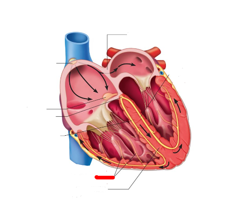
Map the electrical conduction of the heart from start to finish
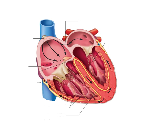
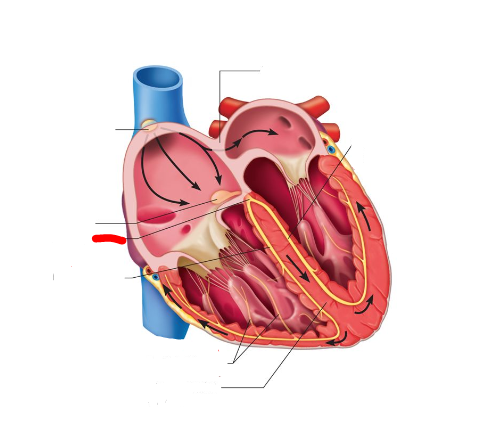
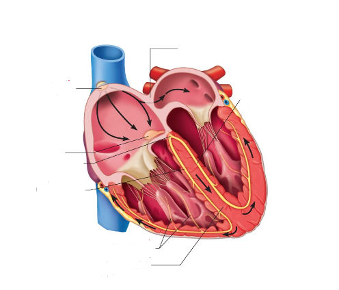
The SA node initiates contraction by depolarizing
That signal spreads across atria causing them to contract before reaching the AV node
The AV node pauses for 0.1 sec to allow for the atria to fully empty
Branching from the AV node, the AV bundle (Bundle of His) and bundle branches transmit signals down to the apex to initiate contraction there
Bundle branches split into Purkinje fibers to stimulate across heart wall

Identify the SA Node

Identify the AV node


Identify the AV bundle

Identify the left bundle branch


Identify the right bundle branch

Identify the Purkinje fibers
P wave
Atrial depolarization initiated by the SA node
QRS Complex
Atrial repolarization and ventricular depolarization (at apex)
T wave
Ventricular repolarization (at apex)
P-R interval
Time between the start of atrial excitation and ventricular ecxcitation
S-T Segment
Period entire ventricular myocardium is depolarized
Pericarditis in an EKG
Characterized by an infection of the pericardium that may present as PR segment depression
What can ST elevation indicate?
A myocardial infarction (heart attack, STEMI)
Normal sinus rhythm
60-100 BPM

Tachycardia
Abnormally fast heart rate at rest, above 100 BPM

Ventricular Fibrillation
Life threatening condition where heart contractions aren’t coordinated.

Bradycardia
Abnormally low heart rate, below 60 BPM

Junctional Rhythm
SA node not functioning, P wave absent. AV node has taken over.

Fetal circulation
Mother supplies oxygen and nutrients to the fetus via the placenta. A baby’s lungs are not in use in utero and are filled with fluid. Specialized structures in use to bypass pulmonary circulation include the foramen ovale and the ductus arteriosis
Foramen ovale
In utero passes some blood from the right atrium to the left atrium. Assists in bypassing pulmonary circulation.
Ductus Arteriosis
In utero carries oxygenated blood from the umbilical cord (connected to the placenta) through the liver and connects to the inferior vena cava. Also passes blood from the pulmonary trunk into the aorta. Assists in bypassing pulmonary circulation.

Identify the ductus arteriosis

What does the ductus arteriosus degenerate into after birth?
Ligamentum arteriosum

What does the foramen ovale close into?
Fossa ovalis

What happens when the foramen ovale and ductus arteriosus don’t properly close?
A hole is left in the heart, oxygenated and deoxygenated blood may mix. This leads to inefficient pumping and may lead to congestive heart failure if left uncorrected. Called patent foramen ovale or patent ductus arteriosus.