pharmacology of antihistamines & steroids/theraputics &prescribing hay fever - week 8
 Call Kai
 Learn
 Practice Test
 Spaced Repetition
 Match
 Flashcards
 Knowt Play
1/49

There's no tags or description
Looks like no tags are added yet.
50 Terms
what Is histamine
Histamine is a biogenic amine made from the amino acid histidine.
Stored mainly in mast cells and basophils, especially in the skin, lungs and gut.
During allergies and inflammation, mast cells release histamine from their granules by degranulation (exocytosis).
In the stomach, enterochromaffin-like (ECL) cells release histamine to stimulate gastric acid secretion.
In the brain, histamine acts as a neurotransmitter.
Glossary
Biogenic amine: A biologically active substance made from an amino acid.
Histidine: An amino acid used to produce histamine.
Basophils: White blood cells involved in allergic and inflammatory responses.
Degranulation: Release of stored substances from cell granules.
Exocytosis: Process where vesicles fuse with the cell membrane and release their contents outside the cell.
ECL cells: Stomach cells that release histamine to stimulate acid production.
Neurotransmitter: Chemical messenger that sends signals between nerve cells.
Key point to remember: Histamine is important in allergic reactions and inflammation, but it also helps regulate stomach acid secretion and brain signalling.
How do mast cells contribute to the allergic response?
Mast cells develop from bone-marrow precursors and live in tissues. Basophils circulate in the blood.
Sensitisation: First exposure to an allergen causes production of allergen-specific IgE antibodies, which attach to mast-cell surfaces.
Re-exposure: The allergen binds and cross-links IgE molecules, triggering mast-cell degranulation.
Mediator release: Histamine and other mediators are released, causing allergic symptoms such as itching, sneezing, swelling and bronchoconstriction.
Important distinction: IgE is an antibody; histamine is a mediator released by mast cells.
Glossary
Sensitisation: The first exposure that prepares the immune system to react to an allergen.
Allergen: A substance that triggers an allergic reaction.
IgE: An antibody involved in allergic responses.
Cross-linking: An allergen connecting two or more IgE molecules on a mast cell.
Degranulation: Release of stored substances from cell granules.
Bronchoconstriction: Narrowing of the airways, making breathing more difficult.
four histamine receptors
H1
H2
H3/H4
All four are G-protein-coupled receptors (GPCRs): they pass signals into cells.
H1
Causes allergic symptoms, such as itching and sneezing.
Increases vascular permeability (fluid leaks from blood vessels), causing swelling.
Causes smooth muscle contraction, including bronchoconstriction (narrowing of the airways).
H2
Stimulates gastric acid secretion in the stomach.
Contributes to cardiac effects, including increased heart rate and force of contraction.
H3/H4
H3 receptors: Regulate neurotransmitter release in the brain and nervous system.
H4 receptors: Involved in immune-cell signalling and regulation of inflammation.
how does histamine produce its effects
a receptor passes the message from histamine into the cell
histamine binds:
histamine attaches to an H1 receptors on the cell surface
the cell receives a signal:
the activated receptor triggers messages inside the cell
a response occurs:
blood vessels become leakier or sensory nerves trigger itching
Key idea: H1 receptor activation contributes to allergic symptoms such as itching and sneezing.
how histamine affects different tissues
blood vessels
Vasodilation causes redness. Greater vascular permeability lets fluid enter tissues, producing swelling.
airways
Histamine can cause bronchial smooth-muscle contraction. Other mediators also contribute to wheeze and airway swelling.
sensory nerves
Activation of sensory nerve endings produces itching and contributes to sneezing.
secretions
Histamine and other inflammatory mediators contribute to watery nasal discharge and congestion.
When effects are widespread, vasodilation and fluid leakage can contribute to hypotension and shock.
the triple response of lewis
a gentle stroke of the skin can trigger a visible response
red line: local vasodilation causes redness
flare: a wider area of redness, partly due to a sensory-nerve axon reflex
wheal: a raised swelling causes by increased vascular permeability
this demonstrates some of histamine’s effects on skin

What are the main allergic conditions caused by histamine release?
Allergic rhinitis: Sneezing, itchy nose, watery nasal discharge (rhinorrhoea) and congestion.
Urticaria (hives): Itchy, raised swellings called wheals.
Allergic conjunctivitis: Itchy, watery eyes.
Anaphylaxis: Rapid, severe systemic allergic reaction affecting the airway, breathing or circulation. Can be life-threatening.
Glossary
Rhinitis: Inflammation of the nasal lining.
Rhinorrhoea: Runny nose.
Urticaria: An allergic skin reaction causing itchy, raised wheals.
Wheals: Raised, swollen patches on the skin.
Conjunctivitis: Inflammation of the membrane covering the eye.
Systemic: Affecting the whole body rather than one area.
Key point: Allergic reactions can range from local symptoms (nose, skin and eyes) to life-threatening systemic reactions (anaphylaxis).
What is the difference between Type I allergy and delayed contact dermatitis?
Type I allergy (immediate, IgE-mediated)
Occurs within minutes of exposure.
Involves IgE antibodies, mast cells and histamine release.
Examples: hay fever, some food allergies and some cases of anaphylaxis.
Delayed contact dermatitis (Type IV hypersensitivity)
Develops hours to days after exposure.
T-cell mediated (does not involve IgE).
Example: allergic reaction to nickel.
Glossary
IgE: Antibody involved in immediate allergic reactions.
Mast cells: Tissue cells that release histamine and other inflammatory mediators.
T-cell mediated: Immune response controlled by T lymphocytes.
Type I hypersensitivity: Immediate allergic reaction involving IgE.
Type IV hypersensitivity: Delayed allergic reaction involving T cells.
Key difference to remember: Type I = IgE + mast cells + minutes. Type IV = T cells + hours to days.
skin reaction or anaphylaxis?
Itchy wheals can occur without anaphylaxis. Always assess the whole patient, not just the skin.
Skin symptoms only
Itching or raised wheals:
Consider urticaria and assess the likely trigger. Reassess if symptoms change.
Possible anaphylaxis
Airway, breathing or circulation symptoms:
For example: throat tightness, wheeze, collapse or marked dizziness. This is an emergency.
Clinical priority: breathing or circulatory compromise requires urgent emergency management
recognizing anaphylaxis
rapid onset of airway, breathing or circulation problem
may involve throat swelling, wheeze, collapse or marked hypotension
skin changes are common but may be absent
this is a medical emergency, not ordinary hay fever
skin rash is not sufficient to diagnose anaphylaxis
how do H1 antihistamines work?
Bind to H1 receptors and reduce their activity. Most act as inverse agonists.
Reduce histamine-related symptoms such as itching, sneezing and watery nasal discharge.
Do not block all inflammation and cannot replace adrenaline (epinephrine) in anaphylaxis.
why does receptor selvitivity matter Glossary
H1 receptor: Receptor that histamine binds to, causing allergic symptoms.
Inverse agonist: Binds to a receptor and reduces its activity, including its baseline activity.
Adrenaline (epinephrine): Emergency treatment for anaphylaxis; helps open airways and maintain blood pressure.
Key point: H1 antihistamines reduce allergic symptoms but do not treat life-threatening anaphylaxis.
why does receptor selectivity matter?
medicines act as particular receptor types, so their effects differ
H1 receptor
allergy symptoms:
Histamine contributes to sneezing, itching and watery nasal discharge
cetirizine reduces H1 receptor signalling to relieve these symptoms
H2 receptor
stomach acid
histamine stimulates acid secretion in the stomach
famotidine reduces H2 receptor signalling to lower stomach acid
first gen vs second gen H1 drugs
first generation:
more likely to enter the brain and cause sedation
often have antimuscarinic effects
second generation
usually less sedating, but not always non-drowsy
often preferred for routine hay fever treatment
Antimuscarinic effects = side effects caused by blocking acetylcholine.
blood-brain barrier
many substances cannot cross freely into the brain
lips solubility is only one factor; transport proteins also matter
central H1-receptor blockade contributed to sedation
why brain exposure matter
first gen H1 drugs
more likely to enter brain by crossing BBB and cause drowsiness
send gen H1 drugs
generally lower brain exposure, but sedation can still occur
BBB- blood-brain barrier
why do antihistamines cause drowsiness?
brain penetration depends partly on physicochemical properties and transport at the BBB
older H1 drugs more readily block central h1 receptors
cetirizine may still cause drowsiness; fexofenadine is generally less sedating
antihistamines that you should be able to recognize
cetirizine
Second-generation H1 antihistamine. Reduces histamine-driven sneezing, nasal itching and watery discharge in allergic rhinitis. Can cause drowsiness
loratadine
second generation; usually less sedating; common OTC choice
chlorphenamine
first generation; sedating and anticholinergic adverse effects are important
Glossary
Anticholinergic = reduces the effects of acetylcholine in the body.
antihistamine formulations and clinical uses
oral tables and liquids
eye drops and nasal sprays
topical or injectable products
in anaphylaxis, antihistamines do not replace intramuscular adrenaline
antihistamine adverse effects
drowsiness and impaired driving, especially with older agents
dry mouth, blurred vision, constipation and urinary retention can occur with antimuscarinic drugs
consider interactions, renal or hepatic impairment and patient-specific risk
“second generation” does not mean “zero side effects”

which patients need extra care with antihistamines?
older people
first-generation drugs may worsen confusion, constipation and urinary retention
kidney or liver impairment
check the individual: some require altered dosing to extra caution
driving and other sedating medicines
ask about sleepiness, alcohol and medicines that reduce alertness
age, pregnancy and interactions
check suitability and approved product information before advising
where do H1 antihistamines help?
nose and eyes
allergic rhinitis and allergic conjunctivitis
skin
urticaria, itching and some insect-but symptoms
other uses
certain older antihistamines are also used for motion sickness or nausea; actions differ between drugs
antihistamine limitations
nasal congestion may respond less well to an oral antihistamine than sneezing and itching
inflammation is driven by several mediators, not histamine alone
for persistent or moderate-to-severe hay fever, an intranasal corticosteroid may give better overall control
where are adrenal hormones made?
Adrenal cortex (outer): Produces steroid hormones.
Zona glomerulosa → Aldosterone
Zona fasciculata → Cortisol
Zona reticularis → Adrenal androgens
Adrenal medulla (inner): Produces adrenaline and noradrenaline (not steroids).
Glossary:
Cortex: Outer layer.
Medulla: Inner layer.
Androgens: Precursors to sex hormones.
Steroid hormones: Hormones made from cholesterol.

glucocorticoids and mineralocorticoids
glucocorticoids
cortisol (hydrocortisone is the same hormone used as a medicine)
anti-inflammatory and immunosuppressive effects
roles in normal metabolism
mineralocorticoids
aldosterone (fludrocortison is a related medicine)
promotes renal sodium and water retention; increases potassium and hydrogen-ion exctretion
helps regulate blood volume, blood pressure and electrolyte balance
where do steroid hormones come from
Steroid hormones are lipid-derived and made from cholesterol.
Share a common four-ring structure.
Adrenal cortex: Produces cortisol and aldosterone.
Testes: Mainly produce testosterone.
Ovaries: Mainly produce oestradiol.
Adrenal cortex: Also produces DHEA and androstenedione, which can be converted into active sex hormones by other tissues.
Glossary:
Lipid-derived: Made from fats.
DHEA & androstenedione: Precursors that can be converted into active sex hormones.

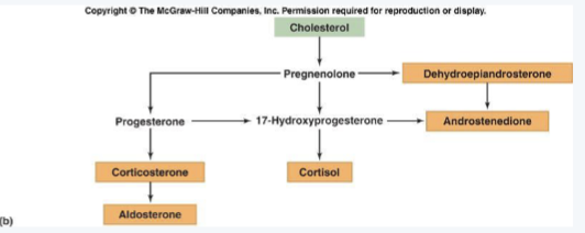
steroid synthesis in the adrenal cortex
the adrenal cortex uses different enzymes to convert cholesterol into steroid hormones
progesterone is a reproductive hormone and is also an intermediate in several steroid synthesis pathways
where are adrenal hormones released?
Aldosterone:
Regulates fluid balance and potassium.
Increased by angiotensin II (low blood volume/pressure) or high blood potassium.
Mainly controlled by the RAAS.
Cortisol:
Released in pulses throughout the day; highest around waking.
ACTH from the pituitary stimulates release, especially during stress.
Adrenal androgens:
Release regulated by ACTH.
DHEA and androstenedione can be converted into testosterone and oestrogens in other tissues.
Glossary:
RAAS: System that regulates blood pressure and fluid balance.
ACTH: Pituitary hormone that stimulates the adrenal cortex.
DHEA / androstenedione: Precursors to sex hormones.
what does cortisol do
glucose and tissues
increase liver glucose production (gluconeogenesis)
reduces glucose uptake in some tissues
promotes protein breakdown and mobilizes fat
stress response
helps maintain blood pressure responses to adrenaline
makes fuels available during illness, injury or other stress
immune regulation
regulates inflammatory signalling
higher therapeutic exposure strongly suppresses inflammation and immune-cell activity
how is cortisol secretion controlled?
The HPA axis (hypothalamic-pituitary-adrenal axis) controls cortisol release.
Hypothalamus: Releases CRH.
Anterior pituitary: CRH stimulates release of ACTH.
Adrenal cortex: ACTH stimulates cortisol production and release.
Negative feedback: Cortisol reduces further CRH and ACTH release.
Important: Aldosterone is controlled mainly by angiotensin II and blood potassium, NOT the HPA axis.
Glossary:
CRH: Hormone that stimulates ACTH release.
ACTH: Hormone that stimulates cortisol release.
Negative feedback: When cortisol levels rise, they signal the brain to reduce further cortisol production.

stress response: what is released and when?
rapid response
adrenal medulla
sympathetic nerves stimulate release of adrenaline and noradrenaline
releases within seconds during an acute threat or stressor
increases heart rate and supports an immediate response
hormonal response
adrenal cortex
pituitary ACTH stimulates the synthesis and release of cortisol
cortisol can rise within minutes of stress. Many effect on gene expression take longer
supports metabolism, blood pressure and regulation of inflammation
stress may be physical (injury) or psychological (anxiety)
cortiol changes throughout the day
cortisol follows a circadian rhythm (roughly 24-hour cycle), with levels usually highest around waking
levels are generally lower during the night, but stress can raise cortisol secretion
this daily pattern matters for cortisol tests and timing replacement therapy

how do glucocorticoids work?

what changes when glucocorticoids reduce inflammation?
fewer cytokines and other inflammatory mediators are produced
fewer immune cells are recruited and activated in affected tissues
swelling redness and irritation decreases as inflammation settles
online H1 antihistamines, glucocorticoids influences several inflammatory pathways
clinical like; topical hydrocortisone reduces inflammation and itching in eczema
where are corticosteroids used?
1. Local treatment (specific areas):
Nasal: Hay fever.
Inhaled: Asthma.
Topical hydrocortisone: Eczema (reduces redness and itching).
2. Systemic treatment (whole body):
- Oral or injected: Inflammatory bowel disease (IBD), rheumatoid arthritis and transplant-related immunosuppression.
3. Hormone replacement:
Hydrocortisone: Replaces cortisol in adrenal insufficiency.
Fludrocortisone: Replaces mineralocorticoids when needed.
Glossary:
Topical: Applied directly to the skin.
Systemic: Acts throughout the body.
Immunosuppression: Reduces immune system activity.
Adrenal insufficiency: When the adrenal glands don't produce enough hormones.
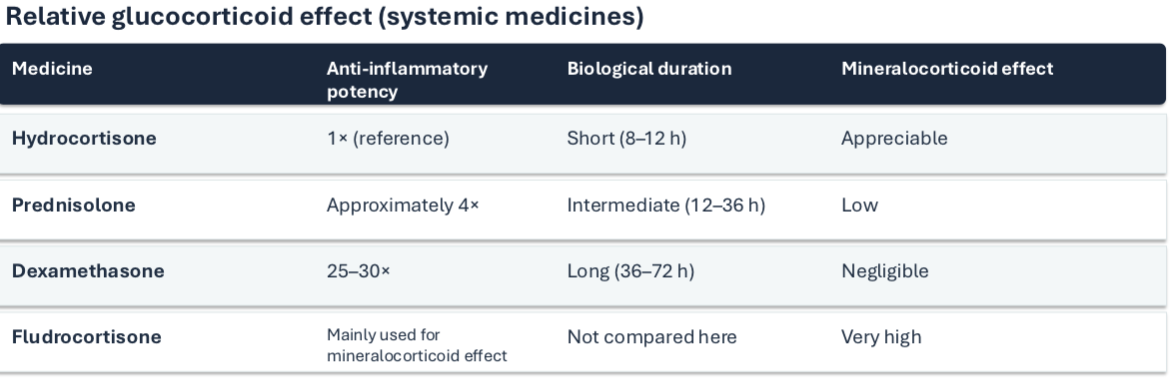
relative corticosteroid potency
corticosteroid adverse effects
risk rises with higher does, longer treatment and greater systemic exposure
metabolic and cardiovascular: increase blood glucose, fluid retention and blood pressure
immune and bone effects: increased infection risk, impaired wound healing and osteoporosis
sleep, mood and appearance can change. “cushingoid” means features such as facial rounding and central weight gain
local products have different risks (eg. nasal and nosebleeds"); substantial exposure can still have systemic effects
why you can’t always stop steroid suddenly
HPA suppression
prolonged systemic glucocorticoid exposure can reduce natural cortisol production
withdrawal risk
abrupt stoppage after relevant exposure can cause adrenal insufficiency
practical point
the need to taper depends on treatment, dose and duration; follow the individual plan
addisons’s disease
Primary adrenal insufficiency: Adrenal glands don't produce enough hormones.
Low cortisol and often low aldosterone.
Symptoms include:
Fatigue
Weight loss
Low blood pressure
Glossary:
- Primary adrenal insufficiency: Failure of the adrenal glands to produce enough hormones.
cushing’s syndrome
Caused by excessive glucocorticoid exposure.
Can result from the body's overproduction of cortisol or long-term use of corticosteroid medicines.
Glossary:
Glucocorticoids: Steroid hormones that regulate metabolism and stress responses.
Cortisol: Main glucocorticoid produced by the adrenal cortex.
Cushing’s disease
A specific cause of Cushing's syndrome.
Caused by an ACTH-secreting pituitary tumour.
Excess ACTH stimulates the adrenal cortex to produce too much cortisol.
Glossary:
ACTH: Hormone that stimulates cortisol release.
Pituitary tumour: Abnormal growth in the pituitary gland.
primary aldosteronism (Conn’s syndrome)
Caused by excess aldosterone production.
Causes:
High blood pressure.
Low potassium.
Metabolic alkalosis.
Glossary:
Aldosterone: Hormone that controls salt, water and potassium balance.
Alkalosis: When the blood becomes too alkaline (less acidic).
why does excess cortisol cause these clinical signs?
more glucose production → high blood glucose; diabetes can develop
protein breakdown → muscle wasting, thin and poor wound healing
altered fat metabolism → central weight gain and a rounded face
immune suppression → greater infection risk
altered bone metabolism _> bone loss and osteoporosis
allergic rhinitis: what are we treating?
typical symptoms:
repeated sneezing
itchy nose, throat or eyes
clear water nasal discharge
blocked nose, sometimes sleep disturbance
questions to ask:
when did it start, and what triggers it?
how severe are symptoms; how is sleep affected?
any wheeze, fever, unilateral symptoms or pain?
which medicines have already been tried?
what can the pharmacist recommend?
non-drug measures
reduces allergen exposure when practical
saline nasal rinse or spray
avoid smoke and other irritants
check pollen information and symptom patterns
drug treatments
second-generation oral H1 antihistamine
intranasal corticosteroid
intranasal antihistamine or combination spray
antihistamine eye drops for itchy eyes
short-term nasal decongestant for a blocked nose
comparing common hay fever treatments

choosing a starting treatment
mild/occasional
a suitable second-generation oral antihistamine or intranasal option may be reasonable
persistent/troublesome
an intranasal corticosteroid often offers better overall nasal control
severe/not controlled
check technique and adherence. consider a combined nasal steroid + antihistamine. if still uncontrolled, refer; selected patients may benefit form allergen immunotherapy
treatment choice to safe prescribing
choose the option
match oral H1 antihistamine or nasal treatment to symptoms and severity
check suitability
check age, pregnancy where relevant, other medicines, conditions and product licence.
explain safe use
agree a review