ANA Exam #3 Bulk Study
 Call Kai
 Learn
 Practice Test
 Spaced Repetition
 Match
 Flashcards
 Knowt Play
1/77

There's no tags or description
Looks like no tags are added yet.
78 Terms
Excitation-contraction coupling
for a skeletal muscle fiber to contract, its membrane must first be excited (stimulated to fire an action potential)

Muscle Fiber Contraction
(image)
Acetylcholine (ACh)
neurotransmitter released at the NMJ
Triad
arrangement of a T-tubule with SR membranes on either side
Step 1
action potential arrives at neuromuscular junction
Step 2
ACh is released, binds to receptors and opens sodium ion channels, leading to an action potential in sarcolemma.
Step 3
action potentials travel along T-tubules
Step 4
thick and thin filament interaction leads to muscle contraction
muscle cells =
muscle fibers
Epimysium
muscle covering/fascia; connects to tendons, bones

Perimysium
covers Fascicles; blood vessels and nerves

All CT layers in muscles
loose CT, usually Reticular; all layers are connected (continuous)
Origin
connection of a muscle to the skeletal system; less moveable side; usually proximal
Insertion
Connection of a muscle to the skeletal system; more moveable side; usually distal
Direct attachments
Epimysium attaches directly to bone or cartilage; no tendon or aponeurosis; ex. brachialis, Gluteus Medius and Minimus origins
Indirect attachments
Epimysium connects to CT, then to bone; very common; stronger, can stretch, smaller to cross over joint; ex. insertions of gluteus medius and minimus, top of skull (aponeurosis, broad + flat)
Retinacula
Specialized structure of strong ligaments that holds tendons down; tendons would lift up (wrist) or slip out (ankle) without them.

Endomysium
covers muscle fibers
Synergists
muscles that work together to produce the same movement; ex. 4 quadriceps muscles → knee extension
Antagonists
opposite motions; muscles that work together to accelerate/decelerate; ex. biceps vs. triceps(elbow flexion/extension), hamstrings vs. quadriceps(knee flexion/extension)
Prime movers
the strongest/most important player in a group of synergists; ex. gluteus maximus (greatest extensor), rectus femoris (knee extension)
Fixator
Stabilizes a joint instead of producing movement; ex. gluteus medius contracting while standing on one foot
Circular fascicle arrangement
locations: external openings, ex. sphincters, facial muscles
Convergent fascicle arrangement
Broad origin, converging toward a single tendon
locations: pectoralis major, latissimus dorsi
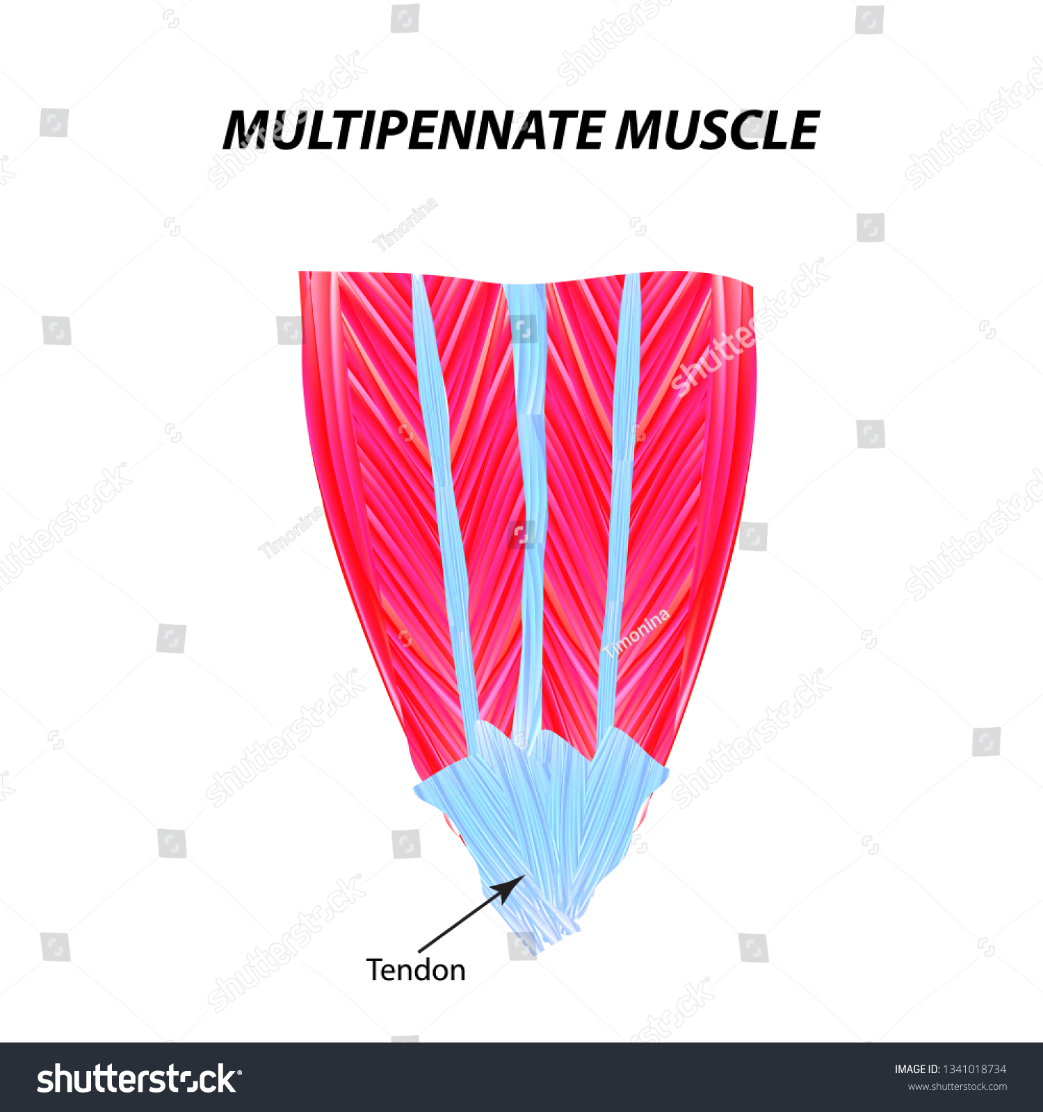
Pennate
fibers branch off a long central tendon
purpose: strength due to multiple angles of pull on same tendon; does not shorten much
Unipennate
fibers branch off in one direction; ex. extensor digitorum longus

Bipennate
fibers branch off in 2 directions; ex. rectus femoris

multipennate
fibers branch off in multiple directions; ex. deltoid
Parallel
very long muscle fibers, can shorten well; not as strong as pennate muscles, less fibers in contact with tendons; ex. Sartorius

Fusiform
similar to parallel, but with enlarged central portion; can produce more force than parallel; ex. biceps brachii

Functional groups of muscles
synergists, antagonists, prime movers, fixators
Fascicles
Covered by Perimysium, together make up a muscle

Muscle fiber (muscle cell)
Covered by Endomysium; together make up a fascicle.
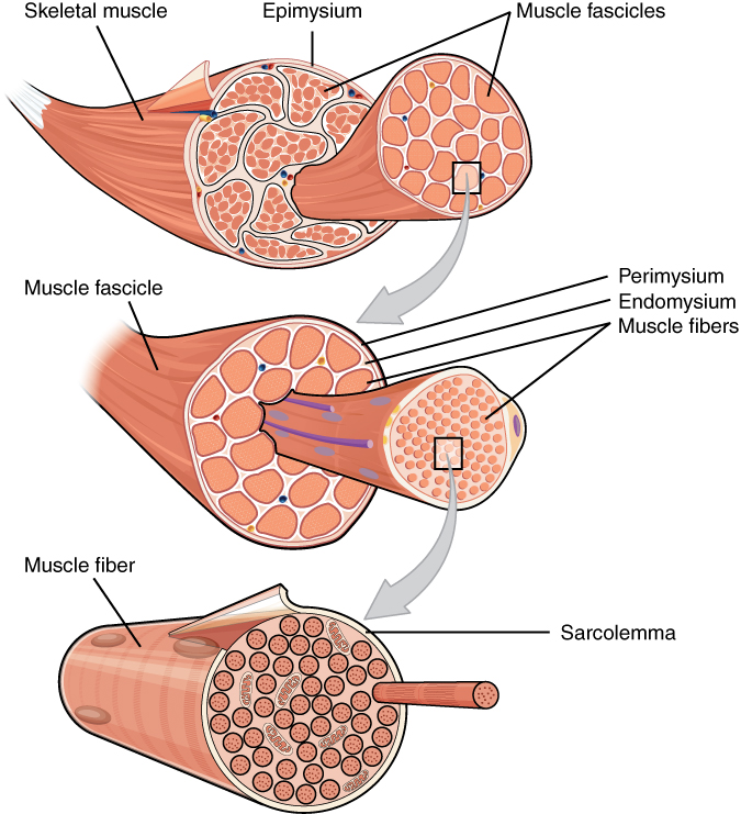
Sarcolemma
cell membrane; found under the endomysium of muscle fibers.

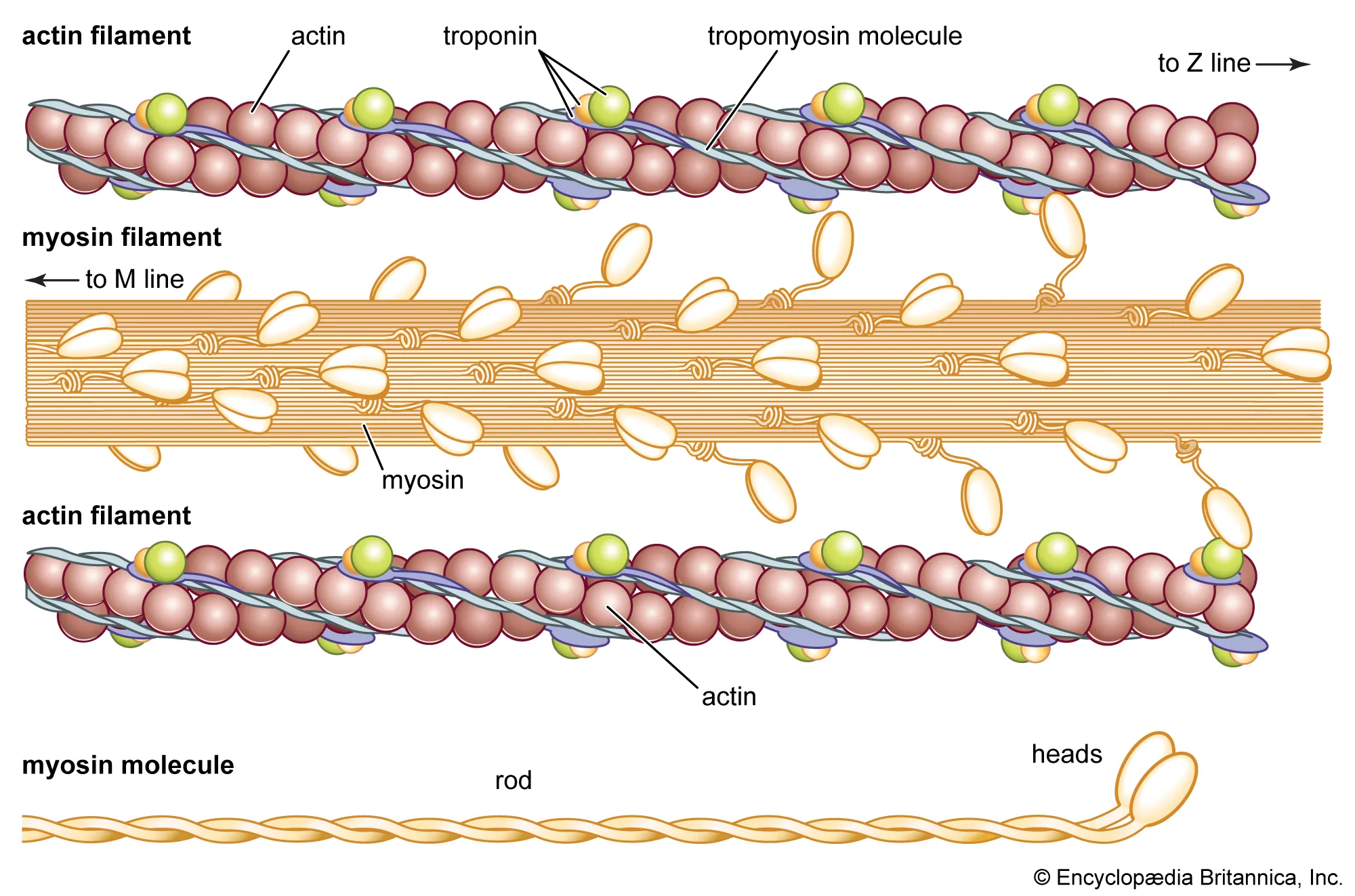
Myofilaments
together make up sarcomeres; contain protein fibers actin and myosin.

Muscles of the Gluteal Region
location: posterior hip
general actions: hip extension, abduction, rotation (medial and lateral), and stabilizing pelvis during locomotion (lifting of one limb when walking or running, etc.)
names: gluteus maximus, gluteus medius, gluteus minimus, tensor fasciae latae, (IT Band, not a muscle but in this region)
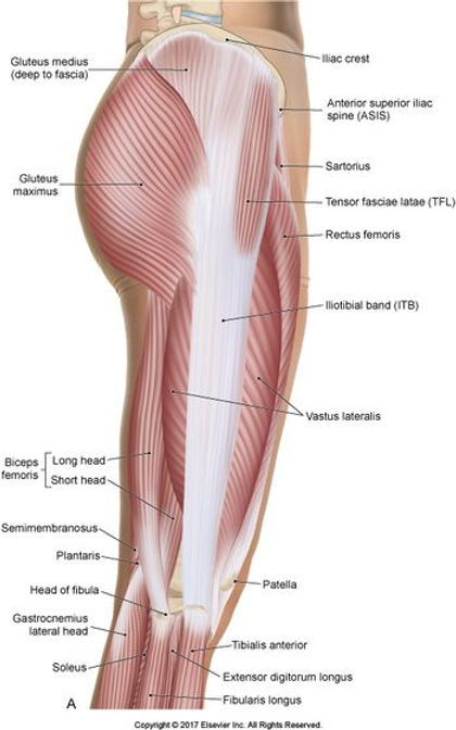
Illiotibial tract (IT band)
Thick CT, joins with fascia latae to affect the knee, provides partial insertion point for gluteus maximus; crosses hip and knee joint
Gluteus Maximus origin and insertion
Origin: posterior ilium, sacrum, and coccyx
Insertion: gluteal tuberosity, IT band
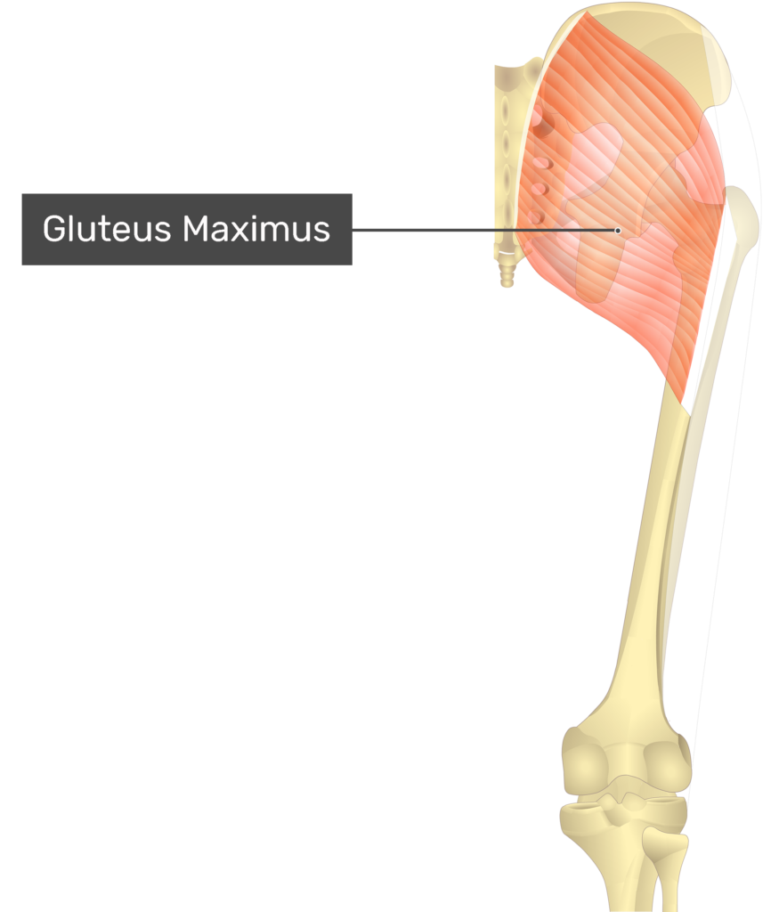
Gluteus Maximus actions
Primary = Hip extension (from a place of flexion)
Secondary = hip abduction, lateral/external rotation of hip
Crosses hip joint
Gluteus Medius origin/insertion
origin: Posteriur Ilium, below Iliac crest
Insertion: Greater trochanter of the Femur
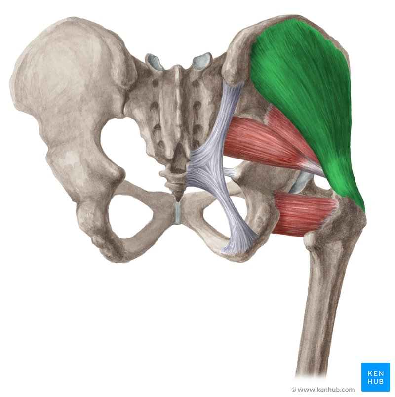
Gluteus Medius actions
(same as minimus) Hip abduction, pelvis stabilization while walking (Trendelenburg’s sign), internal/medial hip rotation
crosses hip joint
Gluteus minimus origin/insertion
Origin: ilium, slightly inferior to medius site
Insertion: greater trochanter of the femur (same as medius)
Gluteus minimus actions
(same as medius) Hip abduction, pelvis stabilization during walking, internal/medial hip rotation
crosses hip joint
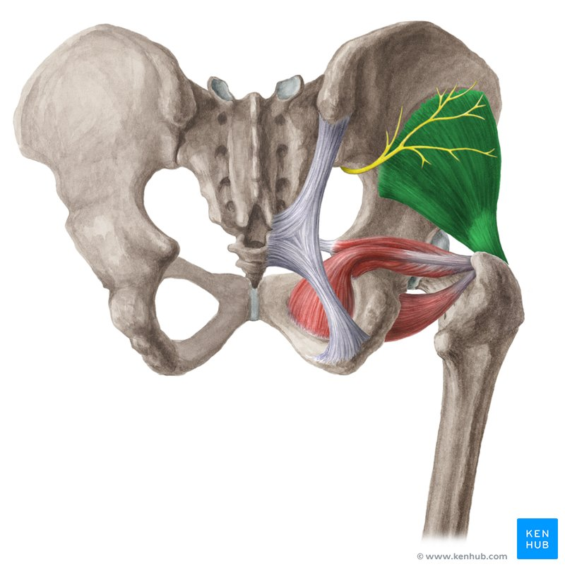
Tensor Fasciae Latae origin/insertion
Origin: Anterior superior iliac spine and Anterior iliac crest
Insertion: IT band

Tensor Fasciae Latae actions
locks knee into extension by tightening the IT tract, hip abduction, hip medial/internal rotation
Crosses: directly hip joint, by extension of IT band, knee joint

Anterior thigh muscles overview
Innervation: femoral nerve
Actions: knee extension, hip flexion

Posterior thigh muscles (AKA hamstrings) overview
3 muscles (medial to lateral): semimembranosus, semitendinosus, biceps femoris
innervation: sciatic nerve
Actions: knee flexion, hip extension
Crosses: hip and knee joints
Tight hamstrings = difficulty bending over, touching toes, walking, etc.; leads to back problems

Medial thigh muscles overview
hip stabilizers and adductors
Pectineus, adductor longus, gracilis, adductor brevis, adductor magnus
innervation: obturator nerve
Action: hip adduction

Iliopsoas (anterior thigh muscle)
made up of 2 muscles (iliacus, psoas major)
origin: T12-L5, Iliac fossa, lateral sacrum
insertion: lesser trochanter of femur
action: primary hip flexor
crosses: hip joint

Quadriceps Femoris (anterior thigh muscles) (origin/insertion)
4 headed muscle(RF, VL, VI, VM); most powerful in the body; quad-hamstring ratio (quads typically stronger than hams)
Origins: RF → at iliac/hip; Vl, VI, VM → at femur
Insertion: patella (tibial tuberosity)
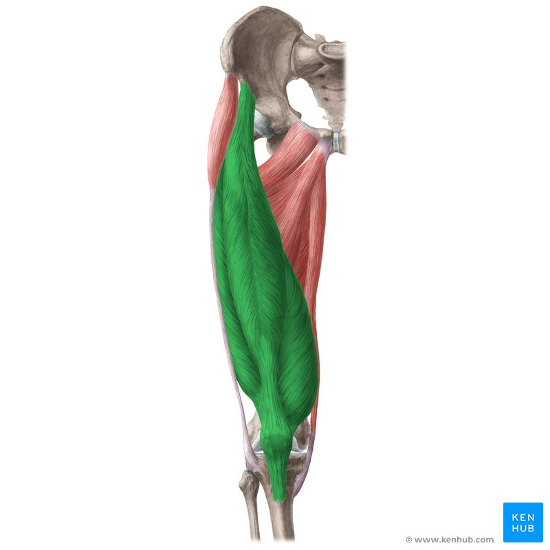
Quadriceps Femoris actions
RF: Hip flexion, kne extension (ex. soccer kick)
VL (most powerful of the V’s): knee extension, knee stabilization
VI: knee extension
VM: knee extension, knee stabilization
VL & VM work together to keep Patella tracking (within its natural groove)

Sartorius (anterior thigh muscle)
longest muscle in the body
Origin: ASIS
Insertion: Proximal-medial tibia (pes anserine)
Actions: hip flexion, hip abduction, lateral/external rotation of hip
Crosses: hip joint primarily

Biceps Femoris (origin/insertion/actions)
has 2 separate heads (“bi”), lateral most hamstring
Short head origin: linea aspera of femur (doesn’t cross hip like other hamstrings)
Long head origin: Ischial tuberosity
Insertion: lateral condyle of tibia and head of fibula
Actions: knee flexion, hip extension, external rotation of the hip
Crosses: knee and hip
Semimembranosus (origin/insertion/actions)
Origin: ischial tuberosity
Insertion: medial condyle of Tibia
Actions: hip extension, knee flexion, internal/medial hip rotation (same as tend.)
Crosses: knee and hip joints


Semitendinosus
Origin: Ischial tuberosity (like memb.)
Insertion: medial, proximal tibia (pes anserine)
Actions: hip extension, knee flexion, internal/medial hip rotation (same as memb.)
crosses: knee and hip joints

Pectineus
innervated by femoral nerve
Origin: superior anterior edge of pubis
Insertion: inferior to lesser trochanter (posterior)
Actions: hip flexion, hip adduction
Crosses: hip joint

Adductor Longus
origin: near pubic symphysis
Insertion: linea aspera (posterior femur)
Action: adduction of hip
Crosses: hip joint

Gracilis
2nd longest muscle
Origin: inferior ramus of pubis, medial to adductor longus
Insertion: pes anserine
Action: hip adduction, knee flexion
Crosses: hip and knee joint

Adductor Brevis
Origin: inferior ramus of pubis (like Gracilis)
Insertion: linea aspera
Action: hip adduction
Crosses: hip joint

Adductor Magnus
(deep to brevis + longus)
Origin: ischial tuberosity
Insertion: linea aspera
Action: powerful hip adduction (ex. soccer kick)
crosses: hip joint
Shoulder joint complex
articulations: glenohumeral joint, sternoclavicular joint, acromioclavicular joint, and scapulothoracic joint
bones: scapula, humerus, sternum, clavicle

Glenohumeral joint

sternoclavicular joint

Acromioclavicular joint

Scapulothoracic joint

Elbow joint complex
articulations: humeroulnar, humeroradial, proximal radialulnar joint
bones: humerus, ulna, radius

proximal radialulnar joint

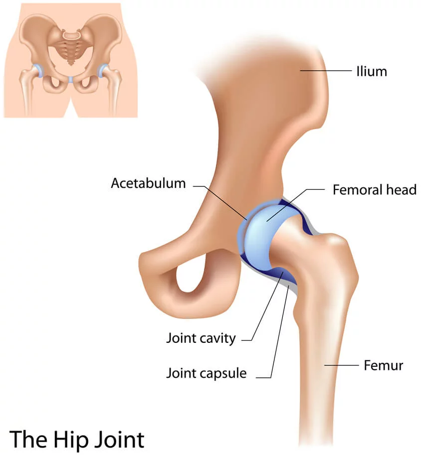
Hip Joint
bones: femur, pelvis (os coxa)
Knee Joint complex
articulations: femur-tibia, fibula-tibia, patella-femur
bones: Tibia, fibula, patella, femur
Fibrocartilage: medial and lateral menisci

Ankle joint complex
articulations: Talo-crural joint, Subtalar joint (talus-calcaneus)
bones: tibia, fibula, talus, calcaneus

subtalar joint

Muscle fiber
a long, thread-like, multinucleated cell filled with smaller subunits called myofibrils, which contain the protein filaments actin and myosin responsible for contraction

Fascicle
bundles of muscle fibers

myofibril
a long, rod-like organelle inside muscle cells that causes muscles to contract

sarcomere
the basic functional and contractile unit of striated muscle tissue

actin
tiny, flexible protein fibers inside cells that provide structural support and help the cell move; forms the thin filaments

myosin
a primary motor protein that forms the thick filaments within muscle fiber sarcomeres